Wed mammary gland code for mcb 10. Breast cancer. Causes, symptoms and treatment. Classification of breast cancer
RCHD (Republican Center for Health Development of the Ministry of Health of the Republic of Kazakhstan)
Version: Archive - Clinical Protocols of the Ministry of Health of the Republic of Kazakhstan - 2007 (Order No. 764)
Mammary gland, part unspecified (C50.9)
general information
Short description
The most common tumor in women, belonging to the classic hormone-mediated oncological diseases; develops in an organ that is part of the reproductive system of the body. These tumors come from epithelial tissue ducts or lobules of the mammary gland - "target" for hormones produced by the ovaries (estrogens and progestins).
On average, about 3,000 breast cancer patients are diagnosed annually in the Republic of Kazakhstan, of which more than 1,380 women die. In particular, in 2005, 2954 cases of breast cancer were registered, which amounted to 19.5 (32.3 in Almaty) per 100,000 population. Mortality at 1 year of life is 10.8%, and 5-year survival is 49.3%.
Protocol code: H-S-008 "Malignant neoplasms of the breast. Breast cancer"
Profile: surgical
Stage: hospital
Code (codes) according to ICD-10:C50 Malignant neoplasm of breast
Classification
Histological classification of breast tumors
It is now customary to use the histological classification of the International Cancer Union (2002, 6th edition).
| A | Non-invasive cancer (in situ): |
| Intraductal (intracanalicular) cancer in situ | |
| Lobular (lobular) cancer in situ | |
| IN | Invasive cancer (infiltrating carcinoma): |
| ductal | |
| Lobular | |
| Mucous (mucinous) | |
| Medullary (cerebral) | |
| tubular | |
| Apocrine | |
|
Other forms (papillary, squamous, juvenile, spindle cell, pseudosarcomatous, etc.) |
|
| WITH | Special (anatomical and clinical) forms: |
| Paget's cancer | |
| Inflammatory cancer |
Most often, patients have invasive ductal cancer (50-70%), then lobular (20%). Ductal cancer is characterized by more frequent spread through the milk ducts, and lobular cancer is characterized by primary multiplicity and bilaterality.
INTERNATIONAL TNM CLASSIFICATION
Currently, the classification of tumors according to the TNM system of the international anticancer union (2002) is used. The stage of cancer is established during the initial examination of the patient, and then specified after the operation (pTNM).
The classification refers only to carcinomas and applies to both the male breast and the female breast.
In the case of primary multiple synchronous tumors in one breast, the tumor with the highest
category T. Synchronous bilateral breast tumors should be classified independently of each other to allow separation of cases by histological type.
The following methods shall be used to evaluate categories T, N and M:
Anatomical areas:
1. Nipple (C 50.0).
2. Central part (C 50.1).
3. Upper inner quadrant (C 50.2).
4. Lower inner quadrant (C 50.3).
5. Upper outer quadrant (C 50.4).
6. Inferior outer quadrant (C 50.5).
7. Axillary tail (C 50.6).
Regional lymph nodes:
1. Axillary (ipsilateral), interthoracic nodes (Rotter) and lymph nodes along the axillary vein and its branches which can be divided into the following levels:
Level I (lower armpit): lymph nodes located lateral to the lateral border of the pectoralis minor muscle;
Level II ( middle part axillary fossa): lymph nodes located between the medial and lateral border of the pectoralis minor muscle, and interthoracic lymph nodes (Rotter);
Level III (apical axilla): apical lymph nodes and nodes located medial to the medial border of the pectoralis minor muscle, excluding those defined as subclavian.
Note. Intramammary lymph nodes are coded as axillary lymph nodes.
2. Subclavian (ipsilateral) lymph nodes.
3. Intramammary (ipsilateral) lymph nodes: lymph nodes in the intercostal areas along the edge of the sternum in the endothoracic fascia.
4. Supraclavicular (ipsilateral) lymph nodes.
Metastases in any other lymph nodes are defined as distant metastases (M1), including cervical or contralateral intramammary lymph nodes.
Under the symbols TNM means: T - primary tumor.
| Tx | Insufficient data to evaluate the primary tumor. |
| T0 | The tumor in the mammary gland is not defined. |
| Tis |
Pre-invasive carcinoma (carcinoma in situ) Tis (DCIS) - ductal carcinoma in situ Tis (LCIS) - lobular carcinoma in situ Tis (Paget) - Paget's disease (nipple) without tumor Note: Paget's disease with a tumor is classified in according to tumor size. |
| T1 | Tumor less than 2 cm in greatest dimension |
| T1mic |
Microinvasion up to 0.1 cm in greatest dimension Note: Microinvasion is defined as the spread of cancer cells over basement membrane limits with lesions less than 0.1 cm If the foci of microinvasion are multiple, the largest one is classified according tothe size of the focus (it is impossible to summarize the sizes of microfoci) Availability multiple foci of microinvasion should be noted additionally |
| T1a | Tumor larger than 0.1 cm but not larger than 0.5 cm in greatest dimension |
| T1b | Tumor larger than 0.5 cm but less than 1 cm in greatest dimension |
| T1s | Tumor larger than 1 cm but not larger than 2 cm in greatest dimension |
| T2 | Tumor more than 2 cm but not more than 5 cm in greatest dimension |
| T3 | Tumor more than 5 cm in greatest dimension |
| T4 |
Tumor of any size with direct extension to the chest wall or skin Note: The chest wall includes the ribs, intercostal muscles, and anteriorserratus muscle, but not pectoralis muscle |
| T4a | Spread to the chest wall |
| T4b |
Swelling (including "lemon peel"), or ulceration of the skin of the breast,or satellites in breast skin |
| T4c | Features listed in 4a and 4b together |
| Т4d | Inflammatory form of breast cancer |
Note: Inflammatory breast carcinoma is characterized by diffuse brown skin induration with an erysipeloid margin, usually without an underlying mass. If a skin biopsy indicates no involvement and no localized, sizable primary cancer, category T is pTX in histopathological staging of inflammatory carcinoma (T4d).
Dimpled skin, nipple indrawing, or other skin changes other than those seen in T4b and T4d may be scored as T1, T2, or T3 without affecting classification.
N - regional The lymph nodes.
| NX | Insufficient data to assess the status of regional lymph nodes |
| N0 | No evidence of metastatic involvement of regional lymph nodes |
| N1 |
Metastases in displaced axillary lymph nodes (e) on the side defeat |
|
N2 N2a N2b |
Metastasis in immobile ipsilateral axillary lymph node (ax) or in a clinically obvious ipsilateral intramammary lymph node(s) In the absence of clinically obvious metastases in the axillary lymph nodes metastasis to axillary lymph node(s) linked to each other or to other structures Metastasis to clinically apparent intramammary lymph node(s) only, with absence of clinically obvious metastasis in the axillary lymph node |
| N3 |
Metastasis in the ipsilateral subclavian lymph node(s) withdamage to the axillary lymph nodes or without them; or in a clinically obvious ipsilateral intramammary lymph node(s) in the presence of clinically obvious metastases in the axillary lymph nodes; or metastasis in the ipsilateralsupraclavicular lymph node(s) with or without axillary or intramammary lymph node involvement |
|
N3a N3b N3c |
Metastasis in subclavian lymph node(s) Metastases in intramammary and axillary lymph nodes |
|
Note. "Clinically overt" means identified as a result of clinical trial or imaging (for except for lymphoscintigraphy) |
M - distant metastases.
pTNM pathohistological classification.
pT - primary tumor.
Histopathological classification requires examination of the primary carcinoma, in the absence of macroscopic tumor at the resection margins. A case can be classified as pT if there is only a microscopic tumor along the edge.
Note. When classifying pT, the size of the tumor is the value of the invasive component. If there is a large in situ component (eg 4 cm) and a small invasive component (eg 0.5 cm), the tumor is classified as pT1a.
pN - regional lymph nodes.
For histopathological classification, examination of one or more sentinel lymph nodes may be undertaken. If the classification is based only on sentinel node biopsy without subsequent axillary node dissection, then it should be designated (sn) (sentinel node - sentinel node), for example: pN1 (sn).
| рN1mi | Micrometastasis (greater than 0.2 mm but not greater than 2 mm in greatest dimension) |
| PN1 |
Metastases in 1-3 ipsilateral axillary lymph nodes (e) and/or ipsilateral intramammary nodes with microscopic metastasesdetected by sentinel lymph node dissection, but not clinically evident |
| pN1a |
Metastases in 1-3 axillary lymph nodes (e), among them at leastone over 2 mm in greatest dimension |
|
pN1b pN1c |
Intramammary lymph nodes with microscopic metastases, identified as a result of sentinel lymph node dissection, but clinically not explicit Metastases in 1-3 axillary lymph nodes and intramammary lymph nodes with microscopic metastases identified by dissectionsentinel lymph node, but not clinically obvious |
| pN2 |
Metastases in 4-9 ipsilateral axillary lymph nodes or clinically obvious ipsilateral intramammary lymph nodes, with |
|
Note. "Clinically non-obvious" means not identified by clinical investigation or imaging (other thanlymphoscintigraphy); "clinically evident" means determined by clinical examination or imaging (excluding lymphoscintigraphy), or macroscopically visual. |
|
| pN2a |
Metastases in 4-9 axillary lymph nodes, among them at least one larger than 2 mm |
| pN2b |
Metastasis to clinically apparent intramammary lymph node(s) absence of metastases in the axillary lymph nodes |
| pN3 |
Metastases in 10 or more ipsilateral axillary lymph nodes; or in ipsilateral subclavian lymph nodes; or in clinically obvious ipsilateral intramammary lymph nodes, in the presence of one or more affected axillary lymph nodes; or in more than 3 axillary lymph nodes with clinically non-obvious microscopic metastases in intramammary lymph nodes; or in ipsilateral supraclavicular lymph nodes |
| pN3a |
Metastases in 10 or more axillary lymph nodes (at least one of which more than 2 mm) or metastases in the subclavian lymph nodes |
| pN3b |
Metastasis to clinically apparent intramammary lymph node(s) if presentaffected axillary lymph node(s); or metastases in more than 3 axillary lymph nodes and intramammary lymph nodes with microscopic metastasis revealed during dissection of the sentinel lymph node, but not clinically obvious |
| pN3c | Metastasis in supraclavicular lymph node(s) |
pM - distant metastases. The rM categories correspond to the M categories.
G histopathological classification
G1- high degree differentiation.
G2 - average degree of differentiation.
G3 - low degree of differentiation.
R classification
The absence or presence of residual tumor after treatment is described by the symbol R. R classification definitions:
RX - the presence of a residual tumor cannot be established.
R0 - no residual tumor.
R1 - microscopic residual tumor.
R2 - macroscopic residual tumor.
Grouping by stages
| Stage 0 | TiS | N0 | M0 |
| Stage I | T1* | N0 | M0 |
| Stage IIA | T0 | N1 | M0 |
| T1* | N1 | M0 | |
| T2 | N0 | M0 | |
| Stage IIB | T2 | N1 | M0 |
| T3 | N0 | M0 | |
| Stage IIIA | T0 | N2 | M0 |
| T1* | N2 | M0 | |
| T2 | N2 | M0 | |
| T3 | N1, N2 | M0 | |
| Stage IIIB | T4 | N0, N1, N2 | M0 |
| Stage IIIC | any T | N3 | M0 |
| Stage IV | any T | any N | M1 |
Note. *T1 includes T1mic (microinvasion 0.1 cm or less in greatest dimension).
|
Tis T1mic T1a T1b T1c T4a T4b T4d |
in situ £2cm £0.1cm > 0.1 to 0.5 cm > 0.5 to 1 cm > 1 to 2 cm > 2 to 5 cm > 5 cm Chest wall/skin chest wall Skin edema/ulceration, satellite nodules on the skin Features characteristic of T4a and T4b Inflammatory carcinoma |
| N1 |
Movable axillary |
pN1mi pN1a pN1b pN1c |
Micrometastases, > 0.2 mm £ 2 mm 1-3 Axillary nodes Intramammary nodes with micrometastasis, detected on sentinel node biopsy, but clinically undetectable 1-3 Axillary nodes and intramammary nodules with micrometastasis detected by sentinel node biopsy, but clinically undetectable |
| N2a |
motionless axillary |
pN2a | 4-9 Axillary knots |
| N2b |
Intramammary- clinically defined |
pN2b |
determined without axillary nodes |
| N3a | Subclavian | pN3a |
³ 10 Axillary nodes or subclavian node(s) |
| N3b |
Intramammary- nye and axillary nye |
pN3b |
Intramammary nodes, clinically defined with axillary node(s) or> 3 axillary nodes and intramammary nodes with micrometastases, which are detected with a biopsy of the sentinel (sentinel node), but clinically undetectable |
| N3c | Supraclavicular | pN3c | Supraclavicular |
Factors and risk groups
Classification of risk factors
1. Factors characterizing the functioning of the reproductive system of the body:
menstrual function;
sexual function;
childbearing function;
lactation function;
2. Hyperplastic and inflammatory diseases of the ovaries and uterus.
Endocrine-metabolic factors due to concomitant and previous diseases:
1. Obesity.
2. Hypertension.
3. Diabetes.
4. Liver disease.
5. Atherosclerosis.
6. Diseases of the thyroid gland.
7. Dishormonal hyperplasia of the mammary glands.
Genetic factors(carriers of BRCA-1 or BRCA-2 genes):
1. Breast cancer in blood relatives (hereditary and "family" breast cancer).
2. Milk-ovarian syndrome (breast cancer and ovarian cancer in the family).
Exogenous factors:
1. Ionizing radiation.
2. Chemical carcinogens, including smoking.
3. Excess consumption of animal fats, high-calorie diet.
4. Viruses.
5. Taking hormones.
Diagnostics
Diagnostic criteria
Complaints(no pathognomonic symptoms characteristic of breast cancer).
There may be complaints about the presence of education in the mammary glands, hyperemia, edema, wrinkling, retraction or protrusion on it, narrowing of the areolar field, etc.
Anamnesis: the presence of oncological diseases in close relatives, the onset of menstruation, the age of the first pregnancy and first birth, the use of OK or HRT, gynecological diseases.
Physical examination
1. Examination of the mammary glands.
On examination, determine:
The symmetry of the location and shape of the mammary glands;
The level of standing of the nipples and their appearance (retraction, deviation to the side);
Skin condition (hyperemia, edema, wrinkling, retractions or protrusions on it, narrowing of the areolar field, etc.);
Pathological discharge from the nipple (quantity, color, duration);
The presence of swelling of the hand on the side of the lesion.
2. Palpation of the mammary glands (in vertical and horizontal positions).
3. Palpation of regional and cervical-supraclavicular lymph nodes (usually performed in an upright position).
Laboratory research
Laboratory studies that must be performed during the initial treatment of the patient before the start of treatment: general analysis blood, blood type, Rh-factor, urinalysis, biochemical analysis blood (urea, bilirubin, glucose), RW (Wassermann reaction), coagulogram, ECG (electrocardiography).
Instrumental Research
X-ray diagnosis is one of the leading methods for detecting breast cancer, especially if the tumor is small and not palpable. Mammography is indicated for all patients with breast cancer.
Examination methods that must be performed by the patient before starting treatment:
1. Puncture biopsy of the tumor with cytological examination or trephine biopsy with determination of the level of expression of ER, PR, Her-2/neu and other genetic factors.
2. Ultrasound examination of the abdominal organs.
3. X-ray examination of the lungs.
4. Osteoscintigraphy (in institutions equipped with a radioisotope laboratory).
5. Ultrasound examination of the mammary glands, regional lymph nodes.
Mammography and ultrasound complement each other, because. mammography may show tumors that are not detected by ultrasound, and vice versa.
Morphological diagnostics:
1. Cytological (puncture) biopsy (fine-needle biopsy).
2. Trephine biopsy or sectoral resection of the mammary gland.
Indications for expert advice.
Required: consultation with a gynecologist.
If necessary, consultation with an endocrinologist, neuropathologist, urologist, radiologist, chemotherapist, and other related specialists according to indications.
List of main diagnostic measures:
1. Determination of hemoglobin.
2. Leukocyte count in Goryaev's chamber.
3. Erythrocyte count for CPK.
4. Determination of ESR.
5. Hematocrit.
6. Calculation of the leukocyte formula.
7. General analysis of urine.
8. Determination of total protein.
9. Cytological examination and histological examination of tissue.
10. Determination of the clotting time of capillary blood.
11. Platelet count.
12. Blood test for HIV.
13. Microreaction.
14. HbsAg, Anti-HCV.
15. Determination of protein fractions.
16. Determination of bilirubin.
17. Coagulogram 1 (prothrombin time, fibrinogen, thrombin time, aPTT, plasma fibrinolytic activity, hematocrit.
18. Determination of residual nitrogen.
19. Determination of glucose.
20. Definition of ALT.
21. Definition of AST.
22. Thymol test.
23. Determination of blood group and Rh factor.
24. Ultrasound of the abdominal organs.
25. Electrocardiography.
26. X-ray of the chest in two projections.
27. Ultrasound of the mammary glands.
28. Mammography.
29. Ductography.
30. Ultrasound of the pelvic organs.
31. Magnetic resonance imaging (MRI) of the breast.
32. Computed tomography (CT) of the breast.
List of additional diagnostic measures:
1. Consultation with a cardiologist.
Differential Diagnosis
| Complaints |
physical data |
ultrasound, mammography |
Morphologicallye signs |
|
| breast cancer |
Availability of education in breast, hyperemia, edema, rugosity, protrusions on it, |
On examination, the presence On palpation the presence of a tumor in the breast, increased regional lymph nodes |
Availability education in dairy gland, calcifications, increase regional lymph nodes |
Presence of cells smear tumors. Conclusion pathologist about availability malignant tumors |
|
Inflammatory breast disease |
Hyperemia, hyperthermia, pain in breast gland, purulent discharge |
On examination, hyperemia On palpation the presence of painful seals in the breast, possible reactive enlarged sometimes lymph nodes |
The presence of a cavity liquid content without clear boundaries |
Availability elements purulent inflammation, leukocytes, neutrophils macrophages, fibroblasts in smears. Histologically - abscess picture, purulent infiltration |
|
Fibroadenoma, cystadenoma MF, localized fibroadenoma toz |
Availability of education in mammary gland, pain |
On examination it is possible MF deformation. the presence of a seal in MF |
Availability education with clear contours, with mammography - the presence of a "rim security" |
The presence of peri- intracanal- cular and mixed fibroadenomas |
|
Cyst mammary gland |
The presence of soft elastic mammary gland, pain, discharge from the nipple |
On examination possible deformation MF. On palpation availability of education soft-elastic consistency in MF |
Availability cavities with liquid content with clear contours |
Presence of a wall cysts, fluid content |
Treatment abroad
Get treatment in Korea, Israel, Germany, USA
Get advice on medical tourism
Treatment
Treatment tactics
Treatment goals: achievement of radicalism of treatment.
Non-drug treatment
Breast cancer is one of the few oncological diseases in which the treatment of all stages is multivariate.
Despite significant progress in the development of new methods for the treatment of breast cancer, surgical intervention is still the main, and in some cases the only method of treating this disease (Ca in situ).
The choice of one or another type of radical operation is determined not only by the degree of spread of the tumor process, but also clinical form, tumor localization, age of patients and some other factors characterizing their general condition.
Recently, increasing importance has been attached to improving the quality of life, which is achieved by performing organ-preserving operations on the mammary gland, as well as reconstructive and restorative operations using local tissues.
Organ-preserving operations for breast cancer provide, along with high rates survival, good cosmetic and functional results. Social and labor rehabilitation of patients after segmental resection of the mammary gland is faster than after mastectomy.
Indications for performing organ-preserving operations on the mammary gland:
The presence of a nodular form of cancer up to 2.5 cm in size;
Lack of multicentricity and multifocality of tumor growth (on mammograms, ultrasound, clinically);
Slow and moderate growth rates, doubling the size of the tumor no faster than 3 months (according to the anamnesis);
Favorable ratio of the size of the breast and tumor to obtain a good cosmetic result of the operation;
Absence of distant metastases;
The presence of single metastases in the axillary region is acceptable;
Reconstructive and restorative operations can be performed with I-III stages Breast cancer at the request of the patient with any localization of the tumor.
A woman should be familiar with all types of surgical interventions.
Radiation therapy technique
Radiation therapy of the mammary gland and areas of regional metastasis (supraclavicular, axillary) is carried out by bremsstrahlung of the accelerator (6 MeV) or on gamma-therapeutic devices (1.25 MeV), and the parasternal zone - by alternating photon and electron beams or only by electron radiation up to 20 MeV depending on the depth of the chain of parasternal lymph nodes.
Irradiation of the parasternal zone with 60C o or only with a photon beam with an energy of more than 4 MeV is fraught with the development of post-radiation pulmonitis, mediastinitis, and pericarditis. Preoperative radiotherapy in many scientific centers the world is not carried out, with the exception of locally advanced breast cancer resistant to neoadjuvant chemotherapy and endocrine therapy.
Postoperative irradiation of the anterior chest wall after mastectomy or irradiation of the remaining mammary gland after radical resection is carried out with a 1.25 MeV or 6 MeV photon beam from tangential fields directed so that no more than 2 cm of lung tissue enters the 100% isodose zone.
tangential fields. Borders:
1. Upper - the level of the sternoclavicular joint (Louis angle); if necessary, the upper limit may be placed higher to include the entire mammary gland.
2. Medial - along the middle of the sternum.
3. Lower - 2 cm below the submammary (transitional) fold.
4. Lateral - 2 cm lateral to the palpable breast tissue, usually along the midaxillary line.
IN postoperative period after mastectomy, the boundaries of the tangential fields are as follows:
1. Upper - the corner of Louis.
2. Medial - midline of the body.
3. Lower - at the level of the submammary fold of the opposite gland.
4. Lateral - middle axillary line.
With atypical localization of the postoperative scar and its location outside the designated boundaries of the irradiation fields, it is recommended to additionally irradiate the scar zone with tissue capture at least 2 cm beyond it. Such irradiation should be carried out with an electron beam or with the help of contact radiation therapy.
supraclavicular field.
Irradiation of the supraclavicular and axillary lymph nodes occurs from the anterior field and the beam is tilted 10-150 to the side of the same name in order to avoid irradiation of the esophagus and trachea.
The upper edge of the field is at the level of the upper edge of the cricothyroid recess.
medial border- the middle of the sternum.
Lateral border - the medial edge of the head of the shoulder; if it is necessary to irradiate the entire armpit, the lateral border should be expanded to the lateral edge of the head of the shoulder, which should be covered with a protective block.
The lower border is in contact with the upper border of the tangential field at the level of attachment of the second rib to the sternum (Louis angle).
The larynx, esophagus, and trachea are always protected with a lead block.
The posterior axillary field is used when it is necessary to irradiate the entire axillary zone.
The medial border of the field is located 1 cm medially from the edge of the chest.
The upper border is the upper edge of the clavicle.
Lateral border - the lateral edge of the head of the shoulder.
The lower border is the same level as the lower edge of the supraclavicular field.
parasternal field. Borders:
The medial edge is the midline of the sternum.
Lateral edge - 4-5 cm lateral to the midline.
The upper edge is the lower edge of the supraclavicular field.
The lower edge is the base of the xiphoid process of the sternum.
When irradiating several adjacent fields, the distance between the boundaries of these fields should be determined depending on the selected type of radiation energy.
The dimensions of the irradiation field are selected individually during pre-radiation preparation using ultrasound, computed tomography, X-ray simulator.
Standard postoperative irradiation is carried out in the usual dose fractionation mode (ROD 2 Gy, SOD 40 Gy) to the mammary gland, chest wall and regional metastasis zones. If there is an electron beam in the institution, in patients undergoing segmental resection, the area of the postoperative scar (ie, the tumor bed) can be additionally irradiated at a dose of 12 Gy.
Adjuvant Therapy for Breast Cancer
Different subtypes of breast cancer have become clearly recognizable based on genetic profile and immunohistochemical demonstration of selected targets (Sorlie, 2001; Regan, 2006). The overall treatment strategy emphasizes the paramount importance of targeted (targeted) therapy, where possible, although the appointment of additional less "target-specific" chemotherapy may be required.
The absolute importance of timely, accurate and reliable histopathological evaluation, including target identification, has become apparent. Therefore, a close alliance between clinicians and pathologists will provide a significant improvement in long-term outcomes.
Further clarification of the terminology concerned the definition of endocrine sensitivity. The three categories of susceptibility described in 2005 remained essentially unchanged, but were made more specific in the 2007 guidelines:
1. Tumors highly sensitive to endocrine therapy (high expression of estrogen receptors (ER) and progesterone (PR) in most tumor cells).
2. Incompletely (insufficiently) sensitive to endocrine therapy tumors (lower expression of ER and/or PR).
3. Tumors insensitive to endocrine therapy (complete absence of both ER and PR).
The degree of endocrine sensitivity varies quantitatively and correlates with an assessment of the risk of relapse to decide whether the appointment of one endocrine therapy will be enough. Although it is not possible to define an absolute threshold for high endocrine sensitivity, still patients at low risk (Table 1) can be considered suitable for endocrine therapy alone, while additional chemotherapy may be required for patients also with highly endocrine sensitive tumors in the presence of intermediate or high risk factors for recurrence, as well as patients with insufficient endocrine sensitivity of the tumor.
Peritumoral vascular invasion must be extensive (i.e., tumor emboli are seen in 2 or more tumor blocks) to be at increased risk;
Some small tumors and histological subtypes may be considered low risk despite the lack of expression of steroid hormone receptors (eg, medullary carcinoma, apocrine carcinoma, etc.);
The level of expression or amplification of HER2 are both risk factors and, at the same time, therapeutic targets.
The proposed algorithm (Table 2) should help in choosing the optimal therapy in the near future.
Three categories of sensitivity are defined:
1. Tumors highly sensitive to endocrine therapy. These are tumors with high receptor expression of both steroid hormone receptors (determined by acceptable immunohistochemical methods).
2. Insufficient sensitivity to endocrine therapy (in the 2005 classification, designated as unclear endocrine sensitivity). These tumors show some expression of steroid hormone receptors, but low levels, or lack of expression of one of the receptors: ER or PR.
3. Tumors insensitive to endocrine therapy. There is no expression of steroid hormone receptors. Although this group is clearly defined as non-responsive to endocrine therapy, it includes tumors of different phenotypes (Sorlie, 2003).
HER2- positivity
There are two technologies for determining HER2-positivity.
Immunohistochemical technique - staining (up to 3+) more than 30% of tumor cells.
An alternative method is the determination of gene amplification by the FISH method (fluorescent in situ hybridization: the ratio of HER2 gene copies to chromosome 17 centromeres is more than 2.2) or CISH method (chromogenic in situ hybridization) (Wolff, 2007).
It has already been clearly shown in a number of clinical trials that the presence of overt immunohistochemical staining (HER2+++) is associated with trastuzumab sensitivity. Theoretically, weaker staining (1+ or 2+), even in the presence of amplification, should be associated with less trastuzumab activity. Study #9831 (Perez, 2007) evaluates this hypothesis, but more large trials of the correlation between specific biological markers and anti-HER therapy are needed.
In 2007, the Panel made minor changes to the risk classification (Table 1).
Peritumoral vascular invasion raises the risk category only if it is extensive (Colleoni, 2007). Complete absence of steroid hormone receptors and amplification or overexpression of HER2 are each considered sufficient to rule out low risk, with the exception of rare forms of tumors, such as medullary or apocrine carcinoma, which usually do not contain these receptors.
As in 2005, the Expert Panel did not accept the so-called "Qncotype Dx™ molecular approach", "Mamma Print™ Gene Expression Profile" as a sufficiently accurate risk categorization test. Both methods are currently being tested in prospective clinical trials (Sparano, 2006; Bogaerts, 2006).
SPECIFIC APPROACHES TO THE CHOICE OF TREATMENT
Local and regional treatment
Surgical therapies presented at the San Gallen conference focused on organ-sparing surgery, technology to locate and remove sentinel lymph nodes to avoid excessive axillary dissection. The rationale for surgical intervention on the mammary gland in the presence of distant metastases was also presented. However, these aspects of surgical treatment were not specifically addressed by the panel of experts.
Some issues of radiotherapy were discussed. It was agreed that the ASCO and EUSOMA guidelines could be used as practical guidelines for planning postoperative radiotherapy (Recht, 2001; Kurtz, 2002).
Modern standards of radiotherapy involve the use of a CT-scanning simulator for radiotherapy planning (especially on the left side of the chest) and the use of a “minimal radiation exposure” technique on the heart (Korreman, 2006).
There was a complete agreement of experts in the refusal of radiation therapy after mastectomy in patients with breast cancer without regional metastases (pNO) with tumors of the category T1-T2. At the same time, slightly more than half of the experts consider it expedient to carry out radiation treatment in the presence of 4 or more affected lymph nodes. Data from the Oxford EBCTCG presented in San Antonio in December 2006 suggest that radiotherapy is appropriate after mastectomy and in women with 1-3 affected lymph nodes.
In patients with involved lymph nodes, it is recommended to include the chest wall and supraclavicular region in the scope of irradiation. The experts agreed that irradiation of the axillary region. should be avoided if a complete axillary dissection has been performed. Most experts prefer to avoid radiation
Therapy (even after organ-preserving operations) in elderly patients who are planned for endocrine therapy. Only a few members of the Panel believe that older patients should follow the standards of radiotherapy if it is indicated.
Many other "innovations" of radiation therapy were not supported by experts: simultaneous (combined) chemotherapy radiation therapy, "partial" radiotherapy only to the tumor bed, shortening the duration of radiotherapy with hypofractionation. The proposal to delay endocrine therapy until the end of radiotherapy is not supported.
SYSTEMIC ADJUVANT THERAPY PROGRAM
As in 2005, the main decision was to determine an acceptable goal-directed (targeted) therapy. For highly sensitive and insufficiently sensitive tumors to endocrine therapy, the choice hormonal treatment will depend on the menopausal status of the patient. It may be difficult to determine it in patients who have just received cytotoxic chemotherapy when deciding on the appointment of aromatase inhibitors. The experts insisted on the mandatory confirmation of postmenopausal status before and during the use of aromatase inhibitors.
Other factors that characterize the body, comorbidities, are also important when choosing a treatment. For example, a history of thromboembolism precludes the use of tamoxifen. The presence of cardiac comorbidity may affect the choice of certain chemotherapeutic agents (anthracyclines) or the possibility of treatment with trastuzumab. Patient age and comorbidity may limit the use of more intensive chemotherapy regimens. Various kinds of expected side effects may influence patients' preferences from one treatment strategy to another.
Endocrine Therapy in Postmenopausal Patients
The well-established high efficacy of third-generation aromatase inhibitors (AIs) has greatly facilitated the choice of an appropriate treatment after a quarter of a century of fairly successful use of tamoxifen (Winer, 2005; Coates, 2007; Coombes, 2007; Goss, 2005; Howell, 2005; Jakesz, 2005). However, most Panel members believe that 5 years of tamoxifen alone remains a reliable adjuvant treatment for some categories of patients. Among the strategies for using AIs, the panel of experts expressed a clear preference for "sequential" endocrine therapy - switching to AIs after 2-3 years of tamoxifen therapy.
A significant minority of the Panel also supported the original use of IA. And a very small number of Panel members favored a "prospective" policy of 5 years of tamoxifen followed by an AI. For patients who have already completed 5 years of tamoxifen treatment, the Panel supports the subsequent additional use of AIs, but only in patients with regional metastases. Initial (up front) use of AIs is more acceptable in patients with a high risk of recurrence or with HER 2-positive breast cancer. It is also reasonable to initially use an AI in patients receiving SSRI antidepressants.
The panel clearly preferred sequential rather than simultaneous administration of cytotoxic chemotherapy and endocrine therapy. The total duration of optimal adjuvant endocrine therapy can range from 5 to 10 years.
Most experts consider it necessary to test for ovarian suppression in "younger" postmenopausal women, although the timing and age of such testing remain unclear.
The panel supports the need to assess bone mineral density prior to initiating an AI and the use of calcium and vitamin D and especially exercise that reduce the risk of bone loss and symptoms associated with the use of AIs.
Endocrine therapy in premenopausal patients
The panel of experts unanimously accepted as the standard of adjuvant endocrine therapy for premenopausal patients with breast cancer or -
- administration of tamoxifen in combination with suppression of ovarian function or
- treatment with tamoxifen alone.
One suppression of ovarian function is considered possible if the patient plans to become pregnant in the future, although the refusal of simultaneous treatment with tamoxifen cannot be fully justified.
The panel supports the use of a gonadotropin-releasing hormone (GHG) analog as a means of suppressing ovarian function. A large majority of experts consider surgical oophorectomy an acceptable option. The method of "turning off" the ovaries depends on the type of disease and other circumstances. Irradiation of the ovaries to suppress them was rejected by most experts. It is important to be aware that in some patients the GH analogue alone may not completely suppress ovarian function (Jimenz-Gordo, 2006).
Although the optimal duration of suppression of ovarian function with HGH analogs remains unclear, most experts believe that such treatment should be continued for 5 years, especially in patients with ER+ breast cancer at high risk of recurrence and/or HER2 (+) disease (Mauriac , 2007).
Again without sufficient evidence, most experts suggest delaying the use of HGH analogues until chemotherapy is completed.
The use of aromatase inhibitors (AIs) as the only endocrine therapy for premenopausal breast cancer patients is considered unacceptable.
The use of AIs against the background of suppression of ovarian function is currently being tested in clinical trials.
And outside of clinical trials, such a combination (AI + HGH analog) is allowed if there are contraindications to the use of tamoxifen. Patients who were premenopausal at the time of diagnosis but became postmenopausal after chemotherapy or during adjuvant endocrine therapy may also receive an AI, but the cessation of ovarian function should be clarified before and during AI administration, since such treatment usually stimulates endocrine-ovarian function.
(Barroso, 2006).
CHEMOTHERAPY
Perhaps the most difficult issue in planning modern adjuvant therapy is the selection of patients with tumors that are highly or insufficiently endocrine sensitive, who, in addition to endocrine therapy, should also receive additional chemotherapy. Signs that indicate the questionable adequacy of endocrine therapy alone include relatively low expression of steroid hormone receptors, metastatic involvement of regional lymph nodes, a high degree of malignancy or a high level of "proliferative" markers, large tumor sizes, and extensive peritumoral vascular invasion. The proposed molecular genetic technologies (Oncotype DXTM, Mamma printTM) for facilitating the choice of therapy were not supported by experts due to the fact that there is still no convincing evidence of their contribution to the planning of therapeutic approaches.
A wide range of chemotherapeutic regimens is considered acceptable, but there is little agreement on a particular "favorite". Most experts support the use of anthracyclines in all patients, including those with HER-positive tumors.
An expert panel considers it appropriate to include DNA damaging drugs in patients with "triple negative" tumors (ER-, PR-, HER2-) (James, 2007). Combinations of cyclophosphamide, 5-fluorouracil and anthracyclines (CAF, CEF, FEC, FAC) enjoy broad Panel support, as does the combination of anthracyclines and cyclophosphamide followed by paclitaxel or docetaxel. Only a few members of the Panel supported high-dose-dense chemotherapy, and high-dose chemotherapy, which requires the supportive use of peripheral blood stem cells, was categorically rejected.
In general, the Panel allows the use of "less intensive" chemotherapy (4 courses of AC or 6 courses of CMF) in patients with highly endocrine sensitive tumors but at high risk of recurrence or in patients with insufficiently endocrine sensitive tumors and HER 2-negative disease. Other regimens are also considered suitable for this group of patients, including the CAF regimen and the combination of docetaxel with AC (TAC regimen).
Most Panel members consider shorter duration of chemotherapy (12-16 weeks) to be appropriate for older patients, and early initiation of such therapy is especially important for patients with receptor-negative tumors (ER-/PR-). In this case, elderly patients with sufficient life expectancy should be offered standard chemotherapy. Although members of the Panel highly appreciate the importance of hematopoietic factors in patients with febrile neutropenia, only a few of them support their routine use. An increased risk of acute leukemia has been reported in elderly patients treated with hematopoietic factors (Hershman, 2007).
However, this information is not from randomized trials, and no such complications have been reported in prospective studies.
Table 3 summarizes the treatment approaches and concepts discussed above.
In 2007, oncologists had two therapeutic targets for targeted (targeted) therapy: steroid hormone receptors (ER / PR) and HER 2. In treatment planning, the risk of disease recurrence plays a secondary role, although the magnitude of the risk should be taken into account in patients with endocrine-sensitive tumors with determination of indications for additional chemotherapy (before endocrine therapy).
Patients with tumors highly sensitive to endocrine therapy, especially in the absence of other adverse prognostic signs (low and intermediate risk of recurrence, HER2-), can successfully receive only endocrine therapy, while those with a high risk of recurrence may require additional chemotherapy.
Decisions about additional chemotherapy should be based on an assessment of the degree of endocrine sensitivity of the tumor, risk factors, and patient preference. Experts emphasize that there are no absolute rules in justifying a treatment decision, which remains the subject of discussion between the patient and the attending physician.
Preoperative systemic therapy
Clinically, one often encounters the most difficult choice of treatment for patients with locally advanced breast cancer. Specific gravity such tumors range from 5% to 40%. Reasons for prescribing neoadjuvant systemic therapy for MBC are:
1. High probability of latent (micrometastatic) spread.
2. The ability to reduce the amount of surgical intervention within the "clean" surgical margins.
3. Ability to assess clinical response to in vivo therapy.
4. Availability of an accurate pathomorphological assessment of the degree of tumor regression.
5. The possibility of special studies of biopsy tumor material before, during and after the completion of the primary systemic treatment.
The goals of this type of systemic treatment are:
1. Achieve tumor regression and conduct radical local-regional treatment.
2. Given the extremely unfavorable prognosis in this group of patients, using systemic therapy to improve long-term results of treatment.
Scheme of neoadjuvant systemic treatment:
Mammography, ultrasound, trephine biopsy with determination of the level of ER, PR, Her 2/neu. 4 courses of neoadjuvant chemotherapy - surgery - 4 courses of adjuvant chemotherapy. If there is no effect after 4 courses of neoadjuvant chemotherapy, it is necessary to change the chemotherapy regimen.
Based on the already routine use of such treatment in large tumors, most Panel members supported the use of preoperative systemic therapy (including chemotherapy and/or endocrine therapy for ER+ tumors) to improve surgical management, including organ-sparing treatment of breast cancer (Kaufmann, 2006; Semiglazov, 2007 ) Estimating the magnitude of the response to neoadjuvant treatment may (according to some members of the Panel) justify prescribing the same treatment in adjuvant regimens. Most Panel members also supported the inclusion of trastuzumab in preoperative treatment programs for patients with HER2-positive breast cancer.
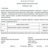
Table 1. Definition of risk categories in patients with operable forms of breast cancer. San Gallen, 2007.
| Risk category | |
| low risk |
No affected lymph nodes (p NO) and all of the following: p T ≤2 cm and grade (G 1) and Expression of ER and PR and No increased expression or amplification of HER 2/neu Age≥ 35 years |
| intermediate risk |
Absence of affected lymph nodes (p NO) and at least least one of the following signs: p T> 2 cm or Presence of extensive peritumoral vascular invasion or Increased expression or amplification of HER 2/neu Age< 35 лет |
|
The presence of single regional metastases (1-3 involved l / s) Expression of ER + / PR + , No overexpression or amplification of HER2/neu |
|
| high risk |
The presence of solitary regional metastases (1-3 lymph nodes involved and no expression of steroid hormone receptors (ER-PR-) or |
| Presence of 4 or more affected lymph nodes |
Table 2. Planning for adjuvant treatment of breast cancer. San Gallen, 2007.
|
highly sensitive to endocrine therapy |
Not enough endocrine sensitive |
Insensitive to endocrine therapy |
|
| HER (-) |
endocrine therapy, additionally chemotherapy for high risk groups relapse |
endocrine therapy, additionally chemotherapy for intermediate and high risk of relapse |
Chemotherapy |
| HER (+++) |
Endocrine therapy + trastuzumab+* Chemotherapy** |
Endocrine therapy + Trastuzumab + Chemotherapy |
Trastuzumab + Chemotherapy |
*Trastuzumab (Herceptin®) is not considered standard of care in women with tumors smaller than 1 cm and without metastatic lymph nodes (pNO), especially in women with highly endocrine sensitive tumors.
**Available clinical trial data do not support the recommendation of trastuzumab without prior or concomitant chemotherapy.
Table 3. Adjuvant treatment depending on therapeutic targets and risk categories. San Gallen, 2007.
| HER 2 (-) | HER 2 (+++) | |||||||||||
|
High endocrine. feels. |
Incomplete feelings. To endocrine. |
Insensible To endocrine. therapy |
High endocrine. feels |
Incomplete feelings. To endocrine. |
Insensible To endocrine. therapy |
|||||||
| low risk | uh | uh | uh | uh | ||||||||
|
Prome- creepy- risk |
x→ |
x→ |
x→ uh |
x→ uh |
x | x |
x→ e+t |
x→ e+t |
x→ e+t |
x→ e+t |
x+t | x+t |
|
x→ |
x→ |
X → |
X → EE |
X → EE |
x |
x→ e+t |
x→ e+t |
x→ e+t |
x→ e+t |
x+t | x+t | |
| high risk |
heh |
heh |
heh |
heh |
x+t | x+t | ||||||
|
x→e |
x→e | x→e | x→e | X | X |
x→ e+t |
x→ e+t |
x→ e+t |
x→ e+t |
x+t x+t |
x+t x+t |
|
X-chemotherapy
E- Endocrine Therapy
T-trastuzumab (Herceptin)
Adjuvant treatment of breast cancer patients according to sensitivity to endocrine therapy
AI - aromatase inhibitors
HT - chemotherapy
Tam - Tamoxifen
SOF - suppression of ovarian function (surgical, radiation therapy,
conservative)
AC - anthracycline + cyclophosphamide
CEF, FEC - cyclophosphamide + epirubicin + 5-fluorouracil
CAF - anthracycline + cyclophosphamide + 5-fluorouracil
Tah - taxanes
Let - letrazole
Exe - exemestane
Ana - anastrozole
TREATMENT FOR DIFFERENT STAGES OF BC
0, stage I
1. organ-preserving treatment.
After organ-preserving surgery, taking into account the expression level of ER, PR, Her-2/neu, one of the types of systemic treatment is prescribed. In the absence of the need for systemic treatment, it is possible to prescribe radiation therapy. Irradiation of the mammary gland is carried out using photon radiation (6 MeV) of a linear accelerator or gamma radiation of a 60Co installation (1.25 MeV) from two tangentially located fields, aimed at ensuring the most homogeneous irradiation of the gland. ROD 2 Gr, SOD 60 Gr. The postoperative area is additionally irradiated at a dose of 12 Gy (2 Gy each). Irradiation by electronic triggering is preferred.
2. radical mastectomy.
With all of the above localizations of the first stage of the disease, it is possible to perform a radical mastectomy with or without restoration of the shape of the gland (at the request of the patient).
Systemic treatment includes: chemotherapy in patients under 50 years of age with invasive forms, hormone therapy with tamoxifen in postmenopausal patients with receptor-positive tumors for 5 years. Patients under 50 years of age with preserved menstrual function: bilateral oophorectomy or LHRH analogues monthly for 2 years while taking tamoxifen.
Patients with negative EP, PR - PCT (CMF or CAF) do not undergo hormone therapy.
Chemotherapy regimens for stages 0 and I:
C.M.F. Bonadonna regimen
Methotrexate 40 mg/m*2 IV 1 day
5FU 600 mg/m*2 IV for 1 day
Repeat every 3 weeks for 6 cycles
Cyclophosphamide 100 mg/m*2 orally 1-14 days
5FU 600mg/m*2 IV 1 and 8 days
Prednisolone 40 mg/m*2 orally 1 and 14 days
Repeat every 4 weeks for 6 cycles.
Doxorubicin 60mg/m*2 IV 1 day
Cyclophosphamide 600mg/m*2 IV for 1 day
II stage
Treatment identical to that in stage I, however, in patients with N0, but with the presence of unfavorable prognostic signs (age under 35 years, negative hormone receptors, positive Her 2-neu status) in the postoperative period, except for the entire breast, with tumor localization in the internal quadrants or the central zone, as well as in all patients with N + (with metastatic lesions of three or less axillary lymph nodes), the parasternal and supraclavicular zones are additionally irradiated from the side of the main focus.
Postoperative RT is carried out in the classical dose fractionation mode (ROD 2 Gy, SOD 30 Gy) after organ-preserving surgery and systemic therapy. The postoperative area is additionally irradiated at a dose of 12 Gy (2 Gy each).
In patients with N+, when four or more axillary lymph nodes are affected and/or when the tumor invades the capsule of the lymph node, in addition to the remaining mammary gland, the parasternal, supraclavicular-axillary zone is irradiated from the side of the lesion.
ALL stage II patients should receive adjuvant systemic chemotherapy (CMF, AC, TAC, AC+T, FAC, CAF, FEC, A+CMF).
At +ER tamoxifen for 5 years.
With -ER - chemotherapy.
Patients with positive Her 2-neu - trastuzumab 8 mg / kg on day 1, every 21 days, 4 mg / kg
Chemotherapy regimens:
cyclophosphamide 100 mg/m*2 orally 1-14 days
5FU 600 mg/m*2 IV 1 and 8 days
repeat every 28 days.
methotrexate 40 mg/m*2 IV 1 and 8 days
5FU 600mg/m*2 IV 1 and 8 days
repeat every 28 days.
repeat every 21-28 days.
5FU 500 mg/m*2 IV 1 and 8 days
doxorubicin 50 mg / m * 2 IV long-term infusion 72 hours 1-3 days.
cyclophosphamide 500 mg / m * 2 in / in 1 day.
repeat 21 if haematological parameters are restored.
Taxotere 75 mg/m*2 IV for 1 day
Doxorubicin 50 mg/m*2 IV 1 day
Cyclophosphamide 500 mg / m * 2 in / in 1 day.
repeat every 21 days.
Cyclophosphamide 600 mg / m * 2 in / in 1 day.
5FU 600 mg / m * 2 in / in 1 day.
Repeat every 21-28 days.
Doxorubicin 60 mg/m*2 IV for 1 day
Cyclophosphamide 600 mg / m * 2 in / in 1 day.
Repeat every 3-4 weeks depending on the recovery of hematological parameters.
doxorubicin 60 mg/m*2 IV 1 day
cyclophosphamide 600 mg / m * 2 in / in 1 day. X 4 cycles.
continue paclitaxel 175 mg/m*2 IV for 3 hours infusion once every 3 weeks for 4 cycles.
Doxorubicin 60 mg/m*2 IV 1 day
Cyclophosphamide 600 mg / m * 2 / in 1 day X 4 cycles.
Continue docetaxel 75 mg/m2 IV once every 3 weeks for cycle 4.
Cyclophosphamide 75 mg/m2 orally 1-14 days
Epirubicin 60 mg/m*2 IV for 1 day
5FU 500 mg/m*2 IV 1 and 8 days every month 6 cycles.
Doxorubicin 75 mg / m * 2 / in 1 day every 3 weeks for 4 cycles.
Cyclophosphamide 600 mg / m * 2 in / in 1 day.
Methotrexate 40 mg/m*2 IV 1 and 8 days
5FU 600 mg/m*2 IV 1 and 8 days
Repeat 8 cycles every 3 weeks.
At stage IIA, the general effects are prescribed in accordance with the table. 4.
Table 4 Absence of metastases in axillary lymph nodes
|
Menstrual status |
low risk |
Intermediate and high risk |
|
Hormone sensitive tumors |
||
|
Menstruating |
Tamoxifen zoladex or diphereline |
Chemotherapy chemotherapy + tamoxifen (with shutdown of ovarian function) |
|
Postmenopause |
Tamoxifen IA |
Tamoxifen or chemotherapy + tamoxifen or AI |
|
Hormone resistant tumors |
||
|
Menstruating |
Chemotherapy |
|
|
Postmenopause |
Chemotherapy |
|
Patients with a positive Her 2-neu - trastuzumab 8 mg / kg on day 1, every 21 days, 4 mg / kg for 1 year. In patients of reproductive age with ER (-) and PR (-) status in combination with PCT (taxanes or CMF, excluding anthracyclines). In postmenopausal patients with ER(+) and PR(+) status in combination with AI, in ER(-) and PR(-) status it is necessary to carry out therapy in combination with PCT (taxanes or CMF, excluding anthracyclines).
In premenopausal women with 8 or more metastatic lymph nodes after completion of 6 courses of chemotherapy and ongoing menstrual function, bilateral oophorectomy or shutdown of ovarian function is indicated by the appointment of LHG releasing hormone agonists (giserelin - 3.6 mg subcutaneously in the abdominal wall every 28 days for 2 years, triptorelin 3.75 mg every 28 days for 2 years) while taking tamoxifen 20 mg per day for 5 years. When the menstrual function stops after 6 courses of PCT, tamoxifen is prescribed at 20 mg per day for 5 years.

Patients with a positive Her 2-neu - trastuzumab 8 mg / kg on day 1, every 21 days, 4 mg / kg, for 1 year. In patients of reproductive age with ER (-) and PR (-) status in combination with PCT (taxanes or CMF, excluding anthracyclines). In postmenopausal patients with ER(+) and PR(+) status in combination with AI, in ER(-) and PR(-) status it is necessary to carry out therapy in combination with PCT (taxanes or CMF, excluding anthracyclines).
Surgical intervention 3 weeks after the end of treatment in the amount of RME according to Maden, radical resection of the mammary gland, organ-preserving or reconstructive plastic surgery.
Surgical treatment. The operational benefit is performed according to the generally accepted technique in the amount of radical mastectomy (according to Madden, Patey). The volume of surgical intervention (mastectomy option) is determined by the prevalence of the tumor process. In all cases, the removal of regional lymph nodes of three levels is indicated: axillary, subclavian, subscapular with their subsequent marking. The tumor should be labeled according to size and location in the quadrants of the breast.
It is possible to perform an immediate or delayed reconstructive surgery (at the request of the patient).
Postoperative radiotherapy. Postoperative RT is carried out in the classical dose fractionation mode (ROD 2 Gy, SOD up to an equivalent dose of 60 Gy). Irradiation fields: supraclavicular, axillary, parasternal, chest wall (at pT3, 4). 61. Erythrocyte mass, cytological or histological verification of the diagnosis, complete blood count (6 indicators), urinalysis, blood for b / chemistry (9 indicators), blood for coagulogram, electrocardiography, fluorography or R-graphy of the lungs, ultrasound mammary glands, regional zones, liver, pelvic organs, mammography. Ductography, magnetic resonance imaging, computed tomography of the mammary glands, determination of hormone levels (ER -, ER +, Her-2-neu), apoptosis, CA15-3 if possible and if indicated.
Information
Sources and literature
- Protocols for the diagnosis and treatment of diseases of the Ministry of Health of the Republic of Kazakhstan (Order No. 764 of December 28, 2007)
- 1. V.F. Semiglazov, V.V. Semiglazov, K.Sh. Nurgaziev. Substantiation of standards for the treatment of breast cancer., 362 pages, Almaty, 2007. 1. Barroso G, Menqcal g. Felix H. Roias-Ruis J.C. et al. Comparison of the efficacy of the aromatase inhibitor letrozole and clomiphene citrate as adjuvants to recombinant folliclestimulating hormone in controlled ovarian hyperstimulation: a prospective, randomized, blinded clinical trial. // Fertil Steril .- 2006.- Vol 86p.1428-1431 2. Bogaerts J, Cardoso F, Buyse M, et al. Gene signature evaluation as a prognostic tool: challenges in the design of the MINDACT trial.//Nat Clin Pract Oncol .-2006.- Vol .3: p.540-551 3. Clarke CA, Glaser SL. Recent declines in hormone therapy utilization and breast cancer incidence: clinical and population-based evidence. // J Clin Oncol.- 2006 .-Vol.24.p 49 4. Coates AS, Keshaviah A, fthurlimann B, et al. Five years of letrozole compared with tamoxifen as initial adjuvant therapy for postmenopausal women with endocrine-responsive early breast cancer: update of study BIG 1-98// J Clin Oncol .-2007-Vol. 25 p.486-492 5. Colleoni M, Rotrnensz N, Peruzzotti G, et al. Prognostic role of the extent of peritumoral vascular invasion in-operable breast cancer. Ann Oncol .-2007 (accepted for publication) 6. Coombes RC, Kilburn LS, Snowdon CF, et al. Survival and safety of exemestane versus tamoxifen after 2-3 years" tamoxifen treatment (Intergroup Exemestane Study): a randomized controlled trial. // Lancet.- 2007.- Vol.349.p.1110-1117 7. Goldhirsch A, Glick JH , Gelber RD et al. Meeting highlights: international expert consensus on the primary therapy of early breast cancer.//Ann Oncol.-2005.-Vol.16.p.1569-1583 8. Goldhirsch A; Cda^es AS, Qelber RD et al.First-select the target: better choice of adjuvant treatments for breast cancer patients.//Ann Oncol.-2006.-Vol.17 p.1772-1776 9. Goss PE, Ingle JNJ Martino S, et al. Randomized trial of letrozole following tamoxifen as extended adjuvant therapy in receptor-positive breast cancer: updated findings from NCIC CTG MA.17 // JNCI Cancer Spectrum.-2005.- Vol.97.p.1262-1271 10. Howell A, Cuzick J, Baum M, et al. Results of the ATAC (Arimidex, Tamoxifen, Alone or in Combination) trial after completion of 5 years" adjuvant treatment for breast cancer. // Lancet.- 2005.- Vol 365.p.60-62 11. Jakesz R, Jonat W, Gnant M, et al. Switching of postmenopausal women with endocrineresponsive early breast cancer to anastrozole after 2 years" adjuvant tamoxifen: combined results of ABCSG trial 8 and ARNO 95 trial. // Lancet.- 2005.- Vol 366.p.455-462 12. James CR, Ouinn JE, Mullan PB et al. BRCA1, a Potential predictive biomarker in the treatment of breast cancer //Oncologist.-2007.-Vol 2.p. 142-150 13. Jimenez-Gordo AM. De Las Heras B. Zamora P. et al.: Failure of Goserelin ovarian* ablation in premenopausal women with breast cancer: two case reports. // Gynecol Oncol .- 2000. - Vol 76 p.126-127 14. Joensuu H, Kellokumpu-Lehtinen PL, Bono P, et al. Adjuvant docetaxel or vinorelbine with or without trastuzumab for breast cancer. // N Engl J Med .- 2006.- Vol 354. p.809-820 15. Kaufmann M, Hortobigyi GN, Goldhirsch A, : et al. Recommendations from an international expert panel on the use of neoadjuvant (primary) systemic treatment of operable breast cancer: an update// J Clin Oncol .- 2006.-Vol 24p.1940-1949 16. Korreman SS. Pedefsen AN. Aarup LR et "al. Reduction of cardiac and pulmonary complication probabilities after breathing adapted radiotehrapv for breast cancer. Int J Radiot // Oncol Biol Phvs .-2006.- Vol 65.p.1375-1380 17. Kurtz J. EUSOMA Working Party. The curative role of radiotherapy in the treatment of operable breast cancer // Eur J Cancer.-2002.- Vol 38.p.1961-1974 18. Mauriac L, Keshaviah A. Debled M Mouridsen H et al. Jtedictors of early relapse in postmenopausal women with hormone receptor-positive breast cancer in the BIG 1-98 trial//Ann Oncol.-2007.-Vol.14 p.320-327 19. Perez EA, Combining adjuvant chemotherapy with biological therapy, St. Gallen. .- //Breast.-2007 .- Vol.16 (Suppl): p105-111 20. Piccart-Gebhart MJ, Procter M, Leyland-Jones B et al., Trastuzumab after Adjuvant Chemotherapy in HER2-Positive Breast Cancer.// N Engl J Medi -2005.-Vol.353p.1659-1672, 21. Recht A. Edge SB. Splin LJ. Robinson PSet al. Postmastectomy radiotherapy: "clinical practice guidelines of, the American Society of Clinical Oncology//. J Clin Oncology.- 2001 - Vol 19.p.1539-69 22. Regan MM, Viale G, Mastropasqua MG, et al. Re-evaluating adjuvant breast cancer trials: assessing hormone receptor status by immunohistochemical versus extraction assays. //JNCI Cancer Spectrum.-2006-Vol. 98 p.1571-1581 23. Romond EH, Perez EA, Bryant J et al. Trastuzumab plus Adjuvant Chemotherapy for Operable HER2-Positive Breast Cancer. // N Engl J Med.-2005.-Vol. 353 p.1673-1684 24. Semiglazov V.F., Semiglazov V.V. Dashyan G.A. et al. Phase 2 Randomized trial of primary endocrine therapy versus chemotherapy in postmenopausal patients with estrogen receptor-positive breast cancer// Cancer. -2007-Vol 110.-p. 244-254 25. Slamon D, BCIRG 006 II interim analysis. San Antonio Breast Cancer Symposium, 2006. http://www.bcirg.org/Internet/BCIRG+at+SABCS+2006/default.htm 26. Sorlie T, Tibshirani R, Parker J, et al. Repeated observation of breast tumor subtypes in independent gene expression data sets. // Proc Natl Acad Sci U S A .- 2003.- Vol.100p.8418-8423 27. Sorlie T., Perou CM, Tibshirani R et al. Gene expression patterns of breast carcinomas distinguish tumor subclasses with clinical implications. // Proc Natl Acad Sci U S A .-2001.- Vol. 98p. 10869-10874 28. Sparano JA. TAILOR: trial assigning individualized options for treatment. // Clin Breast Cancer.- 2006.- Vol.7:p347-350 29. Winer EP, Hudis C, Burstein HJ, et al. American Society of Clinical Oncology technology assessment on the use of aromatase inhibitors as adjuvant therapy for postmenopausal women with hormone receptor-positive breast cancer: status report 2004. // J Clin Oncol.- 2005.- Vol 23: p.619-629. 30 Wolff AC, Hammond ME, Schwartz JN, et al. American Society of Clinical Oncology/College of American Pathologists Guideline Recommendations for Human Epidermal Growth Factor Receptor 2 Testing in Breast Cancer// Arch Pathol Lab Med.-2007.- Vol.131p.18.
Information
Mukhambetov S.M., Scientific Center of Oncology
Attached files
Attention!
- By self-medicating, you can cause irreparable harm to your health.
- The information posted on the MedElement website and in the mobile applications "MedElement (MedElement)", "Lekar Pro", "Dariger Pro", "Diseases: a therapist's guide" cannot and should not replace an in-person consultation with a doctor. Be sure to contact medical facilities if you have any diseases or symptoms that bother you.
- Choice medicines and their dosage, should be discussed with a specialist. Only a doctor can prescribe the right medicine and its dosage, taking into account the disease and the condition of the patient's body.
- The MedElement website and mobile applications "MedElement (MedElement)", "Lekar Pro", "Dariger Pro", "Diseases: Therapist's Handbook" are exclusively information and reference resources. The information posted on this site should not be used to arbitrarily change the doctor's prescriptions.
- The editors of MedElement are not responsible for any damage to health or material damage resulting from the use of this site.
Classification is usually considered according to the TNM system, where the stage of cancer is determined. But other classifications are also used to make a more accurate diagnosis. We will now describe the main ones.
Cancer classification according to ICD 10
- C50 malignant neoplasm of the breast;
- C50.0 nipple and areola;
- C50.1 the central part of the mammary gland is affected;
- C50.2 upper inner quadrant lesion;
- C50.3 lower inner quadrant lesion;
- C50.4 upper outer quadrant lesion;
- C50.5 lower outer quadrant lesion;
- C50.6 axillary region;
- With 50.8 defeat more than one position;
- C50.9 localization of cancer development is not defined;
- D05.0 Lobular carcinoma in situ;
- D05.1 Intraductal carcinoma in situ
Histological classification
A. Non-invasive cancer
- intraductal;
- lobular.
B. Invasive cancer
- ductal;
- lobular;
- slimy;
- medullary;
- tubular;
- apocrine;
- other forms (papillary, juvenile and others).
C. Special
- Paget's cancer;
- inflammatory cancer.
The most common forms of cancer currently being diagnosed are squamous cell carcinoma and Paget's cancer.
Classification by tumor growth rate
The growth rate of the tumor indicates its malignancy, the rate is determined using radiation diagnostics. For example:
- A rapidly growing tumor - this is characterized by the addition of a tumor mass 2 times larger over a period of not more than 2 months.
- Medium growing tumor - this is characterized by an increase in tumor mass by 2 times within 1 year.
- Slowly growing tumor - this is characterized by an increase in tumor mass by 2 times over a period of more than 1 year.
TNM classification
T - primary tumor
- TX - primary unavailable for evaluation;
- TO - there are no signs of a primary tumor;
- Tis - cancer;
- Tis (DCIS) - ductal carcinoma;
- Tis (LCIS) - lobular carcinoma;
- Tis (Paget) - Paget's disease of the nipple, not associated with invasive carcinoma;
- T1 - tumor up to 2 cm in size;
- T2 - tumor size from 2 to 5 cm;
- T3 - tumor larger than 5 cm;
- T4 - a tumor of any size that is spread to the skin, to the chest wall.
N - regional lymph nodes
- NX are regional lymph nodes that cannot be assessed.
- N0 - no metastases in regional lymph nodes.
- N1 - the presence of metastases in the axillary lymph nodes, I.II Level, which are not soldered to each other.
- N2 a - the presence of metastases in the axillary region of the lymph nodes of the I.II level, which are soldered together. (c - internal mammary lymph node in the absence of clinical signs and metastasis in the axillary lymph nodes).
- N3 a - the presence of metastases in the subclavian lymph nodes of level III (c - the presence of metastases in the internal mammary and axillary lymph nodes, metastases in the supraclavicular lymph nodes).
M - distant metastases.
- Mo - the presence of distant metastases is not determined;
- M1 - distant metastases are present.
Types of breast cancer
Hormone dependent
Hormone dependent - a disease such as cancer mammary gland directly depends on the hormonal background of the female body. Today, there are many factors that can cause a hormonal imbalance.
Almost all forms of mammary gland hyperplasia are a consequence of a violation of the properties endocrine system. All this is caused by an increase in the body of estrogen, prolactin and a decrease in progesterone.
Similarly, due to the failure of these hormones, breast cancer begins to develop.
Scientists have proven that a long, non-stop use of hormonal contraceptives is one of the causes of breast cancer. Basically, hormonal agents are included in the complex of treatment of the disease.
Negative breast cancer
Negative breast cancer is one of the severe forms of the disease. Difficult to treat. It is determined only by laboratory methods. It differs from others in that it does not have receptors for the main three proteins - estrogen, progesterone, and a specific tumor protein.
Luminal breast cancer
Luminal breast cancer is divided into 2 types - A and B.
Luminal A. Diagnosed in women during menopause, in 33-41% of cases. This type of cancer cells
- receptors respond well to estrogen and progesterone;
- receptors practically do not respond to the cell growth marker Ki67;
- receptors do not respond to cells specific protein HER2-neu.
This type of cancer is highly treatable. Hormone therapy is used for treatment.
Luminal B. Occurs in women of childbearing age, in a ratio of 15-20% of cases. It is characterized by metastases to the nearest lymph nodes. The disease is very difficult to treat. Basically, it is not possible to stop the growth of cancer cells.
Stages of cancer
There are 4 stages of cancer.
First (initial) stage
She is characterized by:
- tumor size within 2 cm;
- absence of metastases.
Second stage
She is characterized by:
- tumor size 2-5 cm;
- the presence of metastases in the lymph nodes;
- single metastases in distant organs are possible.
Third stage
She is characterized by:
- tumor size more than 5 cm;
- the presence of metastases in the lymph nodes of the axillary region (nodes are determined separately from metastases);
- distant metastases may occur.
Fourth stage
She is characterized by:
- The size of the tumor is large, mainly determined outside the mammary gland. May be accompanied by knots.
- Metastases on both sides in the lymph nodes.
- Multiple metastases in distant organs.
Video: classification of breast cancer
medik-24.ru
Classification of breast cancer
The TNM Classification of Malignant Tumors, adopted by WHO for all malignant neoplasms, determines the stages of breast cancer. Based on the recommendations of leading experts, it has been adapted for oncological mammology with the introduction of detailing.
The TNM classification of breast cancer measures the anatomical grade of a tumor based on its size, spread to the lymph nodes in armpits, neck and chest, and also notes the presence of metastases. This international classification of breast cancer is accepted by the International Association for Breast Cancer and the European Society for Medical Oncology (EUSOMA).
According to TNM classification, cancer mammary glands has the following stages:
- T0 - signs of breast cancer are not detected (not proven).
- Tis (tumor in situ) designation refers to carcinomas and is deciphered as follows: abnormal cells are found in situ (no invasion), localization is limited to the ducts (DCIS) or lobules (LCIS) of the breast. There is also Tis Paget, that is, Paget's disease, which affects the tissues of the nipple and areola of the breast.
- T1 Tumor diameter at its widest point 20 mm or less:
- T1a Tumor diameter > 1 mm, but
- T1b - tumor diameter greater than 5 mm, but less than 10 mm;
- T1c Tumor diameter >10 mm but ≤ 20 mm.
- T2 - tumor diameter > 20 mm, but
- T3 - tumor diameter exceeds 50 mm.
- T4 Tumor of any size and has spread to: chest(T4a), skin (T4b), chest and skin (T4c), inflammatory breast cancer (T4d).

Indicators for lymph nodes:
- NX - lymph nodes cannot be assessed.
- N0 - cancer was not found in the lymph nodes.
- N0 (+) - small areas of "isolated" tumor cells (less than 0.2 mm) were found in the axillary lymph nodes.
- N1mic - areas of tumor cells in the axillary lymph nodes more than 0.2 mm but less than 2 mm (can only be seen under a microscope and are often called micrometastases).
- N1 - cancer has spread to 1-2-3 axillary lymph nodes (or the same number of intrathoracic), the maximum size is 2 mm.
- N2 - spread of cancer to 4-9 lymph nodes: only axillary (N2a), only internal thoracic (N2b).
- N3 - The cancer has spread to 10 or more lymph nodes: to the lymph nodes under the arm, or under the collarbone, or above the collarbone (N3a); on the internal thoracic or axillary nodes (N3c); supraclavicular lymph nodes are affected (N3c).
Indicators for distant metastases:
- M0 - no metastases;
- M0 (+) - there are no clinical or radiographic signs of distant metastases, but tumor cells are found in the blood or bone marrow, or in other lymph nodes;
- M1 - metastases in other organs are determined.
Histological classification of breast cancer
The current histopathological classification of breast cancer is based on the morphological features of neoplasia, which are studied in the process of histological studies of tumor tissue samples - biopsy specimens.
In the current version, approved by WHO in 2003 and accepted worldwide, this classification includes about two dozen large types of tumors and almost as many less significant (rare) subtypes.
The following main histotypes of breast cancer are distinguished:
- non-invasive (non-infiltrating) cancer: intraductal (ductal) cancer; lobular or lobular cancer (LCIS);
- invasive (infiltrating) cancer: ductal (intraductal) or lobular cancer.
These types, according to the statistics of the European Society for Medical Oncology (ESMO), account for 80% of clinical cases of malignant tumors of the mammary glands. In other cases, less common types of breast cancer are diagnosed, in particular: medullary (soft tissue cancer); tubular (cancer cells form tubular structures); mucinous or colloidal (with mucus); metaplastic (squamous, glandular-squamous, adenocystic, mycoepidermoid); papillary, micropapillary); Paget's cancer (tumor of the nipple and areola), etc.
Based on the standard protocol of histological studies, the level of differentiation (distinguishing) between normal and tumor cells is determined, and thus the histological classification of breast cancer allows you to determine the degree of malignancy of the tumor (this is not the same as the stage of cancer). This parameter is very important, since the level of histopathological differentiation of the neoplasia tissue gives an idea of the potential for its invasive growth.
Depending on the number of deviations in the structure of cells, degrees are distinguished (Grade):
- GX - the level of tissue discrimination cannot be assessed;
- G1 - the tumor is highly differentiated (low grade), that is, the tumor cells and the organization of the tumor tissue are close to normal;
- G2 - moderately differentiated (middle grade);
- G3 - low differentiated (high grade);
- G4 - undifferentiated (high grade).
Grades G3 and G4 mean a significant predominance of atypical cells; such tumors grow rapidly and spread faster than tumors with G1 and G2 differentiation.

Experts see the main disadvantages of this classification in the limited possibility of a more accurate reflection of the heterogeneity of breast cancer, since tumors with completely different biological and clinical profiles turned out to be in one group. As a result, the histological classification of breast cancer has minimal prognostic value.
Immunohistochemical classification of breast cancer
Thanks to the use of new molecular tumor markers - the expression of cellular tumor receptors for estrogen (ER) and progesterone (PgR) and the status of HER2 (transmembrane protein receptor for epidermal growth factor EGFR, which stimulates cell growth) - a new international classification of breast cancer has emerged, which has a proven prognostic value and allows for more accurate treatment options.
Based on the state of estrogen and progesterone receptors, the activation of which leads to changes in cells and tumor growth, the immunohistochemical classification of breast cancer distinguishes between hormone-positive tumors (ER+, PgR+) and hormone-negative tumors (ER-, PgR-). Also, the status of EGFR receptors can be positive (HER2+) or negative (HER2-), which radically affects the treatment tactics.
Hormone-positive breast cancer responds to hormone therapy with drugs that lower estrogen levels or block estrogen receptors. As a rule, such tumors grow more slowly than hormone-negative ones.
Mammologists note that patients with this type of neoplasm (which occurs more often after menopause and affects the tissues lining the ducts) have a better prognosis in the short term, but cancers with ER+ and PgR+ can sometimes recur after many years.
Hormone-negative tumors are much more likely to be diagnosed in women who have not yet gone through menopause; these neoplasias are not treated with hormonal drugs and increase faster than hormone-positive cancers.
In addition, the immunohistochemical classification of breast cancer highlights triple positive cancer (ER+, PgR+ and HER2+), which can be treated with hormonal agents and drugs with monoclonal antibodies designed to suppress the expression of HER2 receptors (Herceptin or Trastuzumab).
A triple negative cancer (ER-, PgR-, HER2-), which is referred to as a molecular basal subtype, is typical for young women with a mutant BRCA1 gene; The main drug treatment is carried out with cytostatics (chemotherapy).
In oncology, it is customary to make a decision on the appointment of a treatment based on all the possible characteristics of the disease that each classification of breast cancer makes available to the doctor.
ilive.com.ua
Breast cancer: causes, treatment and prognosis
Malignant tumors in the area of the mammary glands are one of the most serious medical and social problems. According to statistics, the incidence of breast cancer is very high - there are about 1.5 million women in the world with this diagnosis. For about 400 thousand of them, the disease ends in death, which is why it is so important timely diagnosis and treatment of breast cancer.
Breast cancer - what is it
Breast cancer is a common cancer that develops in the glandular tissues of the breast. The disease affects both women and men over the age of 13 years. The pathological process can develop in one lobe or in several at once, on the right, on the left, or in both breasts.breast cancer statistics
Every year, more than 1 million 250 thousand new cases of malignant breast pathology are diagnosed worldwide. In Russia alone, there are 54 thousand such cases annually. Moreover, in many countries there is a tendency towards an increase in the incidence. This is due to several reasons. First of all, the fact that diagnostics has improved and mammographic screening has begun. This makes it possible to detect a tumor in initial stage when she still does not show any symptoms. Doctors advise visiting a mammologist and regularly undergo screening. It is also necessary to regularly conduct self-examination of the mammary glands. The statistics are disappointing - every eighth woman sooner or later faces this serious illness. It is not excluded the disease in males, but this is an infrequent occurrence. According to researchers, the ratio of sick men and women is approximately 1:100. The risk of getting breast cancer increases over time. The majority of women with this diagnosis (77%) belong to the age group over 50. There are few young girls among those suffering from breast cancer - about 0.3%.
Breast cancer ICD-10 code
C50 Malignant disease of the breast. C50.0 Nipple and areola. C50.1 Central part of the mammary gland. C50.2 Upper inner quadrant. C50.3 Lower inner quadrant. C50.4 Upper outer quadrant. C50.5 Infero-outer quadrant. C50.6 Axillary region. C50.8 Spread over more than one of the above areas. C50.9 Location, unspecified. D05.0 Lobular carcinoma in situ D05.1 Intraductal carcinoma in situA detailed classification of breast cancer by stages (TNM) and other classifications are covered on our website.

Causes of breast cancer
The etiology of the disease has not been fully elucidated. So far, no specific etiological factor has been found. A certain role in the beginning of the malignant process is played by the adverse effects of the environment and even the way of life of a woman. But at the same time, no more than 50% of cases of the disease can be associated with known factors.At-risk groups
In modern medicine, the following risk groups for breast cancer have been identified:
Low risk group (1-2 times higher). This includes women in early age used COCs, especially before the first pregnancy. Approximately 35% increases HRT. Also included in this group are women with a history of first pregnancy termination and women whose diets are high in fat (saturated), which increase plasma levels of estradiol.
Medium risk group (increased by 2-3 times). Here the main factors are: late first birth, early menarche, late menopause, other types of cancer in the past, alcohol abuse, infertility, increased body weight, breast proliferative phenomena, obesity after menopause.
High-risk group (increased by 4 or more times). This category includes women over 50 years of age and women whose first-line relatives had this diagnosis. There is also an increased risk in women who have had breast cancer in the past, after exposure to radiation, with proliferative breast diseases with the presence of atypia. A serious factor is the mutation of the BRCA1, BRCA2 genes.
hereditary breast cancer
In some cases, the diagnosis of "hereditary cancer" is established. The criteria for its setting are:
The onset of the disease at a young age. Tumor development in both breasts. The patient has relatives of the 1st and 2nd degree of kinship who have had breast cancer. Multiple neoplasms in the patient or (and) relatives. The presence of specific tumor associations.

To date, scientists have been able to identify several genes that are responsible for predisposition to malignant processes. These are the BRCA1, BRCA2, p53, PTEN genes. The last two are responsible for the family and individual predisposition to Cowden and Lee-Fraumen syndromes. According to studies, up to 40-70% of cases of hereditary breast cancer are associated with mutations in the BRCA1, BRCA2 genes. It turned out that the carriers of these mutations have an extremely high risk of a malignant process in one gland - up to 80%. And the risk of a tumor in the second breast is 50-60%. (In the general population, these figures are 2 and 4.8%). In carriers of the BRCA1 mutation, the peak incidence occurs at 35-39 years, with a BRCA2 gene mutation - at 43-54 years. It is worth noting that patients with a BRCA2 mutation have a better prognosis than those with sporadic cancer or a BRCA1 mutation. Pregnancy and childbirth with mutations, as it turned out, are not protective factors. Thus, women who have given birth with mutations develop cancer before the age of 40 even more often than those who have not given birth (about 1.7 times). And with each subsequent pregnancy, the risk increases. If a gene mutation is detected, treatment should be reviewed.
The treatment of such women has its own characteristics:
Organ-preserving operations are not carried out. Mastectomy is recommended for prevention. Indications for chemotherapy treatment are expanding. In the case of a BRCA1 mutation, oophorectomy is recommended for prophylaxis.
The mechanism of development of breast cancer
To date, the pathogenesis of breast cancer has not been fully studied and all stages of the development of the tumor process have not been precisely defined. In modern medical science, it is customary to distinguish three main stages in the development of the disease: initiation, promotion, progression. Carcinogenesis (malignancy) begins as a result of mutation of proto-oncogenes. They are transformed into oncogenes and promote cell growth. (There is an increase in the formation of mutagenic growth factors or there is an effect on surface cell receptors). When a cell is damaged, estrogens encourage that cell to replicate until the damage is repaired. Estrogens are an obligatory factor in the tumor process, supporting it during the period of promotion. When angiogenesis begins, distant metastases appear. This happens even before clinical manifestations disease, during the first 20 doublings. A malignant breast tumor can form in any of the departments of the mammary gland in the alveoli from secreting cells (lactocytes); in the excretory ducts from the columnar epithelium or non-keratinized stratified squamous epithelium near the nipple.IN general information about breast cancer is currently represented by the postulates of B. Fisher:
Dissemination is chaotic, i.e., there is no strict order for the dispersion of malignant cells. - Malignant cells penetrate into the regional lymph nodes by embolization, and this barrier is not effective. - Of great importance for tumor dissemination is the spread of malignant cells throughout the body with the bloodstream. - Operable breast cancer is a systemic disease. - It is not likely that surgical options have an impact on patient survival. - Up to 25% of patients with unaffected lymph nodes and about 75% of women with affected regional lymph nodes die within 10 years due to distant metastases. - Necessary for breast cancer additional methods providing systemic therapeutic effects.
Symptoms of breast cancer
With this disease clinical picture may be different, this is due to the stage of the tumor process. So, with non-palpable neoplasms, clinical signs are not observed. As the tumor grows further, the classic picture of breast cancer is noted, which will be described when we talk about the physical examination.
Breast cancer: photo


Diagnosis of breast cancer
Collection of anamnesis
Diagnosis of breast cancer begins with a study of the patient's history. In this case, it is necessary to find out when the first symptoms of the disease appeared, in what sequence the changes occurred. That is, they analyze the growth dynamics of the neoplasm, pathological changes in the nipple, areola, breast skin, and enlargement of regional lymph nodes. Also, the doctor should know if the patient has had mammary gland injuries or surgical interventions in the past. In addition, you need to ask if there has been any treatment for diseases of the liver, bones and lungs (areas where distant metastases most often occur) within the last 8 months.Inspection and palpation of the mammary glands
Physical examination plays an important role in diagnosis. On examination, pay attention to the shape of the mammary glands, assess the state of the juice, halos (suspected by retraction, ulceration). It is also necessary to assess the condition of the skin. Signs of possible cancer are redness, swelling, the presence of metastases in the skin. The classic sign is the so-called "orange peel", caused by lymphatic edema in the dermis. An important diagnostic feature is the "platform" symptom, when the skin over the neoplasm becomes rigid. "Umbilization" may occur, where the skin is retracted as a result of infiltration of Cooper's ligaments.
Palpation is most informative during the first phase of the cycle. This method makes it possible not only to detect the presence of a tumor, but also to estimate its size. Also, with the help of palpation, you can study the condition of the lymph nodes and suggest the stage of the disease. In the later stages of cancer development, examination is considered perhaps the most informative diagnostic method. At this stage, one can observe swelling of the skin, tissue infiltration, germination of the neoplasm in the skin of the chest. If the cancerous tumor is located in the region of the transitional fold, X-ray examination it may not be revealed. That is why inspection and palpation are so important in diagnosis. Palpation and inspection give the most accurate results during the first phase of the cycle (5-10 days). However, there are times when they do not work. We are talking about non-palpable neoplasms, the diameter of which does not reach 1 cm. It should also be taken into account that it is also impossible to accurately assess the state of regional lymph nodes using these methods.
Lab tests
As for the dynamic monitoring of the patient's condition, here great importance has a study of tumor markers (CA 153, cancer embryonic Ag, tissue polypeptide Ag).Instrumental methods
Mammography
The main diagnostic method, especially when examining patients of older age groups, is mammography. The sensitivity of such diagnostics is very high, it reaches 95%. With its help, it is possible to accurately determine the diameter of the tumor, in some cases to study the state of regional lymph nodes, to detect tumor nodes that cannot be determined by palpation.
Ductography
Ductography is indicated for patients with suspected intraductal tumor. It helps not only to accurately assess the diameter of the neoplasm, but also shows its distance from the nipple.Pneumocystography
Another informative method is pneumocystography. With its help, the internal structure of the cavity formations is visualized.Ultrasound of the mammary glands
Ultrasound is a common diagnostic method that does not compete with mammography. It allows you to more accurately assess the size of the primary tumor, to study its structure and shape, to identify the nature of its blood supply. One of the most important features of the ultrasound method is the ability to examine regional lymph nodes, thanks to which the diagnosis can be clarified.MRI and X-ray CT
CT and MRI in the diagnosis of malignant breast tumors are used infrequently. These methods are more expensive and have less specificity and accuracy.Biopsy
Completes the diagnosis of breast cancer morphological study. Its results must be obtained before the start of the course of therapy. Material for research is taken using the method of puncture aspiration biopsy. Then the biological and morphological parameters of the cells are studied. The sensitivity of such diagnostics is 98%. Given that the tumor process is always systemic, doctors prescribe complex diagnostics to patients with a study of the state of the liver, bones, lungs, etc.
Differential diagnosis of breast cancer
Nodular breast cancer must be distinguished from nodular mastopathy, breast cysts, Paget's cancer must be differentiated from nipple adenoma. Also of great importance is the differential diagnosis of edematous-infiltrative cancer. It must be distinguished from erysipelas and mastitis.
When formulating an accurate diagnosis, it is necessary to take into account the direction of development of the tumor and the quadrant of the gland. They also take into account the form of growth (diffuse or nodular cancer), the diameter of the neoplasm, the condition of the adjacent tissues, the presence of affected lymph nodes and detectable distant metastases. Let us give an example of the formulation of the diagnosis T2N1M0 (II B degree). This means that the tumor has a diameter of up to 5 cm, in the axillary lymph nodes there are single metastases (up to 3). Distant metastases were not identified.
Breast Cancer Treatment
Breast cancer, or any other malignant tumors are not treated with folk remedies! There is not a single reliable case of healing without medical treatment. Everyday deferment medical care can lead to the spread of the pathological process and death.
Tactics of treatment of breast cancer
When deciding on the tactics of treating a patient, the following prognostic factors are taken into account:
The size of the primary tumor. - Presence of malignant cells in regional lymph nodes. - The degree of malignancy according to histology. - Receptor status. The detection of ER and (or) PR in tumor cells indicates that the tumor is highly differentiated. The sensitivity of tumor cells to hormonal treatment is largely associated with the expression of ER and PR on the cell membrane. In women of different age categories, the content of ER and PR is different. Thus, ER and PR are found in 45% of premenopausal and 63% of postmenopausal patients. The goal of hormone therapy is to reduce the effect of estrogens on tumor cells. If the tumor is hormone-dependent, then its growth slows down. - DNA synthesis activity. It can be judged by the following indicators: the number of DNA of aneuploid tumors; the proportion of cells in the S phase of the cell cycle; overexpression of Ki67, ploidy, thymidine kinase activity. Ki67 is the designation of a special marker that characterizes the ability of a neoplasm to proliferate. This nuclear Ag is expressed in any phase of the cell cycle, with the exception of G0. Therefore, it is a marker of cell population growth. Growth factor receptors or growth regulators - EGFR; HER2/neu. The HER2/neu transmembrane glycoprotein is a tyrosine kinase receptor. When it is stimulated, transcription mechanisms are launched, due to which the acceleration of cell growth and proliferation begins. Studies with experimental models have confirmed that Her2/neu may be the cause of neoplasm resistance to endocrine and chemotherapy. Vascular endothelial growth VEGF promotes endothelial cell proliferation and migration. But at the same time, it inhibits apoptosis (destruction) of these cells (tumor progression and the appearance of metastatic foci are associated with angiogenesis). Endothelial growth factor, which is secreted by platelets, is similar in function and structure to thymidine phosphorylase. It is an enzyme that catalyzes the reverse dephosphorylation of thymidine to thymine and 2deoxyribose1phosphate. Its overexpression promotes rapid growth neoplasms and makes cells resistant to apoptosis induced by hypopsia. Oncogenes BRCA1, BRCA2. Other biological factors are also being actively studied. First of all, this concerns Bcl2, p53, PTEN, CDh2, MS h3, ML h2, ALCAM/CD166. Bcl2 is a rather heterogeneous family of proteins. Some of them, such as Bcl2 and BclXI, slow down apoptosis because they inhibit the release of apoptosis-inducing factor and cytochrome C. At the same time, other proteins (Bad and Bax) have the opposite effect, i.e. induce apoptosis. If damage to the DNA structure occurs, the p53 protein activates the apoptosis mechanism. This prevents the reproduction of cells with a disturbed genetic apparatus. It is known that normal p53 tends to rapidly degrade, so it is very difficult to determine its presence in the nucleus. Mutant p53 prevents apoptosis, which makes cells resistant to chemotherapy and radiation treatments.
Methods of treatment of breast cancer
At the stage of treatment planning, it is advisable to have a consultation consisting of physicians of the following specialties: a surgeon, a radiation therapist and a chemotherapist. For all patients, it is mandatory to examine a gynecologist to exclude metastatic ovarian lesions and before performing oophorectomy as part of complex therapy. Comprehensive treatment of breast cancer involves joint application several methods. A combination of local (surgery, radiation exposure) and systemic treatment (hormone and chemotherapy) is necessary. This allows you to achieve a cure for the patient, or at least a stable remission. If a woman has signs that do not allow to exclude a malignant process, hospitalization is mandatory.Treatment with non-drug methods
The main non-drug method is radiation therapy. As self-treatment radiation exposure is usually not used. It is part of the complex therapy of patients with malignant tumors mammary glands. Radiation is used for adjuvant treatment after conservative surgical procedures with or without medical therapy. It is also resorted to after a radical operation, if there are adverse factors affecting the prognosis. Patients with internal localization of the neoplasm are shown radiation exposure to the parasternal region. If more than three lymph nodes are affected, i.e., there is a pronounced metastasis in the lymphatic system, the regional zones of the lymph outflow are irradiated. start radiation treatment at different times. In some cases, it is carried out immediately after surgery, followed by a course of administration. medicines. In other cases, irradiation is carried out simultaneously with drug treatment or after it, but not more than six months after the operation. In the conservative treatment of cancer, radiation methods are often combined with chemotherapy or hormonal drugs. Such treatment is not an alternative to complex therapy using surgical methods. In patients who have undergone complex treatment, 5 and 10-year survival rates are higher. But in some cases, the choice has to be made in favor of a conservative approach, since for some categories of patients (the elderly, those with other diseases), the operation may be unreasonably risky. So, modern therapy cancer patients should be comprehensive. Treatment is prescribed, taking into account the stage of the malignant process and its nature. An individual approach to each patient is also extremely important, taking into account concomitant pathologies and age.Drugs for the treatment of breast cancer
Chemotherapy is an important component of most cancer treatment programs. The indication for its implementation is not only a certain stage of the disease, but also unfavorable prognostic factors. These factors include: - Neoplasm diameter exceeding 2 cm. - Age up to 35 years. - II-IV degree of malignancy. - Receptor negativity. - Metastatic lesions of the lymph nodes. - Hyperexpression of HER2/neu. To date, physicians have a wide range of chemotherapeutic agents at their disposal. Patients at increased risk of tumor progression are shown anticancer drugs such as: CMF (cyclophosphamide, methotrexate, 5fluorouracil), AC (adriamycin, cyclophosphamide), FAC (5fluorouracil, adriamycin, cyclophosphamide) or a combination of anthracyclines with taxanes (AT). The positive impact of this approach on the survival of patients has been proven. For resectable cancers, chemotherapy before surgery does not increase survival compared to adjuvant chemotherapy. But on the other hand, it helps to reduce the size of the tumor, due to which it is possible to carry out an organ-preserving intervention when the process has a locally advanced character. When a chemotherapy course is combined with the use of agents such as trastuzumab and bevacizumab, an even greater effectiveness of therapy can be achieved. Hormonal treatment as an independent course is used infrequently. But in some cases (receptor-positive tumors in older women), it leads to a long-term remission. Hormone therapy showed the greatest effectiveness in the complex treatment of patients with neoplasms that have steroid hormone receptors.Hormonal treatment for cancer has two directions:
- The use of drugs that compete with estrogen for control of the malignant cell. - The use of drugs that reduce the production of estrogen. According to the mechanism of action, antiestrogenic agents belong to the first group. Tamoxifen is the drug of choice for adjuvant treatment. It competes for receptors in cells with estrogens. In addition, it reduces the number of cells in the S phase, and increases it in the G1 phase. The second group of drugs includes aromatase inhibitors. The mechanism of action of these substances is as follows: there is an inhibition of enzymes responsible for the production of estrogens, due to which the content of endogenous estrogens falls. The most specific are letrozole and anastrozole. These two agents are able to inhibit the conversion of androstenedione and testosterone to estrone and estradiol, respectively. Both groups of drugs are highly effective and are widely used as the first line of hormonal treatment of breast tumors.Surgery as a treatment for breast cancer
For tumors of the breast surgical treatment some:
Radical mastectomy, in which the pectoral muscles are preserved. This procedure is standard. After it, primary mammoplasty can be performed.

Areola-sparing mastectomy. After this intervention, it is also possible Plastic surgery.
Organ-preserving surgery and subsequent radiation treatment.
Tumorectomy, which is combined with radiation and drug treatment. This method is used to treat patients with intraductal cancer in situ. In such cases, the sentinel lymph node must be examined. Some patients undergo intraoperative irradiation (dose 20 Gy).
Long-term medical practice shows that an increase in the volume of surgical procedures does not have a positive effect on the survival of patients. Radical mastectomy is performed when the process is locally advanced or when the tumor is centrally located. During the operation, fiber is removed (axillary and intermuscular, as well as subclavian and subscapular). The pectoral muscles are preserved. Due to the low invasiveness of the procedure, the likelihood of complications (pain, neuralgia, venous insufficiency, lymphedema). In order to prevent mental trauma, one-stage mammoplasty is performed.

At stage 1 of breast cancer, as well as at stage 2a, it is possible to perform an organ-preserving operation. In some cases, it is possible to save the breast even with stage 3 breast cancer (after radiation and chemotherapy). Organ-preserving interventions have a positive effect on mental condition patients and their quality of life. Tumorectomy in elderly women followed by radiation and hormone therapy does not have a serious impact on overall and recurrence-free survival. Modern medicine treats reconstructive interventions as an important stage in complex therapy. The purpose of mammoplasty is to prevent the deterioration of the psycho-emotional state of a woman. This procedure can be primary and delayed.
Breast plastic surgery
To restore the shape of the breast and its volume, two methods are used today:
Endoprosthetics. - Reconstruction using autogenous tissue. For inoperable locally advanced or metastatic cancer, palliative surgery is sometimes performed. They are shown to patients with tumor decay and bleeding. At the same time, the following rules are observed: - If the patient does not have distant metastases and there are still prospects for treatment, the operation should be performed as a radical one. - It is likely that adjuvant therapy will help a patient with a locally advanced tumor to recover, and it can prolong life for a patient with distant metastases. Women who have undergone treatment should undergo a comprehensive diagnosis every six months for 2 years. Thereafter, they are examined once a year.
Plastic surgery after breast removal (before and after photos)


Survival prognosis for breast cancer
To date, doctors have made significant progress in the treatment of breast cancer. But at the same time, the best results are achieved in the initial stages of the disease. This means that the early detection of cancer is the main favorable factor.
Breast cancer stage 1: prognosis of survival
1 degree of breast cancer is characterized by small tumor sizes up to 2 cm, while the prognosis for 5 years is 75-95%, for 10 years - 80%;Breast cancer stage 2: prognosis of survival
The 2nd degree of breast cancer is characterized by tumor sizes up to 5 cm, there may be spread to neighboring lymph nodes, the 5th life expectancy is 50-80%, the 10-year-old is 40-60%.Breast cancer stage 3: prognosis of survival
Grade 3 breast cancer is characterized by a large tumor size of more than 5 cm, surrounding tissues and lymph nodes are affected, a five-year life expectancy is not more than 50%, a ten-year life expectancy is up to 30%.Breast cancer stage 4: prognosis of survival
Stage 4 breast cancer can have different sizes with a large number of metastases, 5-year survival is no more than 10%, 10-year up to 5%.Prevention measures
Methods for the prevention of breast tumors have not yet been developed. It is known that childbirth has a protective effect. A woman who first became a mother after 30 years old draws 2-3 times more likely to get breast cancer than a woman who gave birth before 20, this does not apply to the presence of mutated genes. If a woman has mutated genes, then she can get sick at any age, regardless of pregnancy and childbirth.With mutations in the BRCA I and II genes, bilateral mastectomy and ovariectomy are performed for prevention. Thus, the risk of developing a malignant process can be reduced by more than 90%.
Mammography: what it is, how it is done, decoding the results
Mammography of the mammary glands is included in the standards of preventive measures for early diagnosis breast cancer, upon reaching the age of 40, every woman is required to undergo
Ultrasound of the mammary glands in detail: the norm, how it goes, decoding the results
Ultrasound of the mammary glands is a good way to diagnose changes. In addition, the ultrasound examination is completely painless and does not harm the body. Any training
Detail Discharge from the nipples
Finding discharge from the nipples, most women begin to panic, suspecting that they have a serious illness. In fact, in many cases this phenomenon is not a symptom.
In detail Mastopathy of the mammary glands: what is it, treatment regimens and drugs
Mastopathy of the mammary glands is a common disease in women with a disturbed hormonal background, characterized by the appearance of pain in the chest in the middle of the menstrual cycle.
In detail Diffuse fibroadenomatosis of the mammary glands treatment 
Hormonal preparations for breast enlargement in women
2018 Women's Health Blog.
The mammary gland reflects the complete state of the entire female body. Specialists pay special attention to the chest. A change in the chest is the first sign of the presence various diseases in organism. ICD code 10 indicates the presence of several diseases of the mammary glands. They differ in certain characteristics and are classified according to special codes.
Causes of the disease
Currently, the causes that contribute to the development of breast cancer have been identified. First of all, these include the beginning of the menstrual cycle at an earlier age, or its violation in a more mature period. Women who have not been able to give birth to children are prone to developing this disease.
Lack of natural feeding of the child, late menopause, abuse of bad habits, problems with the musculoskeletal system, disruption of the endocrine system, changes in hormonal levels - these reasons provoke the development of cancer.
It is important to know! At the moment, experts have not identified the presence of cancer cells in the female body, which can be the main provocateur in the development of the disease.
signs
The most important features include the mobility of the mammary glands. They are in an absolutely painless condition, the size of which is insignificant. The development of the tumor begins in the mammary glands, which leads to a violation of their mobility. They change in size, deform, the skin turns red and flaky.
At the very beginning of the disease, the nipple secretes a bright red liquid, which eventually acquires a dark shade. The tumor is benign. However, over time, it may take a different form.
Note! Careful attention should be paid to changes in the breast and unusual discharge.
Examination diagnostics
The doctor at the very beginning conducts diagnostics in order to establish an accurate diagnosis. First of all, he asks the patient how the menstruation goes and with what sensations. Especially at this stage, the regularity of sexual activity, the presence of children and the number of miscarriages are important. Similar gynecological examinations will help to understand this disease. The doctor is especially interested in hereditary predisposition.
With accurate information, a specialist can make a specific diagnosis. The doctor examines the breast and views the mammary glands. Particular attention is paid to the color, breast and secretions that are contained in the nipple of a woman. Typically, the patient must undergo a mammogram, ultrasound, and certain tests.
Treatment
by the most effective way treatment at the earliest stage of the disease are considered special medicines. Be sure to express milk. To do this, you need to purchase a breast pump. However, the ideal option is a procedure that is performed by hand. A woman can carry out the procedure on her own at home.
Strong pain may interfere with the procedure itself. In this case, you need to contact the experts. The doctor prescribes antibiotics that effective action in the fight against this disease.
In rare cases, doctors use surgical methods.
In any case, every woman should visit specialists several times a year and conduct a certain examination. It is better to identify the disease at the earliest possible date, so that later serious complications can be avoided. Proper lifestyle is the best option and preventive measure in the fight against breast cancer.
Mastopathy code according to (ICD 10 N60) is a serious pathology that requires qualified treatment.
Fibrocystic mastopathy of the breast (ICD code 10 N60.1) and similar diseases were coded by the International Systematization of Diseases of the tenth revision. This classification is used by experts all over the world. Thanks to her, a unified official statistics is maintained, which also includes cases ending in death.
- Causes
- Symptoms
Causes
This disease is of benign origin. Its main reason is that female body in excess it produces estrogen (male hormone) and in deficiency progesterone (female hormone). As a result, the formation of a connective type of tissue, an increase in the milk ducts and the epithelium of the alveoli is observed. All this happens locally or diffusely. Along with this, an increase in prolactin, a hormone responsible for milk production, can be observed.

If a woman is not expecting a baby, she also has whitish discharge from the nipples.
Any disturbances in the functioning of the breast can provoke the development of such dangerous disease like cancer.
The most common causes of mastopathy (ICD 10 N60) are:
- inflammatory processes of the breast;
- hereditary predisposition;
- artificial termination of pregnancy;
- unwillingness to breastfeed the baby if there is milk;
- psychological problems (stress, overwork, depression, nervous exhaustion);
- hepatic or kidney failure;

- gynecological diseases;
- late menopause;
- early sexual experience;
- thyroid disease;
- overweight, hyperlipidemia;
- diabetes;
- late pregnancy.
This is a small list of those factors that affect the onset of a disease such as cystic mastopathy. More information can only be provided by a medical specialist dealing with this issue.
Symptoms
Fibrocystic mastopathy of the 10th classification is divided into two subtypes. Fibrocystic mastopathy is characterized by an increase in connective tissue, and its cystic form is characterized by the appearance of neoplasms. At the initial stage, they are very small, you can see them only with the help of specialized equipment.

Over time, the cysts increase so much that they even deform the patient's breasts. If the disease is nodular type, then there is an increase in lymph nodes.
Symptoms of mastopathy (ICD code 10 N60):
- seals are felt during palpation;
- specific discharge of a greenish tint appears from the nipples;
- there are painful sensations of the mammary gland;
- breast enlargement may occur;
- during the second half of the menstrual cycle, the mammary gland thickens. This is due to the stagnation of venous blood.
Only after comparing all the results, evaluation general condition at what stage is cystic mastopathy or its fibrocystic type, the specialist prescribes necessary treatment.
Treatment and preventive measures
Although FCM is benign, however, cancer develops quite often against its background. A timely appeal to a specialist allows you to diagnose fibrocystic breast disease at the initial stages and make the treatment as effective as possible.
In this case, the doctor prescribes a comprehensive treatment, which includes taking hormones, homeopathic substances, vitamins and minerals, as well as folk remedies. It is imperative to abandon destructive habits, normalize sleep patterns. This approach enables the human body itself to actively fight pathologies.
Particular attention is paid to the correct and balanced diet women. Optimally fortified food has a positive effect on the hormonal background, helping to stabilize it, normalizes the activity of all systems and organs of the human body. The presence of antioxidants copes with the transformation of cysts into malignant species.

Cystic mastopathy can be cured if the cause of the hormonal imbalance is found and neutralized. The above methods are effective if you follow all the appointments of medical specialists, steadily following them. In more severe cases, surgical procedures may also be prescribed if more gentle methods do not bring the desired result.
If there is any concern about the presence of fibrocystic mastopathy, a woman should immediately consult such doctors: an oncologist, a mammologist, a gynecologist. It is necessary to visit the gynecologist systematically, especially after thirty years for all the fair sex.
To protect yourself from fibrocystic mastopathy, it is necessary to practice the following preventive measures:
- pay attention to a healthy “diet” (exclude all harmful foods, alcoholic beverages, eat more cereals, fruits and vegetables);
- do not wear synthetic, squeezing underwear. The wardrobe should have bras made of natural fabrics;
- stabilize your own weight;
- adhere to psychological health: a minimum of bad emotions and a maximum of positive ones.
Gormonys.ru
Approaches to the classification and treatment of fibrocystic mastopathy
In accordance with the domestic classification, diffuse and nodular forms of fibrocystic mastopathy are distinguished.
Both types of the disease have similar symptoms, but with nodular, in addition to soreness in the breast tissue, palpation determines dense mobile nodules of various sizes.
In the International Classification of Diseases of the X revision, fibrocystic mastopathy is considered under the heading of benign breast dysplasia (N 60).

- The doctor told how to get pregnant quickly and effectively! Watch before it's gone...
2 Causes
Factors predisposing to the formation of fibrocystic mastopathy:
- late birth (after 30) or their absence;
- more than 3 induced abortions;
- short lactation period;
- lack of regular sexual life;
- relative or absolute hyperestrogenism (increased estrogen levels).

3 Diagnosis and treatment
If you experience chest pain, you should consult a doctor (mammologist, oncologist) and undergo an examination. In Russia, fibrocystic mastopathy is treated by oncologists, not gynecologists, despite the fact that it is a benign pathology. List of required examinations:
- examination and palpation of the breast;
- ultrasound examination of the breast;
- mammography;
- punch biopsy.

In order to visualize changes in the mammary gland, an ultrasound examination (up to 40 years) or mammography (after 40 years, if necessary, at an earlier age) is performed. When identifying nodules it is recommended to puncture them under the control of ultrasound with the study of the material obtained. This is necessary to exclude the malignant nature of the neoplasm.
Treatment of the disease depends on the form. With nodular mastopathy, surgery is performed with the removal of formations. With a diffuse form, it is shown conservative treatment. The basis of therapy is the use of hormonal drugs. The most popular at the moment are products for external use based on natural progesterone (Progestogel, Crinon).
klimakspms.ru
Breast cancer: ICD-10 code, stages of the disease and methods of treatment
We welcome all readers interested in the topic of breast cancer (BC) to our website. Today it is one of the most studied and studied types of oncology. This serious topic is the subject of our article.
We will consider what the disease is, how it is coded by the international classifier and how the pathological process develops.
The concept of cancer
For breast cancer, the ICD-10 code is C50. This group includes a tumor that develops in the SAH zone (areola + nipple), in the central part of the gland and in its various quadrants. Including how C50.8 encodes a lesion that goes beyond the specified limits.
Cancer is understood as an exclusively malignant neoplasm that affects the glandular tissue of the breast. According to WHO, this is the most common form of "female" cancer, affecting girls from 13 years of age and developing in adult women up to 90 years of age.
Causes of the disease
To date, they are unknown. None of the carcinogenic factors have yet been convincingly associated with the development of this disease. Factors that contribute to the development of this type of oncopathology are:
- early onset of menstruation (up to 12 years);
- violation of the cycle;
- the absence of pregnancies, especially ending in childbirth and breastfeeding;
- violation of lactation;
- late onset of menopause (after 55 years);
- long-term use of hormonal drugs;
- abuse alcoholic drinks, smoking;
- GB and atherosclerosis;
- endocrine pathologies (overweight, diabetes);
- history of genital cancer;
- the presence of breast cancer in blood relatives.
 The relationship between the development of breast cancer and getting into human body BLV (bovine leukemia virus). Moreover, this factor is presumably more significant than all the traditional factors listed above. Whether the virus actually causes cancer or provokes the proliferation of existing cancer cells in the body is not known.
But it became known that the lactation protein ELE5, which is partly responsible for lactation, during the development of cancer stimulates the desire of immune cells to the site of tumor growth and the germination of new vessels in this area. What does not destroy the tumor, but helps its growth.
the discovery in the future may lead to the discovery of a revolutionary treatment for this disease. In the meantime, surgery is considered the main method.
TNM classification and disease stages
The tumor is classified according to the degree of prevalence:
- primary (T);
- with damage to regional lymph nodes (N);
- with the presence of distant metastases (M).
The primary tumor may be very small, without any extension into the surrounding tissue. This is the so-called cancer in situ (on the spot), it is marked "Tis". This group includes ductal and lobular carcinoma, Paget's disease.
Larger tumors are classified by stages. It is customary to distinguish 4 main stages of the disease:
- T1 - the neoplasm does not reach 2 cm, does not metastasize, does not grow into the surrounding tissues.
- T2 (a) - this group includes tumors up to 2 cm, germinating to the surrounding tissue. Or localized, but larger neoplasms (2-5 cm in diameter).
- T2 (b) - the tumor does not exceed 5 cm, but metastasizes to regional lymph nodes.
- T3 (a) - the neoplasm grows up to 5 cm or more, can grow into the muscles of the chest. This stage is characterized by discharge from the nipple (brown, bloody), the appearance of sores on the skin, changes in the shape of the breast, retraction of the nipple, lemon peel syndrome and swelling of the tissues of the affected area. There are no regional metastases.
- T3 (b) - the size of the tumor remains the same, but metastases are found in the parasternal, axillary and subclavian lymph nodes.
- T4 - this group includes tumors of any size, if they are accompanied by growth in the surrounding tissues, dissemination to the skin with the formation of nodules and ulcers. At this stage of the pathological process, cancer spreads to the second mammary gland, affects other organs, lymph nodes, and not only nearby ones.
 The process enters the terminal phase. At this stage of development, the disease is practically untreatable.
Signs of breast cancer
Unfortunately, with cancer in situ, there are almost never any symptoms. Except for Paget's disease. Its symptoms mimic psoriasis or eczema.
The first signs of breast cancer include palpable masses:
- mobile;
- practically painless;
- small diameter.
With the development of the tumor process, the neoplasm is fixed in the glandular tissue, its mobility is impaired. The mammary gland changes in volume, deforms, the skin over the growing tissues swells, turns red and flakes. Discharge appears from the nipple at first pink with scarlet streaks, then brown.
Similar symptoms (especially early ones) can be with intraductal (intraductal) papilloma. The tumor is benign, but prone to malignancy. A characteristic difference between the papillary formation is considered to be a decrease in the size of the tumor with pressure on the seal and the release of exudate from the nipple.
Diagnosis of the disease
When making a diagnosis, the examination begins with palpation and examination of the glands. Acceptable hardware methods are:
- various types of mammography;
A biopsy is done to confirm a formidable disease and cytological examination fabrics. 
Treatment
The main method of treatment is surgery. Organ-preserving methods are used, partial resection for small, limited, non-metastatic tumors and complete removal of the affected gland (mastectomy). Partial resection of the mammary gland is usually supplemented with radio-beam treatment. After removal, this disease in many cases does not recur if there is no metastasis.
On this we say goodbye to you, dear readers, until new articles. Visit our website for new information and share it with your friends via social networks.
krasivayagrud.ru
Breast cancer coding according to ICD 10
Oncological processes in the breast in women are quite common, especially after 40 years or at the time of menopause.
- Etiological factors
- Variety of localization
Worldwide, breast cancer is coded C50 in ICD 10, excluding skin cancer of the breast, which refers to skin diseases oncological plan (С43.5-С44.5).
 International classification diseases of 10 readings is a normative document in the diagnosis, treatment and methods of preventing the development of oncological pathology. Statistical data make it possible to analyze regional morbidity, to analyze the performance clinical protocols treatment.
Descriptions of diseases class=”sprite sprite-diseases” title=”Diseases and Syndromes”>
Medical standards. help with
C50 Malignant neoplasm of breast
Inclusions: connective tissue and mammary glands Excludes: skin of mammary glands (C43.5, C44.5)
C50. 0 Nipple and areolaC50.
1 Central part of the breast C50. 2 Upper inner quadrant of the breast C50.
3 Infero-internal quadrant of the breast C50. 4 Upper outer quadrant of the breast C50.
5 Inferoexternal quadrant of the breast C50. 6 Axillary posterior breast C50.
8 Breast disease extending beyond one or more of the above locationsC50. 9 Mammary gland, part unspecified.
We welcome all readers interested in the topic of breast cancer (BC) to our website. Today it is one of the most studied and studied types of oncology. This serious topic is the subject of our article.
We will consider what the disease is, how it is coded by the international classifier and how the pathological process develops.
The concept of cancer
For breast cancer, the ICD-10 code is C50. This group includes a tumor that develops in the SAH zone (areola of the nipple), in the central part of the gland and in its various quadrants. Including how C50.8 encodes a lesion that goes beyond the specified limits.
Cancer is understood as an exclusively malignant neoplasm that affects the glandular tissue of the breast. According to WHO, this is the most common form of "female" cancer, affecting girls from 13 years of age and developing in adult women up to 90 years of age.
Causes of the disease
Oncological processes in the breast in women are quite common, especially after 40 years or at the time of menopause.
- Etiological factors
- Variety of localization
Worldwide, breast cancer in ICD 10 has the code C50, excluding cancer on the skin of the breast, which refers to skin diseases of the oncological plan (C43.5-C44.5).
The International Classification of Diseases 10 readings is a regulatory document in the diagnosis, treatment and methods of preventing the development of oncological pathology. Statistical data make it possible to analyze regional morbidity, to analyze the implementation of clinical treatment protocols.
Etiological factors
A malignant neoplasm in the mammary gland is usually preceded by a number of predisposing aspects and situations.
So most likely breast cancer will manifest itself in a woman who has the following factors:
- advanced age;
- burdened oncological anamnesis;
- injury;
- propensity to mastopathy;
- smoking;
- alcohol abuse;
- overweight;
- radioactive impact;
- early onset of menstruation;
- late birth.
Neoplasm in the mammary gland, like oncology prostate, has a benign initial form of development of the pathological process. These diseases affect women and men in equal percentages.
Symptoms on early stage very meager, having no differences from ordinary inflammation, therefore, it is very problematic to detect stage 1-2 cancer.
Variety of localization
In ICD 10, breast cancer is coded as C50. The number after the dot determines the specific location of the oncology, for example, C50.0 determines the presence of a neoplasm within the nipple and halo, and C50.2 deciphers the location of the tumor in the upper inner quadrant of the gland. In total, 10 official varieties of the possible location of the cancer process have been registered.
Approaches to the classification and treatment of fibrocystic mastopathy
Mastopathy code according to (ICD 10 N60) is a serious pathology that requires qualified treatment.
Fibrocystic mastopathy of the breast (ICD code 10 N60.1) and similar diseases were coded by the International Systematization of Diseases of the tenth revision. This classification is used by experts all over the world. Thanks to her, a unified official statistics is maintained, which also includes cases ending in death.
Titles
Breast cancer in men.

Description
Breast cancer in men accounts for approximately 1% of all breast cancers in women. That is, this disease in men is 100 times less common than in women.

Titles
Russian name: Gemcitabine.
English title: Gemcitabine.
Latin name
Gemcitabinum (Gemcitabini).
chemical name
2-Deoxy-2,2-difluorocytidine (as hydrochloride).
Pharm Group
Antimetabolites.
Pharmacodynamics
Antimetabolites.
pharmachologic effect- antitumor. Pharmacodynamics.
An antitumor agent, an antimetabolite of the pyrimidine analog group, inhibits DNA synthesis. It exhibits cycle specificity, acting on cells in the S and G1/S phases.