Street Cataract Code on the ICD 10. Secondary Cataract Code on the ICD. Diagnostic and Medical Events
Cataract is called the pathology of the eye, which is characterized by the cloud of a lens or its capsules (both fragments can be used immediately), which is accompanied by a significant decrease in human vision.
Ophthalmology specialists denote this pathology with special cipher, according to the requirements of the Ministry of Health all over the world, that is cataract in ICD 10 has code H25 or H26. The difference in ciphers determines the age of the patient, since the first value is inherent in the elderly population, and the second code is characterized in the presence of a disease in young people.
Each of the values \u200b\u200battributing damage to the lens of the eye has several types, which determines the second value in the cipher, for example, cataract elder nuclear (H25.1) or cataract of traumatic genesis (H26.2).
Disease varieties
There are many different types of pathological changes in eye lens. In general, the cataract is considered to be a disease of the elderly, which causes the normal process of physiological aging, and the second value of the code H25, the subsequent after the point, characterizes the exact localization of damage and morphological features.
The lesion of the lens is at a young age occurs much less and always has a certain provocation factor, for example, one of the following:
- injury of organs of vision;
- exposure to radiation;
- long reception hormonal drugs (corticosteroids);
- prolonged eye diseases;
- common diseases of the body;
- toxic lesions;
- work in the production of vibration.
The disease is well amenable to treatment at a young age, but provided timely diagnosis.
The latest technologies in modern ophthalmology are able to stop the progression of pathology and preserve human vision.
Clinical signs
Cataract code for ICD 10 assumes the presence of certain symptoms and subjective sensations in the patient. Complaints related to the following disorders are usually observed. acute view:
- blurry and distortion;
- invalid color perception;
- flashing of stars manifested in the dark;
- patients with farsightedness often marked temporary improvement of the perception of objects near.
Cataract has a property to hit both eyes, but one of the paired organs is more damaged.
RCRZ (Republican Center for Health Development MD RK)
Version: Clinical Protocols MH RK - 2017
Congenital cataract (Q12.0), lens dislocation (H27.1), diabetic cataract (E10-E14 + with total fourth sign 3), other cataracts (H26), cataract uncomfortable (H26.9), senile cataract (H25), Traumatic cataract (H26.1)
Ophthalmology
general information
Short description
Approved
Joint Commission for Medical Services
Ministry of Health of the Republic of Kazakhstan
from September 15, 2017
Protocol No. 27.
Cataract - Any congenital or acquired clouding capsule or lens substance, accompanied by a deterioration of its optical properties.
Input part
Code (s) μb-10:
| MKB-10. | |
| The code | Name |
| H25 | Student Cataract |
| H26 | Other cataracts |
| H28.0. | Diabetic Cataract |
| Q12.0 | Congenital cataract |
| H 26.1 | Traumatic and post-traumatic cataracts |
| H 27.1 | Scrostalika subluxation |
| H 27.1 | Luxury leaky |
Date of development / revision of the Protocol:2013 (revision 2017)
Abbreviations used in the protocol:
Protocol users: Ophthalmologists, a general practitioner, pediatricians, ambulance doctors.
The scale of the level of evidence:
| A. | High-quality meta-analysis, systematic Overview of RKK or large rock with a very low probability (++) systematic error The results of which can be distributed to the corresponding population. |
| B. | High-quality (++) systematic overview of cohort or studies Case-control or high-quality (++) cohort or studies Case control with a very low risk of systematic error or rock with low (+) risk of systematic error, the results of which can be distributed to the corresponding population . |
| C. | Cohort or study case-monitoring or controlled study without randomization with a low risk of systematic error (+). The results of which can be distributed to the appropriate population or rock with a very low or low risk of systematic error (++ or +), the results of which cannot be directly distributed to the corresponding population. |
| D. | A description of a series of cases or uncontrolled research or the opinion of experts. |
| GPP. | Best clinical practice. |
Classification
Clinical classification
By time of emergence:
· Congenital;
· Acquired.
By origin:
· Congenital (intrauterine, hereditary);
· Age;
· Complicated (caused by some diseases of the eye, common diseases of the body, as a result of the side effect of the long-term use of certain drugs or the impact of certain physical or chemical factors);
· Traumatic (in the results stupid or penetrating eye injury);
· Secondary cataracts - Later complication of cataract surgery, developing as a result of the migration of Adamyuk's balls - Elshniga to the optical zone, the rear capsule fibrosis of the crust.
Localization:
· Nuclear;
· Cortical;
· Sound;
· Subcapsular;
· Capsular (front, rear);
· Full.
In stage (age-related cataract):
· Initial;
· Immature;
· Mature;
· Persered (Morganiev).
Separately allocate
Swelling cataract - acute diseaseaccompanied by hypershydration of crustal tissues, the occurrence of secondary facomorphic glaucoma.
Diagnostics
Methods, approaches and diagnostic procedures
Diagnostic criteria
Complaints and anamnesis
· Painless progressive decrease in corrected and non-corrosive visual acuity,
· Pellena before his eyes,
· Distortion of the form of objects,
· Change refraction,
· Detergence of color perception,
· Violation of deep perception, binocular vision.
· With swelling cataract, the presence of sharp strong pain in the eye, with irradiation in the corresponding half of the head.
Physical examination: not.
Laboratory research: not.
Tools:
· Visno: Reducing non-corrected and / or corrected visual acuity;
· Biomicroscopy: the presence of dystrophic changes in the leading segment of the eye, clouding a lens of various intensity, with a pearl tint. With swelling cataract there may be an injection of the eyeball, the edema of the cornea, a small front camera;
· Ophthalmoscopy: Depending on the intensity of clouding, the Eye bottom can not be inspected;
· Honoscopy: Various degree of opening angle of the anterior chamber depending on the features of the anterior chamber, crust thickness;
· Perimetry: in the absence of accompanying pathology of the Eye DNA within the normal range;
· Tonometry: within the normal range in the absence of concomitant pathology (glaucoma). With swelling cataract - an increase in ophthalmotonus;
· A-in scan: Echographic indicators, in the absence of concomitant pathology, there are no pathological echoes;
· EFI: Results depend on the functional state of the retina and the optic nerve;
· Spectral endothelial microscopy and patchhethermetry: the number of endothelium cells (internal protective layer of cornea cells) per quarter. mm. corneal thickness;
· Morphometric retinal analysis: Morphometric parameters of the structures of the Eye DNA;
· Ultrasonic biomicroscopy: anatomy-topographic features of the front segment (thick crystal, lens position, features of the angle of the front chamber, condition rear camera, the state of the zonasular ligaments, etc.).
Indications for consultation of specialists:
In the presence of general pathology, it is necessary to conclude a corresponding narrow specialist about the absence of contraindications to surgical treatment. In mandatory conclusion of a otolaryngologist and a dentist for the absence of chronic foci of infection.
Diagnostic algorithm under Katarakte:

Differential diagnosis
Differential diagnosis and substantiation of additional research:
| Diagnosis | Justification for differential diagnosis | Surveys | Criteria for exclusion of diagnosis |
| Microfakia | With biomicroscopy: a crystal of small diameter. In case of echobiometry, B-scan: echo per lens, but the diameter of the lens is less than normal. Congenital anomaly of a family-hereditary nature. It may be accompanied by the Lowe syndrome (oculotropro-renal), when the lens is not only less, but has a disk form. | ||
| Micro spain | Complaints for low vision, with biomicroscopy - deep front camera, iridodonez | Biomicroscopy, echobiometry, in scan, molecular-genetic analysis | With biomicroscopy: a crystal of small diameter and spherical shape. When echobiometry, scan: echo chart per lens, but the diameter of the lens is less than normal. Family (dominant), no accompanying systemic diseases. |
| Martan Syndrome | Complaints for low vision, with biomicroscopy - deep front camera, iridodonez | The diagnosis of marfana syndrome is based on a family history, the presence of typical diagnostic signs in patients with the results of physical inspection, ECG and ECCG, ophthalmic (biomicroscopy, echobiometry, B-scan) and X-ray examination, molecular genetic analysis and laboratory studies. | Autosomal - dominant disease of the connective tissue, accompanied by a preemptive lesion of the musculoskeletal system, eye, cardiovascular system. When biomicroscopy: expopy of the lens, double-sided, is detected in 80% of cases. The subluxation is more often award, but maybe in any meridian. With echobiometry and on B-scan: Echo signal characteristic of a lens shifted from its place. The lens can be microspheropheri. |
| WEILL - Marchesani syndrome | Complaints for low vision, with biomicroscopy - deep front camera, iridodonez | Based on a family history, the presence of typical diagnostic signs in the results of physical inspection, ECG and ECCG, ophthalmic (biomicroscopy, echobiometry, scan) and X-ray examination, molecular genetic analysis and laboratory research | Rare systemic connecting tissue disease. The opposite of the Martha Syndrome is characterized by a height delay, brachidactile with sedentary joints and mental retardation. Inheritance Autosomal - dominant and autosomal - recessive with biomicroscopy: crusting ectopia double-sided, downward. With echobiometry and on B-scan: Echo signal characteristic of a lens shifted from its place. It is found in 50% of cases among adolescents or at the beginning of 3 decades of life. . |
| Scrostalika subluxation | Complaints for low vision, with biomicroscopy - deep front camera, iridodonez |
Diagnosis The lens is based on the presence of stupid injuries in history, the presence of typical diagnostic signs in the results of physical inspection, ECG and Ehocheg, ophthalmic (biomicroscopy, echobiometry, in-scan) and X-ray examination and laboratory Research |
With biomicroscopy: uneven front camera, presence of deposits for the pupil edge, pseudoexpholia, Irdodenez, berodez. |
| Luxury lens in a vitreous body | Complaints for low vision, with biomicroscopy - deep front camera, iridodonez, in the region of the pupil led | Biomicroscopy, a-in scan | With echobiometry, in-scan: Echosignal from a lifted lens is localized in different departments of the vitreous body |
Treatment abroad
Treat treatment in Korea, Israel, Germany, USA
Get advice on medical examination
Treatment
Preparations (active substances) used in the treatment
| Brinzolamide (Brinzolamide) |
| Bromufenak (Bromfenac) |
| Sodium Hyaluronate (Sodium Hyaluronate) |
| Hypromellose (Hypromellose) |
| DexameThasone (DexameThasone) |
| Dexpanthenol (DEXPANTHENOL) |
| Dextran (Dextran) |
| Diclofenac (Diclofenac) |
| Dorzolamide (Dorzolamide) |
| Levofloxacin (Levofloxacin) |
| Lidocaine (Lidocaine) |
| Moxifloxacin (Moxifloxacin) |
| Nepafenac (Nepafenac) |
| Oxybuprocaina (Oxybuprocaine) |
| Opleoxacin (offloxacin) |
| Proximetacaina (Proxymetacaine) |
| Sulfacetamide (Sulfacetamide) |
| Timolol (timolol) |
| Tobramycin (Tubramycin) |
| Tropikamid (Tropikamid) |
| Phenylephrine (Phenylephrine) |
| Ciprofloxacin (Ciprofloxacin) |
Treatment (ambulatory)
Tactics of treatment on an outpatient level
Tactics of treatment depends on the degree of cloud of lens. With a minor reduction of vision and initial clouds, observation is possible in dynamics with medical treatment with the purpose of slowing the progression of cataracts. In the presence of indications for surgical treatment, direction in day hospital or around the clock hospital.
Non-media treatment:
Mode - III B.
Diet - table №15 (in the absence of related diseases), corresponding to the correction of anteropy.
Medicia treatment: On the outpatient level is carried out at the initial stages of cataracts in order to reduce its progression, with the purpose of drugs that stimulate metabolic processes. As well as for pharmacological support postoperative period With the appointment of anti-inflammatory and antibacterial drugs.
List of basic medicines (having a 100% probability of use):
| Drug group | Mode of application | Level of evidence | |
| Dexamethasone Eye drops | IN | ||
| Levofloxacin Eye drops | Instillations in the conjunctival bag 2 drops 5 times a day 14 days | IN | |
| M-cholinolitician | Tropicid Eye drops | Instillations in the conjunctival bag 2 drops 5 times a day 14 days | FROM |
|
Glucocorticoids for systemic use |
Dexametanone | Subconjunctive | IN |
|
Topless fabric |
Proximetakain Eye drops | Instillations in the conjunctival cavity | IN |
List of additional drugs (less than 100% application probability):
| Drug group | International Non-Patient Name of LS | Indications | Level of evidence |
| Dexametanone |
Subconjunctive and parabulbar injections |
IN | |
| Nonteroid anti-inflammatory drug for local applications in ophthalmology | Broopenak Eye drops | Instillations in the conjunctival bag 2 drops 5 times a day 14 days | FROM |
| Antimicrobial bacteriostatic agent, sulfanimide | Sulfacetamide Eye drops | Instillations in the conjunctival bag 2 drops 5 times a day 14 days | IN |
| Antimicrobial drug of group of fluoroquinolones for local applications in ophthalmology | Moxifloxacin Eye drops | Instillations in the conjunctival bag 2 drops 5 times a day 14 days | IN |
| Antimicrobial drug of group of fluoroquinolones for local applications in ophthalmology | Oplexacing Eye drops | Instillations in the conjunctival bag 2 drops 5 times a day 14 days | IN |
| Means for local applications in ophthalmology | Oxybupridoine eyeballs | IN |
Surgical intervention:
· Facoemulsification of cataracts with or without IOL implantation.
· Femtolaser cataract extraction with implantation intraocular lenses (FLEC with IOL implantation)
· Tunnel extracapsular cataract extraction with or without implantation IOL
Indications:
· Presence of closets of lens
Contraindications:
· The presence of absolute contraindications in the somatic state, the presence of a subluxation of 3-4 degrees and lexing of the lens.
Further maintenance
· For 2 weeks up to 1 month after surgery, instillation of antibacterial and anti-inflammatory drugs;
· If necessary, selection of speaking correction;
· If there is a monitoring of the concomitant disease.
Treatment Efficiency Indicator:
· In the absence of changes in the nervously - perceive eye apparatus and, with proper optical correction, high visual acuity is preserved and disability.
Treatment (hospital)
Tactics of treatment at the stationary level
Treatment for cataract

· Fish + IOL
· Femtolaser cataract extraction with implantation intraocular lenses (FLEC with IOL implantation)
· Tunnel extracapsular cataract extraction with or without implantation IOL

· Facoemulsification of cataracts with IPK and IOL implantation
· Facoemulsification of cataracts with or without an IOL implantation with a rascoleral fixation
· Femtolazer extraction of cataracts with implantation intraocular lens (FLEC with IOL implantation) + VKK
· Tunnel extracapsular cataract extraction with or without IOL implantation with transclural fixation
· Intrakapsular cataract extraction with anterior vitrectomy + IOL implantation with transcluleral fixation
Non-drug treatment:
Mode 4;
Diet: taking into account the presence or absence of concomitant diseases;
The corresponding correction of AMETROPII.
Medical treatment:In the postoperative period, pharmacological support is carried out with the appointment of antibacterial and anti-inflammatory therapy. At high intraocular pressure, dehydration and local hypotensive therapy are prescribed.
· Preoperative preparation
· List of basic medicines (having 100% probability)
| Drug group | Mode of application | Level of evidence | |
| Antimicrobial drug of group of fluoroquinolones for local applications in ophthalmology | Moxifloxacin | Instillations in the conjunctival bag | UD - A. |
| Antimicrobial drug of group of fluoroquinolones for local applications in ophthalmology | Levofloxacin | Instillations in the conjunctival bag | UD - A. |
| Antimicrobial drug of group of fluoroquinolones for local applications in ophthalmology | Ciprofloxacin | Instillations in the conjunctival bag | UD - A. |
| Antimicrobial drug group of aminoglycoside groups for local applications in ophthalmology | Tobramycin | Instillations in the conjunctival bag | UD - A. |
| Glucocorticoids for local applications in ophthalmology | Dexamethasone Eye drops | Instillations in the conjunctival bag | UD - B. |
| Glucocorticoids for systemic use | Dexametanone |
Subconjunctive Parabulbar Intramuscular Intravenous injections |
UD - B. |
| oxybupler + proximetakain | Installing in a conjunctival bag immediately before surgery and during surgery | UD - A. | |
| Non-steroidal anti-inflammatory means | nepafenac 0 + Bromuphrenic + ml, diclofenacanatium + | Instillations in the conjunctival bag | UD - S. |
| M-cholinolitic short-term action, mydriatic | tropicid + phenylephrin | Instillations to the conjunctival bag immediately before surgery | UD - A. |
· List of additional drugs (less than 100% application probability)
| Drug group | International Non-Patient Name of the Medicinal | Mode of application | Level of evidence |
| Regeneration Stimulants, Keratoprotectors | Decantenol * |
Instillations in the conjunctival bag |
UD - S. |
| Drug for moisturizing and protecting the cornea |
sodium hyaluronate DEXTRAN IN GRIZEELLOZ COMBINATION |
Instillations in the conjunctival bag |
UD - S. |
| Local hypotensive products | Timolol + Dorkhzolamide + Brinzolamide | Instillations in a conjunctival bag 2 drops 1-2 times with an increase in intraocular pressure | UD - S. |
| Topless fabric | Lidocaine solution for injection 2 | For parabulbar and subconjunctive | UD - S. |
Surgical intervention:
Extraction of cataracts with or without implantation IOL and WCC:
· Focoemulsification of cataracts with or without implantation of IOL;
· Femtolazer extraction of cataracts with an implantation of an intraocular lens (FLEC with IOL implantation);
· Focoemulsification of cataracts with implantation of WCC and IOL;
· Facoemulsification of cataracts with or without IOL implantation with transcluleral fixation;
· Tunnel extracapsular cataract extraction with or without implantation of IOL;
· Tunnel extracapsular cataract extraction with or without IOL implantation with transcluleral fixation;
· Extracapsular cataract extraction with or without implantation of IOL;
· Intrakapsular cataract extraction with anterior vitrectomy + IOL implantation with transcluleral fixation.
Further maintenance:
· Outpatient observation from an ophthalmologist on time 10 days, 1, 3, 6, 12 months from the date of operation;
· Instillation of antibacterial and anti-inflammatory drugs for 2 weeks to 1 month after surgery;
· If necessary, selection of spectacle correction in terms of 3 months from the date of operation;
· In the presence of a concomitant disease, regular monitoring of the latter.
Treatment Efficiency Indicators:
· No inflammatory response of the eye in the early postoperative period;
· Finding an IOL with its implantation in a capsule bag, in Sulkus or in the front / rear chamber, depending on the chosen model of IOL;
· Restoring the transparency of optical eyes as a result of cataract removal.
Improving visual functions as a result surgical treatment Cataracts are characterized by:
· Improving corrigated visual acuity;
· Improving incorrect visual acuity and decrease in points from glasses;
· Improved ability to read and work near;
· Improving sensitivity to blinding litters;
· Improving deep perception and binocular vision, eliminating the anisometropy and the presence of good functional visual acuity in both eyes;
· Improving color perception.
Improving physical capabilities, as a result of surgical treatment of cataracts are characterized by:
· Increased ability to carry out daily activities;
· Increasing the ability to maintain or resume labor;
· Improving mobility (walking, driving).
Improving mental health and emotional well-being, as the result of surgical treatment can be characterized:
· Improving self-esteem independence;
· Improving the ability to avoid injuries;
· An increase in social contacts and ability to participate in social events;
· Exemption from fear of blindness;
Hospitalization
Indications for hospitalization indicating the type of hospitalization:
Indications for planned hospitalization
· Reducing visual functions, which no longer meets the needs of the patient and operational intervention implies a reasonable probability of eye improved;
· The presence of clinically significant anisometropy in the presence of cataracts;
· Purplification of lens, which make it difficult to optimal diagnosis and treatment of the path of the rear cut of the eye;
· Triple uveitis or secondary glaucoma (Fakolizis, FacianaFilaxia);
· The lens contributes to the closure of the angle of the anterior chamber (facamorphic);
· Subluxation of lens with cataract elements and / or with ophthalmogypertenzia.
Emergency hospitalization testimony (The level of the day hospital, except for the cases to be treated by VTPU):
· Cataract swelling.
Information
Sources and literature
- Meeting Protocols of the Joint Commission on the Quality of Medical Services MD RK, 2017
- 1) American ACADEMY OF OPHTHTHALMOLOGY. Guideline. Cataract in the adult eye. 2001 2) Panchapakesan J, Mitchell P, Tumuluri K, et al. Five Year Incidence of Cataract Surgery: The Blue Mountains Eye Study. Br J Ophthalmol 2008; 87: 168-72 3) Leske MC, WU SY, NEMESURE B, ET Al. Nine-Year Incidence of Lens Opacities in the Barbados Eye Studies. Ophthalmology 2010; 111: 483-90 4) McCarty Ca, Mukesh BN, Dimitrov PN, Taylor HR. Incidence and Progression of Cataract in The Melbourne Visual Impairment Project. Am J Ophthalmol 2011; 136: 10-7 5) Yamaguchi T, NEGISHI K, TSUBOTA K. FUNCTIONAL VISUAL ACUITY MEASUREMENT IN CATARACT AND INTRAOCULAR LENS IMPLANTATION. Curropinophthalmol 2011; 22: 31-6. 6) GUS PI, Kwitko I, Roehe D, Kwitko S. Potential Acuity Meter Accuracy in Cataract Patients. J Cataract Refract Surg 2010; 26: 1238-41 7) 7. Huang Hy, Caballero B, Chang S, et al. Multivitamin / Mineral Supplements and Prevention of Chronic Disease. Evidence Report / Technology Assessment No. 139. (Prepared by The Johns Hopkins University Evidence-Based Practice Center Under Contract No. 290-02-0018.) AHRQ PUBLICATION NO. 06-E012. Rockville, MD: Agency for Healthcare Research and Quality. May 2009. 8) 8. Findl O, Kriechbaum k, Sacu S, et al. INFLUENCE OF OPERATOR EXPERIENT ON THE PERFORMANCE OF ULTRASOUND BIOMETRY COMPARED TO OPTICAL BIOMETRY BEFORE CATARACT SURGERY. J Cataract Refract Surg 2010; 29: 1950-5 9) 9. Eleftheriadis H. Iolmaster Biometry: Refractive Results of 100 Consecutive Cases. Br J Ophthalmol 2011; 87: 960-3 10) Analeyz, Inc. 2010 Survey Practice Styles and Preferences of U.S. ASCRS Members. Available at: www.analeyz.com/. Accessed June 24, 2011 11) Liyanage SE, Angunawela Ri, Wong SC, Little BC. Anterior Chamber Instability Caused by Incisional Leakage In Coaxial Phacoemulsification. J Cataract Refract Surg 2011; 35: 1003-5. 12) BISSEN-MIYAJIMA H. Ophthalmic Viscosurgical Devices. Curropinophthalmol 2011; 19: 50-4. 13) Gimbel HV, Neuhann T. Development, Advantages, And Methods of The Continuous Circular Capsulorhexis Technique. J Cataract Refract Surg 2012; 16: 31-7. 14. Nixon DR. In Vivo Digital Imaging Of The Square-Edged Barrier Effect Of A Silicone Intraocular Lens. J Cataract Refract Surg 2011; 30: 2574-84 14) KOCH DD, Liu JF. Multilamelharhydrodissection in Phacoemulsification and Planned Extracapsular Surgery. J Cataract Refract Surg 2011; 16: 559-62. 15) Peng Q, Apple DJ, Visessook N, et al. Surgical Prevention of Posterior Capsule Opacification. Part 2: Enhancement of Cortical Cleanup by Focusing On Hydrodissection. J Cataract Refract Surg 2010; 26: 188-97. 16) Vasavada AR, Dholakia SA, RAJ SM, Singh R. Effect of Cortical Cleaving Hydrodissection on Posterior Capsule Opacification in Age-Related Nuclear Catarac. J Cataract Refract Surg 2010; 32: 1196-200. 17) Gimbel HV. Divide and Conquer Nucleofractis Phacoemulsification: Development and Variations. J Cataract Refract Surg 2011; 17: 281-91. 18) Packer M, Fine Ih, Hoffman RS, Smith JH. Techniques of phacoemulsification. In: Tasman W, Jaeger EA, EDS. Duane "S Ophthalmology On DVD-ROM. 2012 Edition. Philadelphia, PA: Lippincott Williams & Wilkins, 2012. 19) Mardelli PG, Mehanna CJ. PhacoanAghylacticEndophthalmitis Secondary to Capsular Block Syndrome. J Cataract Refract Surg 2012; 33: 921-2. 124 20) Chang DF, Masket S, Miller KM, et al, ASCRS Cataract Clinical Committee Complications of sulcus placement of single-piece acrylic intraocular lenses: recommendations for backup IOL implantation following posterior capsule rupture J Cataract Refract Surg 2012; 35..: 1445-58. 21) Hoffman RS, Fine Ih, Packer M. SCLERAL FIREATION WITHOUT CONJUNCTIVAL DISSECTION. J CATARACT REFRACT SURG 2012; 32: 1907-12. 22) Rainer G, Stifter E, Luksch A, Menapace R. Comparison Of the Effect of Viscoat and Duovisc on Postoperative Intraocular Pressure After Small-Incision Cataract Surgery. J Cataract Refract Surg 2012; 34: 253-7. 23) Lundstrom M, Wejde G, Stenevi U, et al. Endophthalmitis After Cataract Surgery: A Nationwide Prospective Study Evaluating Incidence in Relation to Incision Type and location. Ophthalmology 2012; 114: 866-70. 24) Fine Ih, Hoffman RS, Packer M. Profile of Clear Corneal Cataract Incisions Demonstrated by Ocular Coherence Tomography. J Cataract Refract Surg 2012; 33: 94-7. 26. Vasavada Ar, Praveen MR, Pandita D, et al. Effect of Stromal Hydration Of Clear Corneal Incisions: Quantifying Ingress of Trypan Blue Into The Anterior Chamber After Phacoemulsification. J Cataract Refract Surg 2011; 33: 623-7. 25) DEMAMO VA, LAI JC, WINOKUR J, ET AL. Visual Outcome and Bacterial Sensitivity After Methicillin-Resistant Staphylococcus Aureus-Associated Acute Endophthalmitis. Am j Ophthalmol 2012; 145: 413-7. 26) Altan T, Acar N, Kapran Z, et al. Acute-Onset Endophthalmitis After Cataract Surgery: Success Of Initial Therapy, Visual Outcomes, and Related Factors. Retina 2012; 29: 606-12. 27) American ACADEMY OF OPHTHTHALMOLOGY. Code of Ethics; Rules of Ethics # 7 and # 8. Available at: www.aao.org/about/ethics/code_ethics.cfm. ACCESSED May 4, 2011. 28) Lemley CA, Han DP. Endophthalmitis: A Review Of Current Evaluation and Management. Retina 2012; 27: 662-80 29) Kernt M, Kampik A. Endophthalmitis: Pathogenesis, Clinical Presentation, Management, And Perspectives. Clinophthalmol 2011; 4: 121-35 30) Wallin T, Parker J, Jin Y, et al. Cohort Study of 27 Cases of Endophthalmitis AT A Single Institution. J Cataract Refract Surg 2011; 31: 735-41.
Information
Organizational aspects of the implementation of the Protocol
List of protocol developers:
1) Bulgakov Almira Abdulhakovna - Candidate of Medical Sciences, doctor of the highest category of JSC Kazakh Research Institute of Eye Diseases.
2) Aigerim Serikbaevna Tower - Candidate of Medical Sciences, doctor of the Higher Category, Director of JSC "Kazakh Research Institute of Eye Diseases" Branch of Astana.
3) Jackeybekov Ruslan Adilovich - Candidate of Medical Sciences, doctor of the highest category JSC Kazakh Research Institute of Eye Diseases »Branch of Astana.
4) Urich Konstantin Aleksandrovich - Candidate of Medical Sciences, doctor of the highest category ROO "Kazakhstan Society of Ophthalmologists".
5) Baigabulov Marat Gendarbekovich - the doctor of the highest category of JSC Kazakh Research Institute of Eye Diseases.
6) Smagulov Gaziza Azhmagiyegievna - Candidate of Medical Sciences, Associate Professor, Head of the Department of Propedeutics of Internal Diseases and clinical pharmacology RGP on PVV "West Kazakhstan State Medical University. M. Ospanova "- Clinical Pharmacologist
Indication for the lack of conflict of interest: not.
Reviewer:
Utelbaeva Zarea Tursunovna - Doctor of Medical Sciences, Professor of the Department of Ophthalmology of RGP at PVV "Kazakh National Medical University. S.D. Asphendiyarova. "
Protocol revision conditions:revision of the Protocol in 5 years and / or when new diagnostic / treatment methods appear with a higher level of evidence.
Attached files
Attention!
- By self-medication, you can apply irreparable harm to your health.
- The information posted on the MedElement website and in MEDElement mobile applications, "Lekar Pro", "Dariger Pro", "Diseases: Therapist's Directory", cannot and should not replace a full-time doctor consultation. Be sure to contact B. medical institutions If there are any diseases or disturbing symptoms.
- The choice of medicines and their dosages should be stated with a specialist. Only a doctor can prescribe the necessary medicine and its dosage, taking into account the disease and the state of the patient's body.
- Website MedElement and Mobile Applications "MedElement (Medleylement)", "Lekar Pro", "Dariger Pro", "Diseases: Directory of Therapist" are exclusively information and reference resources. The information posted on this site should not be used for unauthorized changes to the doctor's prescriptions.
- The editorial office of MedElement is not responsible for any damage to health or material damage resulting from the use of this site.
Cataract - disease characterized by various degrees persistent closets of substances and / or crystal capsules, which are accompanied by a progressive decrease in human visual acuity.
Classification of Cataract Varieties on the ICD-10
H25 cataract is old.
H25.0 cataract is an old initial.
H25.1 cataract elder nuclear.
H25.2 Cataract Elder Morganiev.
H25.8 Cataracts are old old.
H25.9 Cataract elder uncomfortable.
H26 Other cataracts.
H26.0 Cataract Children's, Youth and Prespenny.
H26.1 Cataract traumatic.
H26.2 Cataract complicated.
H26.3 Cataract caused by drugs.
H26.4 Cataract Secondary.
H26.8 Other clarified cataracts.
H26.9 Cataract is unspecified.
H28 cataracts and other lesions of the lesure with diseases classified in other categories.
H28.0 cataract diabetic.
H28.1 Cataract with other diseases endocrine system, disorders of metabolic disorders, nutrition disorders, which are classified in other categories.
H28.2 Cataracts, with other diseases classified in other categories.
The consolidated analysis of the world's data on blindness shows that this disease is especially frequent cause Preventing blindness in economically developed and developing countries. According to WHO, today 20 million is registered in the world. Dopley due to cataracts and it is necessary to perform approximately 3 thousand. Extraction operations for every million people per year. IN Russian Federation The prevalence of cataracts according to the criterion of treatment can be 1201.5 cases per 100 thousand surveyed population. This pathology of varying degrees of severity is detected in 60-90% of persons aged sixty years.
Cataract patients are about a third of those hospitalized in specialized eye hospitals. Such patients account for up to 35-40% of all operations conducted by ophthalmologist surgeons. By the mid-90s, the number of cataract extraction operations per 1000 people in the population was: in the United States - 5.4; In the UK - 4.5. Available statistical data in Russia is very variable, which depends on the region. For example, in the Samara region, this indicator is 1.75.
In the nosological profile of the primary disability due to eye diseases, the catrank faces occupy the 3rd place (18.9%) yielding only to patients with the effects of eye injury (22.8%) and patients with glaucoma (21.6%).
At the same time, 95% of cases of cataract extraction are successful. This operation is generally considered one of the safest and more efficient, among the interventions on the eyeball.
Clinical classification
Due to the inability to find out the causes of the closets of the lens, the pathogenetic classification does not exist. Therefore, the cataracts are customary to classify the time of occurrence, localization and form of clouding, etiology of the disease.
By the time of occurrence, all cataracts are divided into two groups:
congenital (genetically determined) and acquired. As a rule, congenital cataracts do not progress, formerly or partial. At the acquired cataract, there is always a progressive course.
According to the etiological basis, the acquired cataracts are divided into several groups:
Depending on the location of the clouds and according to their morphological attribute, pathology is divided as follows:
According to the degree of maturity, all cataracts are divided into: initial, immature, mature, overripe.
Diseases of the eye and its apparatus (H00-H59)
Excluded:
This class contains the following blocks:
An asterisk marked the following categories:
Disease history
Main: Artifakia of the right eye. The age of immature cork cataract left eye.
Passport part
1. Age: 67 years
2. Nationality: Russian
3. Marital status: widow
4. Education: secondary technical
5. Social status: pensioner
6. Residence: pos.
7. Time of arrival in the clinic:
Complaints of the patient upon admission
Right eye. Complained to a sharp decline in visual acuity, up to the loss of substantive vision (at a distance of 10 cm from the eye, the patient to elasticly disseminate items, both right in front of the eye and in an eccentric position), a feeling of white solid fog. Only light accumulation was maintained (the patient could determine the angle of falling the beam of light).
Left eye
2. Additional
Complaints of increased fatigue; Transient headaches in the occipital part of the head.
For the first time, complaints about the deterioration of vision when reading and working with small details appeared in the patient in 1949, when working at a factory on the line for the assembly of radio-electronic equipment. The patient appealed to the clinic at the place of residence, where the vision correction was conducted - glasses were assigned to work and reading: OD: SPHERA Concavae (-) 3.0 D OS: SPHERA Concavae (-) 3.0 D and recommendations for improving vision. But the patient did not give it the meanings, the points did not wear. In 1984, there was a feeling of moving and fixed "flies" in the right eye, which did not pass throughout the day, the fatigue of the eye when reading; And after a year, similar symptoms appeared in the left eye. The patient again appealed to the clinic, where she was appointed eye drops (The name of the drug was not able to remember the name of reading and work: OD: SPHERA Concavae (-) 4.0 D OS: SPHERA Concavae (-) 3.5 D, but the aftergraded-owner treatment is the feeling of the presence of "flies" before the eyes preserved. In 1990, against the background of multiple damage to the musculoskeletal system, there was a further deterioration of vision in OD - the feeling of pellenes in front of the eyes was joined to the phenomenon of "Flushing"; The state of OS remained appropriate. In September 1997, against the background of strong stress, there was a sharp deterioration in view in the right eye, the loss of substantive vision - the sick with the right eye could not distinguish between the items at a distance of 10 cm; It only could determine the situation of light (Visus OD \u003d 1 /
proctio Lucis Certa). The state of the left eye remained stable. In the direction of the polyclinic, the patient entered the examination at the Eye Department of BMSC Rybakov, where she was diagnosed with age immature cataracts of both eyes and recommended operational treatment Regarding the cataract OD. On March 25, 1998, the patient entered the adult Eye Branch of BMSH fishermen for the planned operational treatment of the OD age cataract. On March 26, 1998, the patient was operating: extracapsular extraction of the age immature cataracts of the right eye apple with an implantation of an artificial lens.
Born in a year in the village of the region, the third child in the family. At the moment of birth, the age of the mother is 27 years old, the Father is 32 years old. In mental and physical development, did not lag behind the peers. With 7 years of age, went to school, had good performance. After the end of the seventh grade entered a vocational school.
Housing conditions during the life of the patient were good. At the moment, there is a nice private house on the village.
I started working from 15 years old, got a lot of specialties. Currently is on a well-deserved rest; Wides a private farm.
Infectious hepatitis, venereal diseases, a history of history denies. As a child, I was sore. Walled diseases regularly in the winter season.
In February 1990, he suffered an injury - a fracture of the upper third of the left forearm.
In September 1990, the fracture of the lower third of the right leg.
In December 1990, a compression fracture of the lumbar spine at the level L 3 -L 4 due to the car accident.
Poland aged 15 years. He married in 22 years. He has two daughters.
The mother of the patient died at the age of 56 years from the uterus cancer. The native brother of the patient died in 1974 from lung cancer. The sister of the patient was operated on about the neoplasms in the uterus. Other diseases transmitted by inheritance in the family of the patient was not detected.
Epidemic history: Contact with infectious patients did not have.
Allergological history: allergic reactions on household, insek, epidermal, pollen derivatives, food allergens and medications not found.
Does not smoke, drugs and alcohol does not use.
The overall condition of the patient is satisfactory, the consciousness is clear, the situation is active. Facial expression calm. Sitting free. The physique is correct. Constitutional type - hypersthenik. Height 157 cm. Weight - 72 kg.
Patient food is increased. The subcutaneous fatty fiber is uniformly distributed, but there is a somewhat excessive deposition on the front surface of the abdominal wall and on the hips.
Skin cover and visible mucous pale pink color. Tour skin and humidity normal. Pathological pigmentation, peeling, rash, vascular stars, xanthomas are absent. Edems are not marked.
Middle lymph nodes, rounded shape, a value of about 1.5 cm, painless, elastic contestation, mobile, are not laughing with surrounding fiber, are palpable. Other groups of lymph nodes: occipital, posterior, parole, submandibular, front, over- and connectible, elbow, inguinal, poning - not palpable.
The muscular system on the upper and lower limbs is developed evenly. The tone and power of the muscles are saved. Pain in palpation and movement, trembling of individual muscles, spastic paralysis Limbs, sluggish paralysis, paresses are absent.
In the study of the bones of the skull, chest, pelvis, the limbs - the bone thickening in the fracture zone (in the lower third of the left forearm and the lower third of the right leg) are revealed. Other deformations, periostitis, curvatures, acromegaly was not detected. The end phalanges of the fingers of the brushes and the stop are not changed. Sustains of the usual configuration. Movements are active and passive in full. Soreness when feeling and motion, crunch, fluctuations, contractures, no ankyloses.
Breast hypersthenic type, epigastric angle greater than 90 0. The protrusion of the above and connectural spaces is not marked. There is no pathological curvature of the spine. The blades are tightly adjacent to the back surface of the chest.
The chest participates in the act of breathing. The lag of one of the half of the chest in the act of breathing is not marked. Breath type mixed. Rhythmic breathing. The depth of breathing is ordinary, CH \u003d 17 per minute. There is no visible participation of breast muscles in the act of breathing. Dyspnea alone was not marked.
When palpation, the chest is painless, elastic. The width of the intercostal intervals of 1.5 cm. Changes in a change in the symmetric sections of the chest (by segments) are not marked.
When carrying out a comparative percussion along topographic lines (okology, secondary, front, middle and rear axillary, bladder and spaded), pulmonary percussion sound is revealed.
With a topographic percussion, the height of the standing of the lungs, the width of the right and left centers, the lower boundaries of the lungs within the normal range; The mobility of the lower edge of the right lung in the middle axillary line is 7 cm, the mobility of the lower edge of the left light high-rated axillary line - 6.5 cm.
With auscultation of the lungs, vesicular breathing is heard. Wars and side breathing noises are absent.
The cardiovascular system
When examining the heart area - the heart robes is not, the heart push is not determined, pathological pulsations in the area of \u200b\u200bthe projection of the heart chambers is not detected.
The top push is visually detected. When palpation - the localization of the top shock: in the V intercotional-mediated lines, an area of \u200b\u200b1.2 cm 2. normal height, strength and resistance. The symptom of "cat purr" is not determined.
With percussion, the boundaries of relative dullness of the heart are not changed.
In five classic points of auscultation, two tones and two pauses are listened. Things clear, clean, rhythmic, normal timbre. Splitting and splitting tones, additional tones, the rhythms of "quail" and "gallop" was not detected. Heart rate \u003d 67 beats per minute. Extra- and intracardial noises are not listened.
When inspecting the front surface of the neck of the visible pulsation of the sleepy arteries is not found.
By filling and the time of the appearance of the pulse pillar, the pulse is the same on both hands. Heart rate \u003d 67 UD / min. The wall of the radial artery is elastic and uniform. Rhythmic pulse, medium filling, medium voltage, soft, small. There are no phenomena phenomena.
The pulse on the temporal, sleepy, rear tibia, the arteries of the rear of the foot is determined. The capillary and vionic pulse is negative.
Digestive and abdominal organs.
When inspecting the oral oral mucosa of pale pink color. Ulcerations, bleeding gums no. Pink language. Harness and almonds unchanged.
The stomach is slightly increased due to excessive deposits of fatty tissue on the front abdominal wall. Actively participates in the act of breathing, is symmetrical. Swimming, visible peristalistic of the stomach and intestines are missing. Expansion of subcutaneous veins, hernial protrusion, signs of divergence of direct abdominal muscles.
When carrying out a surface-based orientation palpation on the exemplary, the front abdominal wall is painless, the symptom of "muscular protection", the symptom of the brush-panumberg and the symptom of fluctuations is absent.
When carrying out a deep sliding topographic methodological palpation of the intestine and the stomach according to the samples - the storage pathologies was not detected. Pancreas (over the throat) is not palpable.
When percussion on the front surface of the abdominal wall, a tympanic chopper sound is determined. The presence of free fluid during the study is not detected.
With auscultation of the belly, the intestinal peristalsis is listened. The noise of friction is not determined.
When examining the area of \u200b\u200bthe liver projection on the front surface of the chest, the right hypochondrium, the epigastric region, limited or diffuse swelling is not marked. Expansion of skin veins and anastomoses, hemorrhages, no vascular stars.
At percussion - the upper boundary of the liver is at the level of V ribs (according to the right ocal, the secondary pulp and the impact lines).
The lower bound is located: according to the right media-free - at the level of the lower edge of the rib arc; on the front median line - on the border of the upper and middle third of the distance between the sword-shaped process and the navel; On the left edge arc - at the level of the VII rib. Sizes of pectopocurlov: 10 * 8.5 * 7.5 cm.
When the liver palpation is the bottom edge of the liver, soft, smooth.
When examining the area of \u200b\u200bthe bubble projection area on the right hypochondrium, no changes were detected: there is no area in the phase of inhalation or fixation.
There are no changes in the area of \u200b\u200bprojection of the spleen. Percussion of the spleen: Longer is 7 cm; The diameter (on perpendicularusedin long) - 5 cm. The spleen is not palpable.
Urinary organs:
Eutile, hyperemia in the lower back area is absent. Palpation on Botkin: the kidneys are not palpable. The symptom of Pasternatsky is negative. Bladder And the ureter points during palpation are painless.
Endocrine system:
The thyroid gland in size is not increased:
longitudinal size - 6.5 cm,
the transverse size is 4 cm.
Consistency elastic. Surface smooth. Painted stages thyroid gland when swallowing.
Giantism, acromegaly, no pathological pigmentation of the skin no. Symptoms of Gref, Mebius, Schedule, Exophthalma is negative. Secondary sexual signs are expressed.
Neriva-mental sphere:
Consciousness is clear. Memory unchanged. Sleep is not broken. The movements are coordinated, the gait is free. Cosulogue and paralysis. Reflexes are saved. The minimum symptoms (rigidity of the occipital muscles) are absent.
Visual acuity and refraction. Visus OD \u003d 0.1 with SPH correction. CONCAVE (-) 5.0 D \u003d 0.2.
When selecting spherical scattering lenses with a greater optical force, it was not possible to achieve complete acuteness of vision. The scattering lens with optical power 5.0 D allowed the patient to see only the second line of Sivzva (V \u003d 0.2); More vision was not correlated. It was found that poor correlatability of vision is not a consequence of astigmatism (when using a dough with a radiant figure, the patient sees all the rays equally clearly - no astigmatism). It can be assumed that low acuity of vision is due to postoperative changes (reducing the transparency of the cornea), and the flow (cataract myopia).
Coloring. Erythroxia - visible items acquire a reddish tint (according to B.S. Belyaev Erythropsy is often observed after the cataract extraction).
Mobility of the eyeball. in full
Eye slot. The width of the eye slit 1.2 cm, the right eye slit is already the left (from mortgage).
Century. Even eyes, the skin is smooth, tense, badly going into a fold. With a calm look right in front of them, the upper eyelids covers the upper corneal segment, and the underly the bottom segment of the cornea (normally the lower eyelid does not reach the limb of 1-2 mm). The eyelashes are briefly shining (weathered to surgical intervention), black; The growth of eyelashes is correct - on upper eyelids In three rows, on the lowest two. The rear edge of the century has a sharper edge (than the front), fits tightly to the eyeball.
Temaful apparatus. The tear nipples are smoothed (due to edema), there is no pronounced gaping of tear points. When pressed to the lacrimal bag, the discharge was not detected.
Conjuncture of the eyelid. Bright red, smooth, not thickened.
Eye Apple conjuncture. Transparent, - a mixed injection of the eyeball is visible. The semi-lord is expressed weak. The tear meat is reddish, clean, somewhat deformed.
Sclera. Red, has a pronounced mixed injection.
Cornea. Even in the upper segment, wet, spherical shape, a horizontal diameter of 12 mm, vertical - 10 mm, transparency of the joint (due to its swelling); In the upper segment, pesting and unreliable, in the lower cornea segment more transparent, wet, shiny, mirror. The sensitivity of the cornea is reduced.
In the transition of the top segment of the cornea in the scler (i.e., the postoperative seams between 10 and 3 hours are visible.
Front camera. Filled with transparent moisture, the depth of the anterior chamber is about 3 mm.
Iris. Green-gray, drawing is represented by balls, lacunas at 3, 8 and 12 hours. With biomicroscopy and ophthalmoscopy in the opposite form on 4, 7, 11 and 2 hours, the root of the iris, the artificial fastener of the lens is visible. The pupil occupies a central position, rounded shape, expanded, the diameter of the pupil is 6 mm, does not respond to light.
Crystalik. With biomicroscopy, there is an artificial, implanted lens. It is transparent, fixed by an artificial supporting apparatus.
Vitreous body. Transparent
Ocular fundus. Disk of optic nerve pale pink, disk contours clear, stroke and caliber vessels is not changed
Intraocular pressure. The pressure tool method was not measured. When evaluating the priestness of the sclera index finger right hand (Evaluation of the density of the eyeball-tensio) - the eye of normal density (t n).
Perimetry in such early periods of the postoperative period (1 day after surgery) due to disadptation and fast fatigue OD was not carried out.
Visual acuity and refraction. Visus OS \u003d 0.1 with SPH correction. CONCAVE (-) 5.5 D \u003d 0.2.
When selecting spherical scattering lenses with a greater optical force, it was not possible to achieve complete acuteness of vision. The scattering lens with optical power 5.5 D allowed the patient to see only the second line of Sivtsi (V \u003d 0.2); More vision was not correlated. It was found that poor correlatability of vision is not a consequence of astigmatism (when using a dough with a radiant figure, the patient sees all the rays equally clearly - no astigmatism). It can be assumed that the low acuity of sight is due to the turbidity of the lens due to the development of age cataracts (cataract myopyization of the eye).
Coloring. Normal trichromasa.
Eye apple position in orbit. Right
Eye slot. Width of the eye slit 1.5 cm
Century. The skin is clean, smooth, easily going into a fold. With a calm look right in front of them, the upper eyelids covers the top segment of the cornea, and the lowest is not reaching the limb of 1 mm. The eyelashes of the Blacksmith; the growth of the eyelashes is correct, - in the upper eyelid in three rows, on the lowest two. The rear edge of the century has a sharper edge (than the front), fits tightly to the eyeball.
Temaful apparatus. Temaric papillas are expressed, the tear dots are gaping on tear nipples. When pressed to the lacrimal bag, the discharge was not detected.
Conjuncture of the eyelid. Clean, smooth, pale pink, not thickened.
Eye Apple conjuncture. transparent. The semi-lord is expressed weak. The tear meat is pink, clean.
Sclera. white blue There are no injections.
Cornea. spherical shape, horizontal diameter 11 mm, vertical - 10 mm, transparent, wet, shiny, mirror, highly sensitive; Between 9 and 10 hours, closer to the periphery of the cornea there is turbidity in the form of "cloud".
Front camera. Filled with transparent moisture, the front chamber (with gonoscopy) is reduced, - the depth of the front chamber is about 2 mm (due to an increase in the size of the lens during cataract, due to its swelling).
Iris. Green-gray, drawing is represented by balls, lacunas for 4, 8 and 12 hours. The pupil occupies a central position, rounded shape, the diameter of the pupil is 3 mm, responds to light.
Crystalik. Transparency is reduced (clouding), with lateral lighting of the area of \u200b\u200bthe pupil, the crystal acquires a gray shade. Light reflexes from the front and rear surface of the lens (Purkin-Sanson figurines) are reduced. Reflex from the eye bottom is weak.
Fiscame body and eye bottom. Due to clouding, the lens is not available for the survey.
Intraocular pressure. When measuring instrumental method (Maclakova Tonometer Weigh 10 g) The pressure is 21 mm RT Art. When evaluating the elusiveness of the right hand (assessment of the density of the eyeball - Tensio) - the eye of the normal density (t n).
Perimetry: To determine the boundaries of the field of view on white, the white color was used, with a diameter of 8 mm (under standard 3 mm). Environmentalizing the object is due to a decrease in visual acuity in the patient.
Field of view of a healthy person
Field of view in this patient (OS)
The narrowing of the field of view does not indicate in this case to the pathology of the retina. Its occurrence is due to a decrease in visual acuity due to closet of lens. The narrowing of the field of view (centralization of vision) may indicate a cortical species of age-related cataracts, when clouding arise, first of all, in the crust of the lens, at its equator, and the central part last time saves transparency.
Preliminary diagnosis
Main:. OD is the flow of the postoperative process after extracapsular of the extraction of cataracts and implantation of artificial lens. OS - Age of Obrozhetakatractarate
Accompanying: not.
Differential diagnosis
This patient has differential diagnosis, it is necessary to carry out:
1. Right Eye: It is necessary to differentiate with the secondary glaucoma, namely trunctive glaucoma .
The general feature of these states is that during atherofakic eye, and at a trunk glaucoma, the seizure of the secondary closed-curved glaucoma occurs in the presence of a functional (or relative) pupil block, which occurs in the eyes with an excessive crust position. In this case, the iris tightly arrives to the front surface of the lens, which makes it difficult to move the movement of the intraocular fluid from the rear eye chamber in the front. The angle of the front chamber is narrowing. During each attack, intraocular pressure increases; Spikes are formed between the iris chamber's irons and the root-skill wall of the angle of the anterior chamber (gonososcopy), the disease becomes chronic. This patient has no signs of a functional pupil unit - there is no decrease in the depth of the anterior chamber (depth \u003d 3 mm) and the narrowing of its angle. There is also a sign of the bombing of the iris. Intraocular pressure is normal. There is also no clinic, characteristic of an acute or subacute attack of the closed-coronal glaucoma - the patient does not complain about pain in the eye, and associated pain in the head, on top of sight, on the appearance of rainbow circles when looking at the light source. Goniinechia (there are no front spikes).
2. Left eye: it is necessary to carry out differential diagnostics between the age immature cataract and developed ( II. ) Stage primary glaucoma . A common feature of these two diseases is that patients complain of reducing visual acuity, on the narrowing of the fields of view (decrease in peripheral vision from the nasal side by more than 10 0). At the same time, when carrying out perimetry in patients with this stage of primary glaucoma, there is a specific sign - the Bierrum scotoma is a buggy scotoma, a somewhat eccentric, which does not happen in a patient with a cataract. In patients with a developed stage of primary glaucoma there are changes in the bottom of the optic nerve (the edge excavation of the optic nerve disk), the lens is not changed. In patients in the stage of immature cataracts there are swelling and swelling of the lens, which reduces its transparency, which causes complaints of the patient to reduce vision; The glaucomatous sequaluation of the optic nerve is absent.
It is also necessary to conduct a differential diagnosis with acute impassability of the artery of the retina.
General symptoms of these diseases is a loss of vision. The level of the WGD does not change (remains normal). With acute obstruction of the central artery of the retina, the decrease in visual acuity occurs suddenly. Due to the sudden cessation of blood flow. This is more often in patients with hypertensive disease suffering from heart defects and chronic infectious diseases; And also in the case of massive injuries and tissue devices.
In a patient, a decrease in visual acuity took place gradually, for 25 years, although traumatic and stress factors contributed to the deterioration of vision. In addition, the patient does not suffer from a hypertonic disease of the IIB-functional class, which can change the imaging of the vascular trophic retinal; The patient did not have the foci of chronic infection.
In addition, an ophthalmological picture is distinguished: with acute obstruction of the central artery of the retina is not white. The close-up background of the retina clearly stands out dark - red central pocket (symptom of the "cherry bone"), the arteries are sharply narrowed, intermittent blood columns are visible in small arterial trunks, the veins are not changed, the panel of the optic nerve disk. This sick picture of the bottom of the left eyeball can not be seen due to the closet of the lens, but the right eye, which had a similar vision reduction clinic during the same period of time, has a picture of a normal ope without changes in vessels and a discovery nerve.
Considering that this patient has no signs of the initial stage of primary glaucoma, as well as secondary (trotting) glaucoma and signs of acute impassability of the central artery of the retina, the main clinical diagnosis remains: an artifaic OD, the flow of charge and the extractation of the cataract extraction and the implantation of artificial lens. Age immature cataract OS.
Examination plan
1. Clinical blood test
2. Blood on RV and HIV infection
4. Visometry
5. Refractometry
6. Biomicroscopy.
7. Ophthalmoscopy.
8. Gonoscopy.
9. Perimetry
Justification of diagnosis
Right eye: Given the complaints of the patient on a sharp decline in visual acuity, up to the loss of substantive vision (at a distance of 10 cm from the eye of the eye, it was painned by distinguishing items, both right in front of the eye and in an eccentric position), a feeling of white solid fog; Only light supply remained (the patient could determine the angle of falling the beam of light); History data from the disease: complaints about impairment of vision when reading and working with small details appeared in the patient in 1949, when working at a factory on the line for the assembly of radio-electronic equipment. The patient appealed to the clinic at the place of residence, where the vision correction was carried out - glasses were assigned to work and reading: OD: SPHERA Concavae (-) 3.0 D and given recommendations for improving vision; In 1984, there were a feeling of moving and fixed "flies" in the right eye, which did not pass throughout the day, the fatigue of the eye when reading; When re-accessing the clinic, read and work glasses are written: OD: SPHERA CONCAVAE (-) 4.0 D; But the feeling of "flying" before the eyes was preserved; In 1990, against the background of multiple damage to the musculoskeletal system, there was a further deterioration of vision in OD - to the phenomenon "Flushing of the Fleas" the joining of the pellets before the eyes; And in September 1997, against the background of severe stress, there was a sharp deterioration in view in the right eye, the loss of substantive vision - the sick with the right eye could not distinguish the items at a distance of 10 cm; It is only for the position of the light source (Visus OD \u003d 1 /
proctio Lucis Certa). In the direction of thePolikiniki, the patient entered the examination at the Eye Department of BMSF Rybakov, where she was diagnosed with age-related cataracts and recommended operational treatment for the cataract OD; On March 26, 1998, an operation was carried out: extracapsular extraction of the age-related immature cataracts of the right eye apple with an implantation of an artificial lens; Based on the objective study data (STATUS OPHTALMICUS): Visus OD \u003d 0.1Scorrection Sph. CONCAVE (-) 5.0 D \u003d 0.2. When selecting spherical scattering lenses with a greater optical force, it was not possible to achieve complete acuteness of vision. The scattering lens with optical power 5.0 D allowed the patient to see only the second string in the Sivzian table (V \u003d 0.2); More vision was not correlated. It was found that poor correlatability of vision is not a consequence of astigmatism (when using a dough with a radiant figure, the patient sees all the rays equally clearly - no astigmatism). It can be assumed that low visual acuity is due to postoperative changes (reducing the transparency of the cornea), and the flow (cataract myopia); Erythropsy - visible items acquire a reddish tint; The width of the eye slit 1.2 cm, the right eye eyelash, than the left (due to edema); Even eyes, the skin is smooth, tense, badly going into a fold. The eyelashes are briefly shining (the eye was prepared for surgery); The conjunctivation of the eyelid is bright red, smooth, not thickened; Eye apple transparent conjuncture - a mixed injection of the eyeball is visible; Red scler, has a pronounced mixed injection; The cornea of \u200b\u200bthe edema in the upper segment, the transparency of the cornea is reduced (due to its swelling); In the transition of the upper segment of the cornea in the scler (that is, on the limb), postoperative seams are visible between 10 and 3 hours; with biomicroscopy and ophthalmoscopy in the opposite form on 4, 7, 11 and 2 hours, the root of the iris, the artificial fastener of the lens is visible; Eye bottom of the optic nerve pale-pink, disk contours clear, stroke and caliber vessels are not changed; When evaluating the fracture of the sclera with the index finger of the right hand (estimate the density of the eyeball-tensio) - the eye of normal density (t n).
Left eye: Given the complaints of the patient for a decrease in visual acuity, the emergence of moving and fixed white "flies" before the eyes, impairment of vision on the periphery (narrowing of the field of view); on rapid fatigue when reading, looking at small objects; Based on the abstract data of the disease, for the first time complaints about the worsening of the OS, when reading and working with small details, appeared in the patient in 1949; The patient turned to the clinic where she was assigned glasses for work and reading: OS: SPHERA Concavae (-) 3.0 D; In 1985, the venue of moving and fixed "flies", which did not pass throughout the day, the fatigue of the eye when reading; When re-accessing the clinic, she has again discharged for reading and working glasses: OS: sphera concavae (-) 3.5 D, outpatient treatment, but the feeling of the presence of "flies" before the eyes has been preserved; The state of the left eye remained stable. In the direction of the polyclinic, the patient entered the examination at the Eye Department of BMSH Rybakov, where she was diagnosed with age-related cataracts of both eyes and recommended operational treatment for the cataract OD; Based on the objective study data (Status Ophtalmicus): Visus OS \u003d 0.1 with an SPH correction. CONCAVE (-) 5.5 D \u003d 0.2 - Riboreferic scattering lenses with a greater optical force failed to achieve complete acuity of vision. The scattering lens with optical power 5.5 D allowed the patient to see only the second string in the Sivzian table (V \u003d 0.2); More vision was not correlated. It was found that poor correlatability of vision is not a consequence of astigmatism (when using a dough with a radiant figure, the patient sees all the rays equally clearly - no astigmatism). It can be assumed that the low acuity of sight is due to the turbidity of the lens due to the development of age cataracts (cataract myopyization of the eye); The skin is clean, smooth, easily going into a fold; Conjunctive age - clean, smooth, pale pink, not thickened; white-blue color scler, no injection; cornea - spherical shape, horizontal diameter 11 mm, vertical - 10 mm, transparent, wet, shiny, mirror, highly sensitive; between 9 and 10 hours, closer to the periphery of the cornea there is a cloudy in the form of "cloud"; The front camera is filled with a transparent moisture, the front chamber (with gonoscopy) is reduced, - the depth of the front chamber is about 2 mm (due to an increase in the size of the lens during the cataract, due to its swelling); The transparency of the lens is reduced (turbidity), with lateral lighting of the pupil area, the crystal acquires a gray shade; Light reflexes from the front and rear surface of the lens (Purkin-Sanson figurines) are reduced; Reflex from the eye bottom is weak; the vitreous body and the eye bottom due to clouding of the lens are not available for the examination; intraocular pressure - when measured by the instrumental method (Maclakova Tonometer weighing 10 g) the pressure is 21 mm RT Art; When evaluating the elusiveness of the right hand (evaluation of the density of the eyeball - Tensio) - the eye of the normal density (T n); With perimetry - the narrowing of the fields of view.
So you can set basic clinical diagnosis . Artifakia of the right eye. The age immature cork (or gray) cataract of the left eye.
CLINICAL DIAGNOSIS
Main: Artifakia of the right eye. Age immature cataract left eye.
Accompanying: not
Treatment plan:
1. Mode II.
2. Table shared (diet number 15)
- Antibiotic wide spectrum actions:
RP. SOL. Chloramphenicoli 0.25% - 10 ml
D.s. Eye drops. 1-2 drops 3 times / day (OD)
- to improve peripheral blood circulation:
RP. Tab. Xantinoli Nicotinatis 0.15
S. 1 tablet 3 times a day
- Since the 4th day, it is necessary to maintain a pronounced mydriasis due to beginning to this time (according to M.L. Krasnov and V.S. Belyaev) phenomena of postoperative iridocyclitis - locally:
D.s. 2 drops in OS 3-4 times a day
Fisotherapy- (OS):
1. Microwave therapy
2. Endonazal electrophoresis with vasodilatory drugs
3. Elektroson
After 3 months (not earlier - in order to avoid the appearance of astigmatism), the removal of the supramid seams is shown.
Diary of observation
Complaints of the patient: right eye. active complaints does not impose; left eye. complaints about reducing the visual acuity, the emergence of moving and fixed white "flies" before the eyes, impairment of vision in the periphery (narrowing of the field of view); On the fast fatigue of the eye when reading, looking at small items.
The overall condition of the patient is satisfactory, the consciousness is clear, the situation is active. Facial expression calm. Patient food is increased. Skin cover and visible mucous pale pink color. Tour skin and humidity normal. Middle lymph nodes, a rounded form, a value of about 1.5 cm, painless, elastic consistency, movable, are not laughing with surrounding fiber, are palpable. Other groups of lymph nodes are not palpable.
The chest participates in the act of breathing. Breath type mixed. Rhythmic breathing. The depth of breathing is ordinary, CH \u003d 17 per minute. When performing a comparative percussion on topographic lines, a pulmonary perfoor sound is revealed. With auscultation of the lungs, vesicular breathing is heard. Wars and side breathing noises are absent.
With auscultation, cordial tones are clear, clean, rhythmic, normal timbre. Splitting and splitting tones, additional tones, the rhythms of "quail" and "gallop" was not detected. Extra- and intracardial noises are not listened. Rhythmic pulse, medium filling, medium voltage, soft, small. Heart rate \u003d 72 ice / min. Hell \u003d 120/80 mm Hg. Art.
When inspecting the oral cavity is a pink language, a throat and almonds without change. The bloating, the visible peristalistic of the stomach and intestines is absent. When palpation, the front abdominal wall is painless, the pathological symptoms of the morbus absentia are missing. The methods of percussion and auscultation of pathological symptoms of the gastrointestinal tract. There is no chalk disorders. Pathology from the organs of the urogenital system was not identified, urination disorders are not.
When examination of the eyes:
The patient's condition deteriorated somewhat. The character of the patient's complaints remains the same, but there is an increase in cough and an increase in the number of deployed in the morning when waking up (when lifting from bed) of sputum, which, with words of the patient, has a whitish yellowish color. The attacks of a sharp shortage of air (the attack of suffocation is 1-2 times a day and once at night, usually at 5-6 am). At the time of the attack, the patient notes a heartbeat, an approached pain in the heart area, not irradiating and passing after its end. Hell \u003d 120/80, heart rate \u003d 82 UD / min, CH - 24 / min.
The patient is appointed additionally B2-agonist short action (Salbutamol) and recommendations on the correct use of inhalers are given: to produce the last intake of inhalation drugs immediately before bedtime, and when the harbingers attacks suffocate to make 1-2 extraordinary inhalations of sympathomimetics
The patient's condition has improved. The number of attacks declined (1 attack of suffocation during the day, 2-3 night attacks per week). The frequency of cough attacks decreased. Still when coughing there is a discrepanation of sputum mucosa; Heartbeat and pain in the heart area remains at the time of the attack of suffocation. Genthes are preserved, bumping with acidic content, soreness during palpation and percussion in epigastria, weakness, dizziness. According to the testimony of individual picfluometry, the patient continues in the "Red Zone".
Hell \u003d 120/75, heart rate \u003d 80 ot / min, CH - 19 / min. The patient is recommended to continue the prescribed treatment.
Cataract is a violation of transparency (partial or absolute) lens. It is relatively often observed in people over 45 years. It is characterized by a decrease in visual acuity from the periphery to the center.
Code of ICD-10: Lens Diseases (H25-H28)
What happens Cataract?
Cataract is classified for localization:
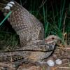
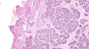
In the photo types of cataracts: 1 - front polar, 2 - zonular, 3 - zonular in flashing lighting, 4 -
starter, 5 - initial elder in flashing light, 6 - immature senile, 7 - mature old, 8 - Morganiev, 9 - secondary
- front polar;
- rear polar;
- vernoe;
- cork;
- nuclear;
- total;
- zonular;
In addition, cataract is divided into natural Cataract (hereditary) and acquired (senile, traumatic).
Symptoms
There are 4 period of the course of the disease:

1. Beginner
Miscellaneous develops unnoticed for the patient. The patient complains of a veil in front of the eyes, the bonding of burning items (moon, light headlights), flies. When viewing the eye, with an extended pupil on its black background, the wrinkles of gray color are noticeable.

The tops of the clouds are fixed to the center, and the base is to the periphery. Lounge lens along the periphery does not worsen visual sharpness. Because of this, this period proceeds asymptomatic, and the patient does not observe impairment of vision.
This phase reaches from month to several years.
2. Immature (swelling)

At this stage, patients complain about an unexpected decrease in visual acuity. In the study of the patient's eye, it is noticeable that the crystal is enlarged, thickened and is located in the area of \u200b\u200bthe pupil, has a gray-white color with a pearl tint. Nevertheless, the clamp itself also saves transparency itself, actually because with lateral lighting, permissible, the presence of a moon-shadow falling from the iris on the muddy layers of the lens.
This stage can last long, and then go to the following.
3. Ripe

At this stage, complete diffuser closer lens is performed. The patient does not see objects at all, it is capable of hardly recognizing the direction of light sources, since the light is left. At this stage, operational interventions are carried out. However, if the lens is not removed, then the cataract is moving to the next one.
4. Overripe
 Patient with overripe cataract form
 The dense cortical substance of the lens is slowly diluted and turns into a milk-shaped mass, inside which crystal base floats.
The lens decreases, the front camera of the eye is deepened, the heat of the iris occurs. Patient's vision is 0%.
These processes are irreversible. In this stage, complications may occur: an increase in intraocular pressure, the gap of the crust capsule.
The reasons
The causes of this disease can be: diabetes, High Degree Myopia, Professional Diseases, Radiation Therapy.
Diagnostics
Cataract diagnostics are contained in the main 2 methods:
- Determination of visual acuity.
- Ophthalmoscopy in straight and lateral projections.
 Diagnosing cataracts by ophthalmoscopy
 Treatment
When cataract, it is necessary to send a patient to the operational pulling into a lens and replacement to an artificial lens. Nevertheless, at each stage, treatment is different.
When a beginner and immature stage, conservative therapy is carried out (Eye drops: Quinaks, Katova). They prescribe 1-2 drops 2 times a day for a long time.
 Eye drops taken with initial symptoms Cataracts
 With a mature stage of cataracts actually operations to remove the lens and installation in its place artificial lenses. The operation is carried out actually in the provided stage, as in the overripe of a large risk of complications.
 Treatment of cataracts by the method of crust of the eye
 In the overripe stage of the cataract, the disclosed extraction is carried out. Cataracts, or deep eye enucleation.
 Cataract surgery
 Forecast
The forecast after a timely manner of crust change operations is extremely favorable for the patient. The patient from the first day notes an improvement in view. The acuity of vision is restored for 15 days. Next, if necessary, for the best visibility, it is possible to choose points.

However, throughout this time, the patient must take care of possible injuries to eliminate the likely complications. infectious diseases and sports. In the case of redness of the mucousa, an emergency visit to an ophthalmologist.
Complications
The patient rarely, however, an acute attack of glaucoma can happen, which is accompanied by pain in the eye and head, sickening at times vomiting, up to the loss of consciousness.

In addition, general complications are likely:
- hypotension;
- hypertensive crisis;
- brain circulation disorder;
- acute urine delay;
- mental disorders;
- other urgent states.
With their occurrence, it will be necessary immediately for help to an ophthalmologist.

Prevention
For the prevention of cataracts, it was necessary 2 times a year to cut the course of vitamins A, E, B, R. Also avoid injury to the eye, wearing sunglasses on a sunny day and in the misty, observe a protein diet.
Cataract, this is a relatively frequent phenomenon in people over 45 years old. And with the present medicine, the surgery for replacing the lens is carried out with ophthalmologists in 15 minutes, and they do not basically have any complications. The patient 2 hours after the operation can go home.
Although this disease and dangerous, however, has extremely favorable results. Due to the operation, patients can return vision by 80%. Which is quite good, despite the fact that the vision is 0% before operation.
In contact with
RCRZ (Republican Center for Health Development MD RK)
Version: Archive - Clinical Protocols MD RK - 2007 (Order No. 764)
Cataract Unclean (H26.9)
general information
Short description
Cataract - partial or complete turbidity of the substance or a crystal capsule, leading to a decrease in visual acuity up to its full loss.
Protocol code: P-S-013 "Cataract"
Profile: surgical
Stage: PMSP
Code (codes) on the ICD-10:
H25 senile cataract
H26 Other Cataracts
H28.0 Diabetic Cataract
Q12.0 Congenital Cataract
Classification
By time of emergence: Congenital acquired.
On the etiological factor:
1. Age.
2. Complicated (effect of uveitis, glaucoma, myopic disease).
3. Traumatic (contusion, after penetrating injuries).
4. Radi.
7. Cataracts with systemic pathology (diabetic, tetanic, motonic).
6. Cataracts arising from contact with toxic substances (Tea, naphthalene, thallium, trinitroxyol, dinitrophenol, nitrocracy) or drugs of drugs (GKS, sulfonamides, allopurinol, bisulfan, salt of gold, haloperidol, thiuridazine).
7. Secondary (postoperative clouding preserved during the operation of the rear capsule of the lens due to its fibrous change and the growth of subcapsular epithelium).
8. Congenital cataract.
Localization:
1. Front and rear polar.
2. Spit-shaped.
3. Sound.
4. Nuclear.
5. Cork.
6. Total.
7. Rear cup-shaped.
8. Polymorphic.
9. Vernoe.
In stage (age-related cataract):
1. Initial.
2. Immature.
3. Mature.
4. Overripe.
Risk factors and groups
1. Patients receiving GCS (systemically or locally).
2. Patients with diabetes mellitus.
3. Hereditary pathology (Martan syndrome, Wilson-Konovalov disease, a myotonic dystrophy (Steiner's disease), Down syndrome, trisomy of 13 and 15 pairs of chromosomes).
4. Hypoparatyoid state.
5. Patients with arterial hypertension.
6. Also the risk of development congenital cataract Increases the presence of pregnant syphilis, toxoplasmosis, cytomegalovirus infection.
7. Eye injuries.
8. Smoking.
Diagnostics
Diagnostic criteria
Complaints and history: A painless progressive reduction in visual acuity, developing gradually, paddle in front of the eyes, distortion of the form of objects.
Physical examination: Assessment of acuity of vision, outdoor inspection, biomicroscopy with a slit lamp, measuring VGD.
Laboratory research: Overall urine analysis, general blood test (ER, HB, L, leukoformula, eR, platelets, coagulation), Calla study on eggs worm, blood sugar, alt, asthma, tank. Analysis from a conjunctival cavity.
Tools: When inspection using a slit lamp, pile, grayish or greenish-brown. The reflection of the eye bottom at ophthalmoscopy is faded, weak or absent.
Indications for consultation of specialists: ENT, dentist, neuropathologist, pediatrician.
List of basic diagnostic measures:
1. Consultation ophthalmologist.
2. Check of visual acuity.
3. General analysis blood (6 parameters).
4. Bomicroscopy eye.
5. Honoscopy.
6. Perimetry.
7. Ophthalmoscopy.
8. Uzi eyes.
9. Tonometry.
List of additional diagnostic activities:
1. Consultation therapist.
2. Pediatrician consultation.
3. Consultation of the endocrinologist.
Differential diagnosis
|
Tumors (Retinoblastoma) |
The crystal is transparent, the tin formation of vascularized, white color will be determined behind the lens |
|
Retina detachment, retinopathy premature |
The crystal is transparent, with ophthalmoscopy - the retina is removed, fibroids changed, shorten. |
|
Persise primary vitreous body |
IN vitreous body Determined by a vitryoretinal litigation fixed to ZN |
| Cate Disease |
In the macular zone - subretinal dystrophic hearth, with an exudative retinal detachment |
|
Extensive chorus coat columus |
Defect of the vascular shell of varying degrees of length in the rear segment |
|
Intraocular inflammation (toxocamographs, metastatic endophthalmite) |
Chorioretinal focus in the rear segment, with clear boundaries, with inflammation there may be stules, with perifocal inflammation and exudation in the vitreous body. |
Treatment abroad
Treat treatment in Korea, Israel, Germany, USA
Get advice on medical examination
Treatment
Tactics of treatment
Treats of treatment: Preventing the further development of cataracts, preservation of visual acuity.
When progressing cataracts is a timely direction for surgical treatment.
Non-drug treatment: no.
Medicia treatment
Drug therapy should be used only at the initial stages of clouding lens in order to improve metabolic processes in lens tissues. It is not enough effective in clouds in the backyard of the lens, but gives a certain effect when long use and cloues in the front department.
Taurine is buried in a conjunctival bag 3 times a day for a long time.
Vitamin drops, as well as eye drops containing the amples, bury 3-5 times a day.
Information
Sources and literature
- Protocols for the diagnosis and treatment of diseases of the Ministry of Health of the Republic of Kazakhstan (Order No. 764 of 28.12.2007)
- 1. American ACADEMY OF OPHTHTHALMOLOGY. Guideline. Cataract in the adult eye. 2001 2. ICO International Clinical Guidelines. Cataract (Initial Evaluation) 2005. 3. EBM Guidelines. Cataract. 2004. 4. ALBERTA MEDICAL ASSOCIATION. Clinical Practice Guideline. Surgical and Non-Surgical Management of Cataract in the Otherwise Healthy Adult Eye. 2005. 5. American ACADEMY OF OPHTHTHTHALMOLOGY. Cataract (Initial and Follow-Up Evaluation). 2003. 6. National Guideline Clearinghouse. Clinical Practice Guideline for the Management of Cataract Among Adults. 2005.
Information
List of developers: Kramoshenko Yu.S. Eye eye diseases of the Republic of Kazakhstan.
Attached files
Attention!
- By self-medication, you can apply irreparable harm to your health.
- The information posted on the MedElement website and in MEDElement mobile applications, "Lekar Pro", "Dariger Pro", "Diseases: Therapist's Directory", cannot and should not replace a full-time doctor consultation. Be sure to contact medical facilities in the presence of any diseases or disturbing symptoms.
- The choice of medicines and their dosages should be stated with a specialist. Only a doctor can prescribe the necessary medicine and its dosage, taking into account the disease and the state of the patient's body.
- Website MedElement and Mobile Applications "MedElement (Medleylement)", "Lekar Pro", "Dariger Pro", "Diseases: Directory of Therapist" are exclusively information and reference resources. The information posted on this site should not be used for unauthorized changes to the doctor's prescriptions.
- The editorial office of MedElement is not responsible for any damage to health or material damage resulting from the use of this site.