Stereotactic radiation therapy. Stereotactic body radiation therapy (SLTT) Stereotactic radiation therapy for stomach cancer
S. I. Tkachev, S. V. Medvedev, D. S. Romanov, P. V Bulychkin, T. V. Yur'eva, R. A. Gutnik, I. P. Yazhgunovich, A. V. Berdnik B.
The emergence of innovative technical developments: three-dimensional planning, the use of a multi-leaf collimator modeled by the intensity of radiation therapy, and more advanced fixation methods have significantly increased the ability to accurately deliver and escalate the dose of ionizing radiation to the selected volume. This has changed the understanding of the role of radiation therapy in the treatment of metastatic liver disease. The data of foreign authors indicate the possibility of achieving 95% of local control one year after the stereotactic radiotherapy, 92% - after two years (and 100% for tumors less than 3 cm in size) with the development of radiation injuries of the third and higher degree in only 2% of cases. In 2011, after the technical re-equipment of the FSBI RONTs them. N.N.Blokhina RAMS, in clinical practice Local stereotaxic radiosurgery (SBRS) has been introduced to treat patients with metastatic liver disease. The technique allows creating a high dose of ionizing radiation locally in a metastatic tumor node and causing destruction of the tumor. This promising trend in the treatment of metastatic liver cancer has significantly expanded the possibilities of combination therapy. The article provides a review of the literature on the treatment of metastatic liver damage; we also publish the results of the use of stereotaxic radiosurgery in thirty-five patients with metastatic liver damage and a clinical case of the successful application of this technique in a somatically burdened patient.
Key words: metastatic liver damage, stereotactic radiosurgery, local control.
Contact Information:
S. I. Tkachev, S. V. Medvedev, D. S. Romanov, P. V. Bulychkin, T. V. Yurieva, R. A. Gutnik, I. P. Yazhgunovich, A. V. Berdnik, Yu. B. Bykov - Radiological Department, Department of Radiation Oncology (Head - Prof. S. I. Tkachev) FSBI RONTs them. N. N. Blokhin, RAMS, Moscow. For correspondence: Romanov Denis Sergeevich, [email protected]
Introduction
During autopsy, metastatic foci in the liver are found in 30% of patients with cancer. For the treatment of patients with multiple metastatic liver disease (more than three foci), systemic and / or regional drug therapy is preferable. In patients with limited liver damage, it is possible to use local treatment methods, such as: surgical resection, radiofrequency thermoablation, chemoembolization, radioembolization, cryodestruction, ethanol administration,
microwave coagulation, laser thermal destruction, electrolysis of metastases. Each of these approaches has its own advantages and disadvantages, but only stereotaxic radiation therapy can be used if there are contraindications for the use of the above techniques.
For a long time, radiation therapy was considered an unpromising method for the treatment of metastatic liver damage. The use of such a technique as total irradiation of the liver has not proved to be effective and safe, as, for example, irradiation of the entire brain in the case of metastatic
OF MALIGNANT TUMOURS
response of this body. With the improvement of the scientific and technological base of radiation therapy: the emergence of new technologies for administering a dose of ionizing radiation, planning systems, verification of plans for external radiation therapy, imaging, fixation of patients, the development of radiobiology, radiation oncologists have received a formidable weapon in the fight against metastatic liver damage - stereotaxic radiosurgery of neoplasms of the indicated organ.
Stereotactic radiosurgery
In the 90s of the last century, the first works on the feasibility of local stereotactic body radiation surgery (SBRS) for single (up to 3 foci) liver metastases appeared in foreign literature.
In connection with the biological characteristics of metastatic liver lesions in colon cancer, patients in this group were allocated to a separate subgroup. The gold standard for local treatment of liver metastases, in particular, colorectal cancer metastases, is liver resection. Several large studies show a 50% overall survival rate five years after surgery. Historically, it was considered possible to perform a liver resection in situations where it is possible to completely remove a limited number of metastases with a negative resection margin of more than one centimeter and a liver volume remaining after the operation sufficient for the organ to function adequately (at least 30% of the total functional liver volume). If these criteria are followed, resection is possible in 30-40% of patients who need it. At the moment, it is possible to simultaneously remove more than seven metastases from the liver; it was found that the width of the negative resection margin does not affect the local control and patient survival. In large centers dealing with this problem, the risk postoperative complications and mortality is reduced to minimum values. Moreover, repeated resections for recurrent liver cancer are quite safe.
and offer the same survival benefits as a first-time resection. Unfortunately, patients with synchronous bilobar, large, localized in inconvenient for surgical intervention, metastases and extrahepatic manifestations of the disease, those in whom resection does not leave the necessary 30% of the liver, patients over seventy years old and burdened somatically are often recognized as unresectable, and following this logic, unbreakable. In addition, there are no randomized trials comparing the effect after resection versus conservative non-surgical local therapy in operable patients.
The emergence of innovative technical developments (3D planning, multi-leaf collimator, intensity modulated radiation therapy (IMRT), improved fixation methods), which significantly increased the ability to accurately deliver ionizing radiation to a selected volume, and therefore deliver a higher dose to tumor volume, have changed the concept of the role of radiation therapy in the treatment of metastatic liver disease. An option for high-precision radiation therapy in which the ablative dose is delivered in 1-5 fractions is called stereotactic radiation therapy. When used extracranially, this type of radiation therapy is called stereotactic body radiation surgery (SBRS). In accordance with the ASTRO definition, SBRS involves the delivery of large doses of ionizing radiation with high conformity and a sharp dose gradient in the surrounding normal tissues in a small number of fractions (from two to six) to tumors located outside the brain.
There are many publications regarding the use of SBRS for the treatment of liver malignant lesions, which demonstrate encouraging results. The earliest of them date back to 1994-1995. In this paper, the researchers report the first results of SBRT on 42 extracranial tumors.
in 31 patients. 23 patients underwent radiation therapy for liver metastases (14 patients) or hepatocellular carcinoma (9 patients). Most of the patients had solitary tumors in the liver, lungs and retroperitoneal space. Their volumes of subclinical tumor spread (CTV - clinical target volume,) ranged from 2 to 622 cm3 (with an average in 14.2 Gy), were supplied in 1-4 fractions. The researchers noted local control in 80% of cases during the subsequent life of patients, which lasted from 1.5 to 38 months. In addition, the disappearance or reduction of tumors in size was noted in fifty percent of cases. The median follow-up period was 10 months for patients with hepatocellular carcinoma (values from 1 to 38 months) and 9 months for patients with metastatic liver disease (values from 1.5 to 23 months).
In 1998, the same research group reported on the experience of using stereotaxic radiosurgery for the treatment of primary malignant and metastatic liver tumors, the SOD ranged from 15 to 45 Gy, given in 1-5 fractions. Fifty patients with 75 tumors were treated. Volumes treated ranged from 2 to 732 cm3 (with an average of 73 cm3). During follow-up with an average value of 12 months (values ranged from 1.5 to 38 months), stabilization of the process was recorded in about 30% of cases, about 40% of tumors decreased in size and 32% completely regressed. Four (5.3%) tumors were interpreted as local failures. Unfortunately, average duration life was only 13.4 months (with values from 1.5 to 39 months) with a predominance of causes of death from progressive liver cirrhosis or extrahepatic progression of the underlying disease.
doses of 20 Gy (two fractions) or 15 Gy (three fractions). Within the follow-up period from 13 to 101 months, local control was achieved over all recurrent tumors with complete regression of metastases in two cases. Only one patient had a local progression of the disease in the form of damage to two lobes of the organ, which was preceded by extrahepatic spread of the disease. One patient subsequently died from non-oncological causes in the absence of signs of the underlying disease, two died from generalization of the malignant process, and one patient at the end of the study was in remission for 101 months after undergoing stereotactic radiosurgery.
Dawson et al. performed SBRT in 16 patients with hepatic metastases and 27 patients with primary hepatocellular carcinoma using 3D-conformal radiation therapy at an average dose of 58.5 Gy (28.5 to 90 Gy) at 1.5 Gy per fraction twice a day. There was one case of grade III RILD and no treatment-related deaths. In a more recent study by Dawson et al. simulated the possibility of complications from normal tissues for the development of RILD within 4 months after conformal radiation therapy for liver metastatic lesions or intrahepatic hepatobiliary tumors. The study demonstrated a significant effect of volume and average single focal dose on predicting the development of RILD in multivariate analyzes. Other significant predisposing factors for the development of RILD were primary diseases liver (cholangiocarcinoma and hepatocellular carcinoma versus metastatic lesion) and male sex. It was noted that these patients were also receiving concurrent topical chemotherapy and bromodeoxyuridine use (versus fluorodeoxyuridine) was also associated with an increased risk of developing RILD. There were no cases of RILD development when an average total focal dose of less than 31 Gy was applied to the liver.
In 2001 Herfarth et al. conducted a study that examined the effect of
Possibilities of stereotactic radiosurgery in the treatment of patients with metastatic liver disease
the effectiveness of using SBRS in 37 patients with 60 lesions in the liver. The absorbed dose was 26 Gy, and the tumor size varied from 1 to 132 cm3 with an average value of 10 cm3. All patients tolerated treatment well, SBRS did not result in any significant side effects... Eleven patients reported intermittent loss of appetite or mild nausea between one and three weeks after the end of treatment. None of the treated patients developed clinically detectable radio-induced liver disease. As a result of SBRS over 5.7 months (ranged from 1 to 26.1 months), fifty-four out of fifty-five (98%) tumors showed a positive effect, according to computed tomography performed after 6 weeks (22 cases of disease stabilization, 28 cases of partial answer and 4 cases of complete answer). The local positive effect was 81% within 18 months after the end of treatment.
Wulf et al. reported the results of SBRS in five patients with primary liver cancer and 39 patients with 51 hepatic metastases performed at the University of Wurzburg. Twenty-eight tumors were assigned to the so-called "low dose" group in three 10 Gy fractions (27 patients) or four 7 Gy sessions (1 patient). In addition, there was the so-called “high dose” group, in which patients were subjected to SBRS with single doses of 12-12.5 Gy in three fractions (19 patients) or 26 Gy per fraction (9 patients). Median follow-up was 15 months (range 2 to 48 months) for primary liver cancer and 15 months (range 2 to 85 months) for patients with metastatic liver disease. In all cases of primary malignant liver disease, a positive effect was achieved, including true stabilization. Among fifty-one metastases, 9 cases of local recurrence were noted in terms of three to 19 months. A delimiting significant correlation was noted between the total radiation dose and local control indicators (p = 0.077) with local control indicators at 86% and 58% after 12 and 24 months.
in the "low dose" group versus 100% and 82% in the "high dose" group, respectively. Not a single case of the development of radiation damage III or higher degrees of RTOG-EORTC. In multivariate analysis, high dose versus low dose was the only significant predictive factor for local control indicators (p = 0.0089). Overall survival at one and two years among all patients was 72% and 32%, respectively. The authors conclude that SBRS of primary malignant diseases and metastatic liver tumors is an effective local treatment method without significant complications for patients who were refused surgery.
In a study by Hoyer et al. , the results of the use of SBRS in the treatment of colorectal cancer metastases are presented. Sixty-four patients with a total of 141 metastases of colorectal cancer in the liver (44 patients) or lung (20 patients) were exposed to SBRS in three 15 Gy fractions for five to eight days. The median follow-up was 4.3 years, and after two years, local control rates were 86%. Radiation toxicity in most cases was moderate, however, there were three cases of serious adverse events and one death. The researchers concluded that SBRS for inoperable metastatic colorectal cancer is not inferior to other methods of local ablation of metastases.
Somewhat later, Schefter et al. reported preliminary results of a multicenter Phase I study of SBRS in patients with liver metastases. Patients had one to three liver metastases, with a maximum tumor diameter of less than six centimeters and adequate liver function. Some of the patients underwent SBRS at a total dose of 36 Gy in three fractions. Another part of the patients received higher doses of radiation up to 60 Gy in three fractions. At least 700 milliliters of healthy liver tissue should have received a total dose of less than 15 Gy. The dose-limiting toxicity was the manifestation of grade III acute radiation damage to the liver or intestines or any manifestation of acute radiation damage.
denium IV degree. No patient had any dose-limiting radiation injuries, so the radiation dose was raised to 60 Gy in three fractions. Twelve of the 18 patients were alive at the time of study by the investigators with a median of 7.1 months after enrollment.
The study was continued in 2006 by Kavanagh et al. reported the results of a phase I / II analysis of a prospective study of SBRS for the treatment of metastatic liver disease. In this case, the study included patients with no more than three tumors with a maximum diameter of less than six centimeters. The total focal dose was 60 Gy in three fractions over three to fourteen days. In 2006, interim results of SBRS were published in 36 patients: 18 from the first phase and 18 from the second phase. Among 21 patients with follow-up periods ranging from six to 29 months, there was only one case of development of third-degree RTOG radiation damage associated with performed SBRS, which occurred in the subcutaneous tissues. Not a single case of grade 4 radiation toxicity has been reported. The researchers noted that for 28 lesions over eighteen months, the positive effect, including true stabilization, was 93%.
In 2009, Rusthoven et al. published the results of a multicenter (conducted from August 2003 to October 2007 in 7 medical institutions) phase I / II study of SBRS use in patients with metastatic liver disease. The study included patients with 1-3 liver metastases and the maximum size of individual nodes less than 6 cm. The initial level of bilirubin, albumin, prothrombin and APTT, and the activity of liver enzymes were taken into account. Chemotherapy was not allowed for 14 days before and after SBRS. For 49 metastatic foci, local control rates were 95% (one year after SBRS) and 92% (two years after SBRS). In 2% of patients, radiation injuries of the third and higher degrees were detected with a median of 7.5 months after stereotaxic radiosurgery. The two-year rate of positive local effect for metastases up to 3.0 cm in diameter was
100%. This is the highest reported benefit rate, despite a 2-year survival rate of 30%. The authors conclude that the method of stereotaxic radiosurgery with a total dose of 60 Gy in three fractions is both safe and effective for treating patients with the number of hepatic metastases from one to three.
Van der Pool et al. in 2010 presented a study in which 20 patients with metastatic liver disease underwent SBRS at doses ranging from 30 to 37.5 Gy in three fractions. One hundred percent indicators of a positive local effect were obtained one year after the treatment. After two years, this figure dropped to 74%, with an average survival rate of 34 months. Among the radiation injuries, one case of rib fracture and 2 cases of elevated hepatic enzymes of the III degree are highlighted as long-term consequences of radiation therapy.
Also published in 2010 was the results of a prospective study by Goodman et al. , in which 26 patients with malignant liver tumors (19 of them with metastatic lesions) underwent SBRS in one fraction in the amount of 18-30 Gy. Local response rates at 12 months were 77%. The two-year survival rate for patients with metastatic liver disease was 49%.
In 2011, after technical re-equipment, at the Federal State Budgetary Institution of the Russian Oncology Center named after V.I. NN Blokhin Russian Academy of Medical Sciences in clinical practice for the treatment of patients with metastatic lesions of the liver introduced the technique of local stereotaxic radiosurgery (SBRS). The technique allows creating a high dose of ionizing radiation locally in a metastatic tumor node and causing destruction of the tumor. This promising trend in the treatment of metastatic liver cancer has significantly expanded the possibilities of combination therapy.
From August 2010 to July 2013 at the radiological department of the N.N. N.N.Blokhin RAMS SBRS performed on thirty-five patients with liver metastases of tumors of various histological structures. A single focal dose ranged from ten to twenty gray, radiosurgery was performed
Possibilities of stereotactic radiosurgery in the treatment of patients with metastatic liver disease
for three sessions within 5-7 days. Two patients did not submit data from the follow-up examination; in two more cases, local progression was recorded. Seven patients showed complete tumor regression, thirteen - partial, and eleven - stabilization of the treated lesions. In five patients, new metastatic lesions were subsequently identified in untreated areas of the liver. The median follow-up was 17 months. In none of the cases were early and late grade III-IV radiation injuries recorded; the incidence of grade II radiation injuries was 9%.
Conclusion
Only with the availability of modern equipment and technologies, there are prospects in the use of stereotaxic radiosurgery in the treatment of patients with metastatic liver disease. This technology is a real alternative to other methods of local impact on metastatic formations. The cited data of foreign authors, as well as the experience of the radiological department of the N.N. NN Blokhin RAMS testify to the high efficiency and safety of this technology, even in those patients who are denied other methods of treatment.
Clinical case
Patient A. 65 years old. Cancer sigmoid colon, metastatic liver disease, T4N1M1, stage IV.
06.07.10 the patient underwent palliative resection of the sigmoid colon. 07/29/10 - left-sided hemihepatectomy, resection of the right lobe of the liver.
On histological examination - adenocarcinoma.
After the operation, 8 courses of chemotherapy were carried out.
In August 2011, according to ultrasound data from 15.08.11, the progression of the disease in the form of solitary metastasis in the remaining part of the liver was revealed.
Up to 11/17/11, 7 courses of chemotherapy were carried out.
According to CT data from 10/26/11 between the portal and right hepatic veins, an education up to 2.7x2.5 cm is determined, in segment VII the focus is up to 0.9 cm (Fig. 1).
According to MRI data from 12/14/11, in the resection zone in the S5-S8 segments, the node is up to 1.8 cm, closely adjacent to the portal vein. In segments S6-7, a node up to 0.5 cm is determined.
From 12/21/11 to 12/27/11, a course of stereotaxic radiosurgery was carried out for both lesions in the liver using the IMRT technique, ROD 15 Gy, 3 times a week, SOD 45 Gy.
The patient was fixed using an individual vacuum mattress,
the verification of the irradiation program was carried out using the technology of computed tomography in a conical beam on the linear accelerator table in the treatment position.
According to CT data from 15.05.12, a new lesion appeared in the S6 liver up to 1.7 cm in size. Two lesions subjected to stereotactic radiotherapy are not visualized (Fig. 2).
Subsequently, the patient received treatment in South Korea. In July 2012 and February 2013, a radio frequency
ablation of the lesion in the S6 liver. The patient noted an increase in body temperature for a long time, an abscess was found at the site of metastasis in the S6 liver. On August 21, 2013, a surgical intervention was performed: in the areas of the liver available for visualization without signs of a malignant process, in the area of the resected focus in the S6 segment - tumor cells along the edge of the resection.
At the moment, the patient is alive. According to the survey data from August 2013, no signs of the disease were found.
Literature
1. Hoyer M., Swaminath A., Bydder S., et al. Radiotherapy for liver metastases: a review of evidence. Int J Radiat Oncol Biol Phys. 2012. V. 82 (3). P. 1047-57.
2. Lax I., Blomgren H., Naslund I., et al. Stereotactic radiotherapy of malignancies in the abdomen. Methodological aspects. Acta Oncol. 1994. V. 32 P. 677-683.
3. Poston G. J. Surgical strategies for colorectal liver metastases. Surg Oncol 2004. V. 13. P. 125-36.
4. de Haas R. J., Wicherts D. A., Flores E., et al. R1 resection by necessity for colorectal liver
metastases: is it still a contraindication to surgery? Ann Surg. 2008. V. 248 (4). P. 626-37.
5. de Jong M. C., Mayo S. C., Pulitano C., et al. Repeat curative intent liver surgery is safe and effective for recurrent colorectal liver metastasis: results from an international multi-institutional analysis. J Gastrointest Surg. 2009. V. 13 (12). P. 2141-51.
6. Potters L., Kavanagh B., Galvin J. M., et al. American Society for Therapeutic Radiology and Oncology (ASTRO) and American College of Radiology (ACR) practice guideline for the performance of stereotactic body radiation therapy.
Possibilities of stereotactic radiosurgery in the treatment of patients with metastatic liver disease
Int J Radiat Oncol Biol Phys. 2010. V. 76. P. 326-332.
7. Blomgren H., Lax I., Näslund I., Svanströ
m R. Stereotactic high dose fraction radiation therapy of extracranial tumors using an accelerator. Clinical experience of the first thirty one patients. Acta Oncol. 1995. V. 33. P. 861-70.
8. Blomgren H., Lax I., Goranson H., et al. Radiosurgery for tumors in the body: clinical experience using a new method. J Radiosurg. 1998. V. 1. P. 63-74.
9. Gunvén P, Blomgren H, Lax I. Radiosurgery for recurring liver metastases after hepatectomy. Hepatogastroenterology. 2003. V. 50 (53).
10. Dawson L. A., McGinn C. J., Normolle D., et al. Escalated focal liver radiation and concurrent hepatic artery fluorodeoxyuridine for unresectable intrahepatic malignancies. J Clin Oncol. 2000.
V. 18.P. 2210-2218.
11. Dawson L. A., Normolle D., Balter J. M., et al. Analysis of radiationinduced liver disease using the Lyman NTCP model. Int J Radiat Oncol Biol Phys. 2002. V. 53 (4). P. 810-821.
12. Herfarth K. K., Debus J., Lohr F., et al. Stereotactic Single-Dose Radiation Therapy of Liver Tumors: Results of a Phase I / II Trial. Journal of Clinical Oncology. 2001. V. 19. P. 164-170.
13. Wulf J., Guckenberger M., Haedinger U., et al. Stereotactic radiotherapy of primary liver cancer
and hepatic metastases. Acta Oncol. 2006. V. 45 (7). P. 838-47.
14. Hoyer M., Roed H., Hansen A. T., et al. Phase II study on stereotactic body radiotherapy of colorectal metastases. Acta Oncol. 2006. V. 45. P. 823-830.
15. Schefter T. E., Kavanagh B. D., Timmerman R. D., et al. A phase I trial of stereotactic body radiation therapy (SBRT) for liver metastases. Int J Radiat Oncol Biol Phys. 2005. V. 62. P. 1371-1378.
16. Kavanagh B. D., Schefter T. E., Cardenes H. R., et al. Interim analysis of a prospective phase I / II trial of SBRT for liver metastases. Acta Oncol. 2006. V. 45. P. 848-855.
17. Rusthoven K. E., Kavanagh B. D., Cardenes H., et al. Multi-institutional Phase I / II trial of stereotactic body radiation therapy for liver metastases. J Clin Oncol. 2009. V. 27. P. 1572-1578.
18. van der Pool A. E., Mendez Romero A., Wunderink W., et al. Stereotactic body radiation therapy for colorectal liver metastases. Br J Surg. 2010. V. 97. P. 377-382.
19. Goodman K. A., Wiegner E. A., Maturen K. E., et al. Dose-escalation study of single-fraction stereotactic body radiotherapy for liver malignancies. Int J Radiat Oncol Biol Phys. 2010. V. 78. P. 486-493.
Stereotactic radiosurgery (SRS) is a field of radiation therapy that involves the use of high-precision radiation. CPX was originally used to treat tumors and other pathological changes in the brain. Currently, radiosurgical techniques (called extracranial stereotaxic radiotherapy, or stereotaxic body radiotherapy) are used to treat malignant neoplasms of any location.
Despite its name, CPX is not a surgical procedure. The technique implies high-precision delivery of high-dose radiation to the tumor bypassing healthy adjacent tissues. This is what distinguishes CPX from standard radiation therapy.
When performing stereotaxic radiosurgery, the following technologies are used:
- Three-dimensional imaging and localization techniques that allow you to determine the exact coordinates of the tumor or target organ
- Devices for immobilization and careful positioning of the patient
- Clearly focused beams of gamma rays or x-rays that converge on a tumor or other pathological formation
- Image-guided radiotherapy techniques that track the position of the tumor throughout the entire irradiation cycle, thereby increasing the accuracy and efficiency of treatment
Three-dimensional imaging techniques such as CT, MRI and PET / CT are used to determine the location of a tumor or other pathological focus in the body, as well as their exact size and shape. The images obtained are essential for planning treatment, during which the beams of rays approach the tumor from a variety of angles and at different planes, as well as for carefully positioning the patient on the treatment table during each session.
As a rule, stereotaxic radiosurgery is performed simultaneously. However, some experts recommend multiple sessions of radiation therapy, especially for large tumors larger than 3 to 4 cm in diameter. A similar technique with the appointment of 2-5 treatment sessions is called fractionated stereotactic radiotherapy.
CPX and extracranial stereotaxic interventions represent an important alternative to open surgical procedures, especially for patients who are unable to undergo surgery. In addition, stereotaxic interventions are indicated for tumors that:
- Located in hard-to-reach places for the surgeon
- Are located near vital organs
- Change their position during physiological movements, such as breathing
Radiosurgical procedures are used in the following cases:
- for the treatment of many brain tumors, including:
- benign and malignant neoplasms
- primary and metastatic lesions
- single and multiple tumors
- residual tumor foci after surgery
- intracranial lesions and tumors of the base of the skull and orbit
- for the treatment of arteriovenous malformations (AVMs), which are clusters of reshaped or enlarged blood vessels... AVMs disrupt the normal blood flow of nervous tissue and are prone to bleeding.
- For the treatment of other neurological conditions and diseases.
Extracranial stereotactic radiotherapy is currently used for malignant and benign tumors of small to medium size, including tumors of the following localizations:
- Lungs
- Liver
- Abdomen
- Spine
- Prostate
- Head and neck
CPX is based on the same principle as for other methods of radiation therapy. In fact, the treatment does not eliminate the tumor, but only damages the DNA of the tumor cells. As a result, cells lose their ability to reproduce. After the performed radiosurgical intervention, the size of the tumor gradually shrinks over a period of 1.5-2 years. In this case, malignant and metastatic foci decrease even faster, sometimes within 2-3 months. If CPX is used for arteriovenous malformation, then within several years there is a gradual thickening of the vessel wall and complete closure of its lumen.
What equipment is used for stereotaxic radiosurgery?
There are three main methods of carrying out stereotaxic radiosurgical operations, in each of which certain devices serve as a radiation source:
- Gamma knife: 192 or 201 beams of highly focused gamma rays are used to irradiate the target organ. The Gamma Knife is excellent for treating small to medium sized intracranial lesions.
- Linear Accelerators are devices that are widely used around the world and are used to deliver high-energy X-rays (photon beams). Suitable for the treatment of large tumor foci. The procedure can be performed once or in several stages, which is called fractionated stereotaxic radiosurgery. The equipment is manufactured by various manufacturers who produce linear accelerators under different names: Novalis Tx ™, XKnife ™, CyberKnife®.
- Proton therapy, or heavy particle radiosurgery - is now performed only in some centers in North America, but the availability and popularity of treatment has continued to grow lately.
Which specialists are involved in performing stereotaxic radiosurgery? Who operates the stereotaxic radiosurgery equipment?
Stereotaxic surgery requires a team approach. The treatment team includes a radiation oncologist, a medical physicist, a dosimetrist, a radiologist / radiology technician, and a nurse in the radiology department.
- The team is led by a radiation oncologist and, in some cases, a neurosurgeon who oversees the treatment process. The doctor determines the boundaries of the area of radiation exposure, selects the appropriate dose, evaluates the developed treatment plan and the results of the radiosurgical procedure.
- The results of the examination and the images obtained are assessed by a radiologist, which makes it possible to identify a pathological focus in the brain or other organs.
- A medical physicist, together with a dosimetrist, uses special computer programs to develop a treatment plan. The specialist calculates the radiation dose and determines the parameters of the beam of rays for the most complete effect on the pathological focus.
- The radiologist and / or radiological technician is directly involved in performing the radiosurgery. The specialist assists the patient on the treatment table and controls the equipment from a shielded room. The radiologist, who can communicate with the patient through a microphone, observes the procedure through the viewing window or using video equipment.
- The nurse of the radiology department helps the patient during and after the procedure and monitors his condition, assessing the occurrence of side effects of treatment or other undesirable phenomena.
- In some cases, a neurologist, neurosurgeon, or neuro-oncologist is involved in the treatment to help select the most appropriate treatment for tumors or other brain lesions.
How is stereotaxic radiosurgery performed?
Radiosurgical treatment with the Gamma Knife system
Radiosurgical treatment using the system Gamma knife consists of four stages: setting a fixing frame on the patient's head, visualizing the position of the tumor, drawing up a treatment plan using a computer program, and the procedure of the irradiation itself.
At the beginning of the first phase, the nurse sets up the intravenous infusion system. drugs and contrasting material. After that, the neurosurgeon performs anesthesia of the scalp at two points on the forehead and two points on the back of the head, and then, using special screws, fixes a special rectangular stereotaxic frame to the skull. This prevents unwanted head movements during the procedure. In addition, a lightweight aluminum frame serves to direct the movement of gamma rays and focus them on the tumor.
During the second stage, magnetic resonance imaging is performed, which makes it possible to determine the exact position of the pathological area in relation to the fixing frame structure. In some cases, computed tomography is done instead of MRI. In the treatment of arteriovenous malformation, angiography is also prescribed.
During the next phase, which lasts about two hours, the patient rests. At this time, a group of attending physicians analyzes the images obtained and determines the exact position of the tumor or pathologically altered artery. With the help of special computer programs, a treatment plan is developed, the purpose of which is to optimally irradiate the tumor and maximize the protection of the surrounding healthy tissues.
At the beginning of the last stage of treatment, the patient lies down on the couch, and the frame frame is fixed on his head. For convenience, a nurse or technologist offers the patient a pillow under the head or a special mattress made of soft material and covers it with a blanket.
Before starting treatment, the staff moves to the next room. The doctor monitors the patient and the course of treatment using a camera installed in the treatment room. The patient can communicate with the medical staff using a microphone mounted in the frame.
After all the preparations, the couch is placed inside the Gamma Knife apparatus and the procedure begins. The treatment is completely painless, and the device itself does not make any sounds.
Depending on the model of the Gamma Knife and the treatment plan, the procedure is performed simultaneously or divided into several small sessions. The total duration of treatment is 1 to 4 hours.
The end of the procedure is announced by a bell, after which the couch returns to its original position, and the doctor removes the fixing frame from the patient's head. In most cases, the patient can go home immediately after the procedure.
Radiosurgical treatment with a medical linear accelerator
Radiosurgical treatment with linear particle accelerator proceeds in a similar way and also consists of four stages: installation of a fixing frame, visualization of the pathological focus, planning of the procedure using a computer program and the actual irradiation.
Unlike the Gamma Knife, which remains stationary during the entire procedure, the beams of rays enter the patient's body at different angles while continuously rotating a special device called a gantry around the couch. If the radiosurgical procedure is performed using the CyberKnife system, then a robotic arm-manipulator rotates around the patient's couch under imaging control.
Compared to the Gamma Knife, the linear accelerator generates a larger beam of rays, which makes it possible to uniformly irradiate large pathological foci. This property is used in fractionated radiosurgery or stereotaxic radiation therapy using a movable fixation frame and is a great advantage in the treatment of large tumors or neoplasms near vital anatomical structures.
Extracranial stereotactic radiotherapy (ESRT)
The ESRT course usually takes 1-2 weeks, during which 1 to 5 treatment sessions are carried out.
Before radiotherapy, as a rule, fiducial marks are placed in or near the tumor. Depending on the localization of the pathological formation, this procedure, during which from 1 to 5 marks are installed, takes place with the participation of a pulmonologist, gastroenterologist or radiologist. Usually this stage is carried out on an outpatient basis. Not all patients require orientation marks.
At the second stage, radiotherapy is simulated, during which the doctor chooses the most appropriate way to direct the path of the beam of rays relative to the position of the patient's body. At the same time, immobilization and fixation devices are often used to accurately position the patient on the couch. Some devices are quite firmly fixing the patient, so the doctor should be notified of the presence of claustrophobia in advance.
After creating a personal fixation device, computed tomography is performed to obtain a picture of the area that will be affected by the radiation. CT is often "four-dimensional", which involves the creation of images of a target organ in motion, such as breathing. This is especially important for lung or liver tumors. After the scan is complete, the patient is allowed to return home.
The third stage of ESRT involves developing a treatment plan. At the same time, the oncologist-radiologist works in close cooperation with a medical physicist and a dosimetrist, which makes it possible to approximate the shape of the beam of rays as accurately as possible to the parameters of the tumor. Radiotherapy planning may require an MRI or PET / CT scan. Through a dedicated software medical staff evaluates hundreds of thousands of different combinations of radiation beams to select the most appropriate parameters for a given case.
The delivery of radiation during ESRT is carried out using a medical linear accelerator. The session does not require any restrictions on food or liquid intake. However, many patients are prescribed anti-inflammatory medications, anti-anxiety medications, and anti-nausea medications before the procedure.
At the beginning of each session, the position of the body is fixed using a prefabricated device, after which an X-ray is taken. Based on the results, the radiologist corrects the patient's position on the couch.
After that, the actual radiation therapy session is performed. In some cases, additional X-rays are required to monitor the position of the tumor during the session.
The duration of the session can be about one hour.
Does the patient require special training for stereotaxic radiosurgery?
Stereotactic radiosurgery and ESRT are usually performed on an outpatient basis. However, short hospitalization may be required.
The doctor must notify the doctor in advance of the need to accompany the patient home by a relative or friend.
You may need to stop eating and drinking 12 hours before your session. It is also important to ask your doctor about drug restrictions.
The doctor should be advised of the following:
- About taking medications by mouth or insulin for diabetes mellitus.
- About availability allergic reactions on intravenous contrast materials, iodine or seafood.
- About the presence of an artificial pacemaker, heart valves, defibrillator, clips for cerebral aneurysms, implanted pumps or ports for chemotherapy, neurostimulators, eye or ear implants, as well as any stents, filters or coils.
- Claustrophobia.
What to expect during stereotaxic radiosurgery?
Radiosurgical treatment is similar to a conventional X-ray examination because it is impossible to see, feel or hear the X-rays. An exception is radiation therapy for brain tumors, which can be accompanied by flashes of light even with closed eyes. The session of radiosurgical treatment itself is absolutely painless. It is important to inform your doctor about the appearance of pain or other discomfort, such as back pain or discomfort when applying a fixing frame or other immobilization devices.
When removing the fixation frame, there may be slight bleeding, which is controlled with a bandage. Sometimes there are headaches that can be treated with medication.
In most cases, after completing radiosurgical treatment or ESRT, you can return to your normal life in 1-2 days.
Side effects of radiation therapy are the result of both direct exposure to radiation and damage to healthy cells and tissues around the tumor. The number and severity of RTVC adverse events depend on the type of radiation and the dose prescribed by the doctor, as well as on the localization of the tumor itself in the body. Any side effects that arise should be discussed with your doctor so that he can prescribe the appropriate treatment.
Early side effects occur during or immediately after stopping radiation therapy and usually resolve within a few weeks. Late side effects appear months or even years after radiation therapy.
Typical early side effects of radiation therapy are fatigue or tiredness and skin problems. The skin at the site of exposure to radiation becomes sensitive and reddens, irritation or swelling appears. In addition, itching, dryness, flaking and blistering of the skin are possible.
Other early side effects are determined by the area of the body that is affected by the radiation. These include:
- Hair loss in the area of radiation
- Ulceration of the mucous membrane oral cavity and difficulty swallowing
- Loss of appetite and indigestion
- Diarrhea
- Nausea and vomiting
- Headache
- Soreness and swelling
- Urinary disorders
Late side effects are rare and occur months or years after radiation therapy, but they persist long-term or permanently. These include:
- Changes in the brain
- External changes spinal cord
- Lung changes
- Kidney changes
- Colon and rectal changes
- Infertility
- Joint changes
- Edema
- Oral changes
- Secondary malignancy
Radiotherapy carries an extremely low risk of developing new malignant tumors. Following treatment for cancer, it is very important to follow a regular check-up schedule with an oncologist, who assesses for signs of recurrence or the appearance of a new tumor.
Radiotherapy techniques such as ESRT allow radiation oncologists to maximize the harmful effects of radiation on the tumor, while minimizing the impact on healthy tissues and organs and limiting the risk of side effects of treatment.
The CYBERKNIFE Center is located at the Grosshadern University Hospital of Munich. It is here that since 2005 the treatment of patients has been carried out using the latest development in the field of medicine called CYBERKNIFE (Cyberknife). This unique equipment is the safest and most effective of all methods of treating benign and malignant neoplasms.
This technique of radiosurgery is comparable to a surgeon's scalpel, but it is also different - first of all, the result is invisible immediately, and occurs after a few weeks - due to focused targeted irradiation of the tumor, its cells die off gradually, therefore, after 2-3 months with large tumors, an additional session can be prescribed , then this course of treatment is called fractionated stereotactic radiotherapy. In fact, the treatment does not eliminate the tumor, but only damages the DNA of the tumor cells, as a result of which the cells lose their ability to reproduce. After the performed radiosurgical intervention, the size of the tumor gradually shrinks.
Who is radiosurgery indicated for?
Radiosurgical treatment by the stereotaxic method is effective and safe for the following diagnoses:- With tumors of the brain and spinal cord, benign and malignant, including more than 3-4 cm in diameter, as well as:
- Primary and metastatic lesions of the brain, lungs and liver
- Single and multiple tumors
- Residual tumor foci after surgery
- Intracranial lesions and tumors of the base of the skull and orbit
- Recurrent brain cancer
- For cancerous tumors located in places that are difficult to access for surgery
- With neoplasms located next to vital organs
- For soft tissue tumors that change their position during physiological movements, such as breathing
- For treatment arteriovenous malformations(AVM), which are collections of altered or dilated blood vessels.
It is also used for a number of benign (non-cancerous) brain tumors such as
- Acoustic neuromas
- Skull base meningiomas
- Pituitary adenomas
- Chordomas
Benefits of radiotherapy in Israel
Israeli clinics have everything the latest equipment available in the world for the treatment of cancer. Radiation stereotactic therapy is carried out on various accelerators and gives the patient advantages in non-invasive treatment and in high efficiency, statistics indicate that almost half of cancer patients at one stage or another are prescribed radiation therapy as the first or second line of treatment, which indicates its relevance practically for every second cancer patient.So, only some the advantages of cancer treatment in Israel using radio-beam therapy:
- Radiotherapy allows you to treat brain tumors (cancer) without craniotomy. The presence of a mode of super-high dose rate and high accuracy of the positioning system makes it possible to "remove" the tumor without surgical intervention in one irradiation session.
- One treatment session replaces 3 weeks of radiation therapy
- There is no need for long-term hospitalization - in most cases, the patient is discharged home on the day of therapy
- Unique precision of impact on the tumor, in which healthy tissues are practically not exposed to radiation
- The possibility of intraoperative radiation therapy immediately after removal of the tumor on its bed, thus local radiation therapy completes the operation, as a result of which shortens the treatment process.
Convalescence!
Stereotactic radiotherapy for oncological diseases is one of the effective methods treatment of oncological diseases organized by our center. Stereotactic radiosurgery (SRS) takes place (despite the name) without a surgical scalpel, this radiation therapy technology does not “cut out” the tumor, but damages the DNA metastases. Cancer cells lose their ability to reproduce, and benign formations are significantly reduced in 18-24 months, and malignant ones much faster, quite often within 60 days.
The following cancers are treated with stereotactic radiotherapy:
- pancreatic, liver, and kidney cancer;
- tumors of the brain and spinal column;
- cancer of the prostate and lungs.
CPX provides extreme accuracy of the impact on the affected organ, without the danger of damaging adjacent tissues and organs. The accuracy of radiation delivery is based on the following components of the stereotaxis technology:
localization using three-dimensional visualization allows you to establish the exact coordinates of the tumor (target, target) in the body;
equipment for fixing the patient in a stationary position during the procedure;
sources of gamma or X-ray radiation allowing to focus the rays directly on the pathology;
visual control of the delivery of radiation to the affected organ before the procedure, correction of the direction of the rays during the procedure.
Stereotactic radiotherapy as an alternative to invasive surgery
Invasive surgery involves penetration to pathology through healthy organs and tissues, that is, intervention through the skin, mucous membranes and other external barriers of the body, respectively, damaging them. For tumors and various anomalies of blood vessels located near vital organs or pathologies deep in the brain, intervention is undesirable.
Stereotaxis treats pathologies minimally affecting adjacent tissues, it is mainly used in the treatment of neoplasms of the brain and spine, but it is also used in the treatment of arteriovenous diseases. Radiation exposure to arteriovenous malformations (AVMs) leads to their compaction and disappearance within several years.
The absence of injuries allows using the stereotaxic technique not only in neurosurgery, but also in studies of the work of the deep structures of the brain.
Stereotactic technique (from Greek: "stereos" - space, "taxis" - location) provides the possibility of low-traumatic access to all parts of the brain, and is a complex technology for the treatment of cancer based on radiotherapy, mathematical modeling, and the latest achievements of neurosurgery.
SLTT and CPX are modern high-precision radiation therapy techniques with the targeted use of high doses of radiation. CPX and SLTT are almost the only alternative for patients who may not undergo surgery, as well as for malignant and benign tumors that:- localized in places that are difficult to access for surgical intervention;
- poorly located relative to vital parts of the body;
- can move;
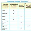
Application of SLTT
For the treatment of small (up to 6 cm) isolated malignant neoplasms in:- lungs: in the vast majority (up to 95%) it is possible effective use SLTT. This applies to both primary and secondary lung cancer.
- liver: primary and secondary with a tumor size of up to 6 cm in 90-100% of cases, SLTT is effectively treated.
- spine: 80-90% of paravertebral tumors succumb therapeutic effect SLTT.
- organs and tissues of the genitourinary system.
- inoperable cancer;
- metastases formed after application.
CPX application:
- small brain tumors;
- dysfunction of the brain.
Pros of SLTT and CPX:
- It is a non-invasive treatment that reduces side effects.
- Spot irradiation can reduce damage to healthy tissue to a minimum.
- In terms of effectiveness, SLTT and CPX are not inferior to surgical methods.
Limitations of SLTT and CPX:
- They require the use of high-precision technology, which is not available in every medical center.
- Quite high cost.
Stages of SLTT and CPX
- Consultation with an oncologist
- Simulation of radiation with the aim of adjusting the beam in relation to the location of the tumor and the location of the patient's body.
- CT of the site of the forthcoming exposure. For areas in the lungs and liver in Israel, a four-dimensional CT scan is used, which tracks the movement of the tumor during breathing. Volumetric visualization of the shape, location of the tumor, as well as related physiological characteristics used to plan upcoming therapy.
- Drawing up a therapy plan: selection of the shape of the beam, the number of sessions, if necessary, additional imaging of the tumor: MRI, PET.
- The actual radiation therapy session using a linear accelerator (LINAC). The patient is rigidly fixed in order to avoid accidental body movements: the radiation beams must hit exactly the specific area at different angles. In Israel, a patient-friendly Body Frame retainer is used. In addition, when working with a tumor. Located in areas where it moves when the patient breathes: lungs, abdomen etc., the technique of synchronization with breathing is used, when irradiation is performed only on inhalation / exhalation for the most targeted hit of the pathology area and preservation of undamaged tissues. The duration of the session is about 40 minutes.
- During therapy, fluoroscopy helps to simultaneously check the effectiveness of treatment and make adjustments if necessary.