Trigeminal neuropathy mcb. Methods for the treatment of trigeminal neuropathy Inflammation of the trigeminal nerve
Trigeminal neuralgia is a chronic neurological disease, with characteristic paroxysmal attacks of pain in the face of an excruciating shade. For the first time a description of trigeminal neuralgia was given at the end of the 18th century.
Information for doctors. According to ICD 10, trigeminal neuralgia is coded under the code G50.0 (paroxysmal facial pain syndrome). The diagnosis indicates the localization by branches, the stage of the disease (exacerbation, remission, etc.), the course of the disease, the frequency of attacks and the severity pain, the presence of sensory disorders.
Causes
For a long time, there was no single view of the causes of neuralgia. However, it has now been established that in the vast majority of cases the disease is caused by a neurovascular conflict - a vessel passing next to the trigeminal nerve. Sometimes the disease is caused by the narrowness of the bony canals in which the branches of the trigeminal nerve are located.
Sometimes the disease develops after an illiterate operation to remove a tooth. Is developing inflammatory process, which in turn goes to the trigeminal nerve. Much less often, the cause of neuralgia is traumatic injury to the bones of the facial skull, leading to direct trauma to the branches of the nerve. Sometimes herpes zoster, a brain stem tumor, multiple sclerosis.
Symptoms
Classically, the disease manifests itself in the form of attacks of shooting, burning pain in the face along the innervation of a certain branch of the nerve (most often the second, less often the third and very rarely the first branch). Pain attacks are often accompanied by autonomic reactions. It can be lacrimation, discharge of discharge from the nose, fever, profuse sweating, etc.
Despite the pronounced intensity of the pain, the attack is most often not accompanied by a cry, because additional jaw movements aggravate the pain. More often there is a pain tic in the form of contraction of facial muscles, a suffering expression appears on the face. Also, sometimes there is a reflex movement of the patient's hands to the face during an attack, but the slightest touch can, on the contrary, provoke pain.
In the interictal period, all patients are afraid of new pain sensations. As a result, people most often try to avoid provoking factors. This is manifested in the fact that a person does not chew on the affected side, does not brush his teeth, do not wash, men may not shave.
With a long course of the disease, changes in sensitivity almost always occur. Initially, there is hyperesthesia (increased sensitivity), ultimately, constant aching pain in the innervation area develops, while hyperesthesia can be transformed into hyposthesia and the presence of numbness.
Author's video
Diagnostics
Diagnosis of the disease is usually straightforward and is based on complaints, anamnesis data and a general neurological examination. In a neurological examination, attention is drawn to the frequent decrease in the corneal reflex on the side of the lesion, the presence of trigger zones on the face that reliably cause an attack, a change in skin sensitivity, and trismus.
Additionally, in some cases, it is possible to conduct an MRI or MSCT examination of the brain. Neuroimaging is aimed at excluding oncopathology of the brain, the presence of neuromas of the cranial nerves, and determining the places of narrowness of the nerve canals.
Treatment
Treatment is subdivided into drug treatment of trigeminal neuralgia, physical therapy, general prophylactic measures, and surgical treatments.
 Drug treatment includes the appointment of anticonvulsants. Finlepsin (carbamazepine) is most often used in small doses, 2-3 times a day. Also, in parallel, patients are prescribed funds to improve microcirculation (pentoxifylline), neuroprotection (B vitamins). With prolonged use, side effects may occur, a decrease in effectiveness, therefore, patients in such cases are transferred to other drugs (thebantine, lyrics, valproic acid preparations, etc.), or the treatment with GABA drugs (phenibut, pantogam, etc.) ...
In all cases, it is possible to carry out physiotherapy measures. The most commonly used technique of medicinal electrophoresis with novocaine is similar to the Berganier half mask. Less commonly used are magnetic fields, laser therapy.
For general prophylaxis, all patients are advised to thoroughly sanitize all foci of chronic infection, to cure dental pathology (caries, etc.). It is recommended to refuse to take excessively hot or cold food, it is recommended to avoid hypothermia, the development of viral infections.
In some cases, especially with a long course of the disease, the presence of pronounced depression, it is necessary to consult a psychotherapist. However, a neurologist can also prescribe antidepressants.
In case of ineffectiveness of the therapy, surgical treatment is necessary in the conditions of the department maxillofacial surgery... Surgeons perform medicinal nerve blocks, perform osteoplastic operations on the nerve canals in order to expand them. If these measures are ineffective, alcoholization of the nerve is necessary (destruction of the nerve fiber alcohol solutions), or transection of the nerve.
ICD-10 was introduced into health care practice throughout the Russian Federation in 1999 by order of the Ministry of Health of Russia dated 05/27/97. No. 170
A new revision (ICD-11) is planned by WHO in 2017 2018.
As amended and supplemented by WHO
Processing and translation of changes © mkb-10.com
Trigeminal neuralgia - treatment and symptoms
Facial trigeminal neuralgia is often manifested by seizures with severe pain symptoms, as well as other signs. If treatment is not carried out, then in the future the manifestations of the disease will worsen, which can lead to various complications.
In another way, trigeminal neuralgia is called Trousseau's tick. This nerve element is designed to control the facial and chewing muscles. With neuralgia, symptoms of intense pain usually appear in the places where the nerve branches pass.
Photo 1. What is the trigeminal nerve responsible for?
The disease is often diagnosed in people of working age; young children rarely get it. It is worth noting that, in comparison with other pathologies that the peripheral nervous system may suffer, trigeminal neuralgia causes the most unpleasant symptoms. In addition, the complexity of treatment is influenced by a wide variety of causes that can cause pathology and its progression. In addition, difficulties often arise already at the stage of diagnosis, since the signs of neuralgia are similar to other disorders. nervous system.
Causes
The variety of factors due to which trigeminal neuralgia appears is quite large. They are divided into two groups and belong, respectively, to exogenous, that is, external, and endogenous, that is, internal. Usually, the disease occurs as a result of:
- Serious mechanical injury to the facial area or skull
- Hypothermia
- Abnormalities in the blood supply to the areas where the vessels approach the trigeminal nerve. Quite often these are the consequences of atherosclerosis, abnormalities in the structure of blood vessels, the presence of an aneurysm, etc.
- Impaired metabolism
- Stem stroke
- Various chronic diseases
- The appearance of cancerous or benign neoplasms
- Multiple sclerosis
- The existence of cystic phenomena in the area where the nerve endings pass. The reason for such formations is often incorrect or insufficient dental, ophthalmological and other treatment.
Photo 2. Scheme of innervation
Symptoms
In most cases, almost three-quarters, the trigeminal nerve undergoes neuralgia on the right side. Rarely enough, the defeat covers both sides at once. The disease develops during periods of exacerbation and remission, when its symptoms either intensify or disappear. The most favorable period for exacerbations is autumn and spring, which is associated with an increase in humidity along with a low temperature. It must be remembered that if the ailment is not treated, then bilateral manifestations cannot be avoided in the future.
Trigeminal neuralgia manifests itself various symptoms, consider in detail each of their groups.
Painful
Pain symptoms are the most common and almost always occur. Their character is intense and sharp, they are not always present, manifesting themselves in seizures. Often, during an exacerbation, a person prefers not to move at all, so as not to provoke an increase in pain, and is in this state until the pain subsides. Many people compare these manifestations with electric shock, which was launched over the body. Usually an attack of trigeminal neuralgia lasts a few minutes, but it will usually repeat dozens or even hundreds of times a day, which negatively affects the strength and psychological state the patient.
The location of the pain usually corresponds to the location of the branches of the nerve on the face. However, in some situations, symptoms are present throughout the face and it is not possible to identify their specific location, since there is irradiation between the nerve branches. If the patient does not take action and treatment is not carried out, then the area of pain will steadily increase.
Quite often, pain appears after physically provoking trigger sites. In such cases, a simple push is enough to trigger an aggravation. Trigger areas on the face include the area:
- Corner of the mouth
- Brows
- Dorsum of the nose
- Inner surfaces of the cheeks
Motor functions
Quite often, trigeminal neuralgia leads to muscle spasms in the face. These symptoms gave the second name of the disease, which is also called pain tic. With an exacerbation, there is an uncontrolled contraction of the muscle structures responsible for chewing, the circular eye muscles and others. Usually manifestations are noticeable on the side on which pain symptoms and neuralgia are observed.
Reflexes
The presence of reflex disorders that may relate to the mandibular, superciliary and corneal zones cannot be independently identified. Only a neurologist can do this when examining a person.
Vegetative and trophic
These symptoms are hardly noticeable at an early stage of the disease, they appear in more advanced cases directly during an exacerbation and consist in:
- Severe blanching or redness of the skin of the face
- Increased lacrimation and salivation
- Runny nose
- With an advanced stage, swelling of the facial area, falling eyelashes, dry skin are often observed
Symptoms at a later stage
- Painful sensations change character from paroxysmal to permanent
- The entire half of the face hurts right away, where trigeminal neuralgia is present
- Anything can cause pain syndrome - loud sounds, bright light, as well as memories of the pathology itself.
Diagnostics
If there are signs of trigeminal neuralgia, the main of which is pain in the facial area, then it is important not to postpone a visit to a neurologist for a diagnosis, since in the absence of treatment the disease progresses very quickly. The first step is to assess symptoms and take a history. The second mandatory step is a neurological examination, checking reflexes and sensitivity of various parts of the head. If the disease is in remission, then its diagnosis becomes much more difficult. In such conditions, pathologies can only be detected with the help of an MRI of the head.
Additional diagnostic methods include:
- Dental examination, as often neuralgia occurs against the background of tooth damage, poor-quality installation of dentures, and so on. In this case, you can identify the cause using a panoramic X-ray of the head, which will help identify a pinched trigeminal nerve.
- Electromyography, which shows the patency of electrical impulses along the nerve endings.
- Complete blood count, which excludes or confirms the viral origin of neuralgia
Treatment
Depending on the degree of damage to the trigeminal nerve and, accordingly, how pronounced its neuralgia is, treatment is prescribed by various methods:
- Physiotherapy
- Medication
- Operative intervention
The first two methods are usually combined, and the third is used in the absence of the effect of conservative therapy.
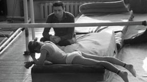
Photo 3. Treatment with physiotherapy
With physiotherapy, treatment is carried out:
- Dynamic currents
- Electrophoresis
- Laser therapy
- Phonophoresis
Medication therapy includes an appointment as prescribed by a doctor the following drugs with anticonvulsant and antispasmodic effects:
- Finlepsin. This remedy is used quite often, as it is a very effective anticonvulsant that relieves neuropathic pain.
- Carbamazepine
- Baclofen
- Gabapentina
- Sodium oxybutyrate
- Trentala
- Nicotinic acid
- Vitamna B
- Glycine
If conservative treatment the result could not be achieved, then surgical treatment is prescribed, which can be carried out by the method:
- Percutaneous balloon compression
- Microvascular decompression
- Glycerin injections
- Radiofrequency ablation
- With ionizing radiation, due to which the trigeminal nerve undergoes partial destruction in the affected area
- Using ionizing radiation to destroy the affected nerve.
ICD-10 code - G50 Disorders of the trigeminal nerve
Effects
A neglected condition and the lack of treatment for trigeminal neuralgia, in addition to annoying and exhausting symptoms, turns into the following consequences and complications:
- Partial or complete paralysis of the facial muscles
- Hearing loss
- Pronounced facial asymmetry
- Deep damage to the nervous system
Photo 4. Consequences for the face
The highest rate of development of negative consequences is observed in groups of age patients - more often women than men - who have cardiovascular diseases and metabolic problems.
Prevention
As we noted above, the nature of the development of trigeminal neuralgia is internal and external. It is impossible to influence many internal factors, for example, it is impossible to correct congenital narrow channels, but external causes, which most often lead to pathology, can and should be influenced.
In order to avoid nerve disease, you should:
- Do not expose the head and especially the facial area to hypothermia, which is especially critical in winter
- Avoid head injury
- Do not start and respond in time to various pathologies that can provoke the neuralgia in question, an example is caries, sinusitis, tuberculosis, herpes, etc.
If it so happened that a person has a disease, then in no case should he be allowed to take its course. You must definitely contact medical institution and fully undergo the treatment that will be prescribed.
Treatments for trigeminal neuropathy
The trigeminal nerve, which is quite often called ternary, is located on the head on two sides, is responsible for the innervation of its half of the face, joins the brain in the cerebellum region, and on the face is divided into three main branches, which are the orbital nerve, maxillary and mandibular.
Its functions are varied: it is simultaneously a motor, sensory and autonomic nerve fiber that controls the muscles of the face, registers sensitivity and controls the work of various glands.
Like any other human organ, it is prone to certain diseases: neuralgia, neuritis or neuropathy of the facial nerve.
What is neuropathy
Neuropathy is a disease of the fibers of the peripheral nervous system (all the nerves of the human body, with the exception of the spinal cord and the brain, which are responsible for transmitting signals to organs from command centers and vice versa, as well as for their implementation).
In neurology, according to the severity of their damage, several types of diseases are distinguished: neuralgia, neuritis and neuropathy.
Neuralgia is a reversible disease that is characterized by pain and dysfunction of the affected nerve due to its excessive irritation under the influence of negative factors without changes or damage to its structure.
Neuritis can occur from neglected neuralgia or arise as an independent disease, in which, for the same reasons, the nerve fiber begins to break down and lose its functions up to a complete loss of working capacity. Neuritis can be stopped, but cannot be reversed, because in adults, nerve cells are unable to multiply and repair nerve tissue. Sometimes a neurosurgical operation is possible to suture a nerve or partial restoration of functions due to the formation of new neural connections by surviving cells.
Neuropathy is synonymous with neuritis. It damages the nerve itself or its myelin sheath (an electrically insulating sheath similar to the insulation of an electric cable, which is designed to protect nerve impulse, which is a simple electrical signal) with a violation of the duties of nerve tissues: motor activity, sensitivity, autonomic functions (unconscious control of the brain or spinal cord by the glands and internal organs).
The generally accepted classification of diseases ICD-10 includes this disease, which has the international code G51 with four subparagraphs:
- 0 Facial paralysis or Bell's palsy is one-sided paralysis of the face.
- 1 Inflammation of the knee node.
- 2 Rosslolimo-Melkersson syndrome - swelling of half of the face, lips, tongue, or cheilitis (blanching of the lips, folds with cracks, the formation of a red border that can pass to the skin around the mouth) sometimes folds of the tongue appear.
- 3 Clonic hemifacial spasm of facial muscles of half of the head.
What Happens to the Facial Nerve
During neuropathy of the facial nerve, due to negative influences, either the myelin sheath or its neuronal structure is damaged.
With this disease, following symptoms caused by the failure of the nerve fiber:
- Weakness or paralysis of the facial muscles for which he is responsible.
- Difficulty swallowing, chewing, speaking.
- Decrease in taste sensations of the receptors of the tongue and aggravation of hearing, due to the fact that paresis of the parotid muscles can tighten the eardrum more.
- Loss of sensation or discomfort, even pain on the affected side.
- Lachrymation or drooling.
- Sometimes trigeminal neuropathy manifests itself in the form of trigeminal neuropathy, when the main symptomatology is pain. The pain is characterized by short lumbago in the innervation of the affected facial nerve, provoked by the usual irritation: washing, talking, brushing teeth, etc.
Neuropathy of this nerve with incomplete recovery from an illness can leave behind some complications:
- Restriction of movement of the facial muscles.
- Synkinesias are simultaneous contractions of two or more facial muscles at once due to the fact that they are now regulated by the same nerve process.
- Crocodile tear syndrome - lacrimation during absorption of food, since the lacrimal and salivary glands also begin to be controlled by one process.
How dangerous is the disease?
By itself, neuropathy of the facial nerve is not dangerous to human life, although it has an extremely unpleasant aesthetic appearance and causes quite severe discomfort in the patient, complicating his existence.
However, this disease can be caused by very serious reasons that are dangerous to the life and health of the patient, therefore, if symptoms occur, you should immediately undergo an examination and begin treatment in order to eliminate the danger to life and prevent a complete loss of nerve function.
Why does it arise?
Trigeminal neuropathy occurs in about 25 out of 10,000 people, while the likelihood of developing the disease is higher between the ages of 10 and 40, and is not divided by gender.
The manifestations are quite varied:
- Infectious lesions.
- Inflammation of the nerve tissue itself or of the surrounding muscles or membranes.
- Toxic damage to nerve tissues.
- Hypothermia.
- Purulent otitis media.
- Lack of vitamins or other substances.
- Multiple sclerosis is the destruction of the myelin sheaths of the neurons in the brain.
- Inflammation of the glands near the ear.
- Tavma of the head.
- Tumors.
- Lymphomas are childhood tumors of overgrown nerve cells.
- Heredity, expressed in a very thin canal of the facial nerve.
Often, ternary neuropathy is caused by diabetes mellitus, pregnancy and arterial hypertension(persistent pressure increase).
Diagnostics
Diagnosis is carried out by a neurologist, who examines the symptoms and directs them for further examination, which consists of the following procedures:
- Electromyography - checking the patency of nerve tissues in order to find out the severity of the injury and the specific area of the injury.
- A blood test to detect an inflammatory process.
- Brain tomography.
- Sometimes an ultrasound of tissue or x-rays may be needed.
Treatment
With pathologies of the facial nerve, treatment should be started as soon as possible, as this can help to avoid irreversible changes in its structure. For therapy, drugs, physiotherapy, surgery or traditional methods are used.
Medical treatment consists in the use of the following drugs:
- Corticosteroids (steroid hormones) to reduce swelling and inflammation.
- Drugs that improve blood circulation in the capillaries.
- Drugs that normalize neural conduction.
- B vitamins and others.
- Eye drops and ointments to eliminate drying out due to incomplete closure.
- Acute neuritis may require anesthetics.
- CMV therapy, UHF - relieve edema.
- Ultrasound therapy, laser infrared therapy, phonophoresis - improve regeneration.
- Electrophoresis using nicotinic acid, ultratonotherapy, massage, paraffin applications - improve blood circulation.
- Darsonvalization is designed to stimulate and improve the direct nutrition of the nerve.
- Myelectrostimulation - increases conductivity.
- Therapeutic muscle gymnastics - restores facial expressions.
Treatment folk methods, selected by a neurologist, is preferable for neuralgia. With neuritis, they are only an auxiliary effect and require mandatory consultation with a doctor, since neuropathy of the facial nerve can have extremely serious causes.
Surgical methods are used in extreme cases, when neuropathy does not go away for more than a year, has tumor or other reasons requiring surgical intervention, and also when nerve function is completely lost.
For chronic neuralgia or neuritis, a spa treatment is recommended.
With the correct treatment, it is possible to restore all functions of the ternary nerve, both immediately and after a certain period of time up to a year, depending on the severity of the disease. If the disease is too severely neglected, the above consequences may remain.
ICD 10. Class VI (G50-G99)
ICD 10. Class VI. Diseases of the nervous system (G50-G99)
Lesions of individual nerves, nerve roots and plexuses (G50-G59)
G50-G59 Disorders of individual nerves, nerve roots and plexuses
G60-G64 Polyneuropathies and other disorders of the peripheral nervous system
G70-G73 Diseases of the neuromuscular synapse and muscles
G80-G83 Cerebral palsy and other paralytic syndromes
The following categories are marked with an asterisk:
G55 * Compression of nerve roots and plexuses in diseases classified elsewhere
G73 * Disorders of the neuromuscular synapse and muscles in diseases classified elsewhere
G94 * Other brain disorders in diseases classified elsewhere
G99 * Other disorders of the nervous system in diseases classified elsewhere
Excludes: current traumatic lesions of nerves, nerve roots
and plexus - see nerve injuries by areas of the body
G50 Disorders of the trigeminal nerve
Includes: lesions of the 5th cranial nerve
G50.0 Trigeminal neuralgia Paroxysmal facial pain syndrome, painful tic
G50.1 Atypical facial pain
G50.8 Other disorders of trigeminal nerve
G50.9 Disorder of trigeminal nerve, unspecified
G51 Disorders of the facial nerve
Includes: lesions of the 7th cranial nerve
G51.0 Bell's palsy. Facial paralysis
G51.1 Inflammation of the knee knot
Excludes: postherpetic inflammation of knee node (B02.2)
G51.2 Rossolimo-Melkersson syndrome Rossolimo-Melkersson-Rosenthal syndrome
G51.3 Clonic hemifacial spasm
G51.8 Other disorders of facial nerve
G51.9 Disorder of facial nerve, unspecified
G52 Disorders of other cranial nerves
G52.0 Lesions of the olfactory nerve Lesion of the 1st cranial nerve
G52.1 Disorders of the glossopharyngeal nerve The defeat of the 9th cranial nerve. Glossopharyngeal neuralgia
G52.2 Defeats vagus nerve... Lesion of the pneumogastric (10th) nerve
G52.3 Disorders of the hypoglossal nerve Damage to the 12th cranial nerve
G52.7 Multiple lesions of the cranial nerves Cranial nerve polyneuritis
G52.8 Disorders of other specified cranial nerves
G52.9 Disorder of cranial nerve, unspecified
G53 * Disorders of cranial nerves in diseases classified elsewhere
Inflammation of the ganglion of the knee node
Trigeminal neuralgia
G53.2 * Multiple cranial nerve disorders in sarcoidosis (D86.8 +)
G53.3 * Multiple neoplastic cranial nerve lesions (C00-D48 +)
G53.8 * Other disorders of cranial nerves in other diseases classified elsewhere
G54 Disorders of nerve roots and plexuses
Excludes: current traumatic lesions of nerve roots and plexuses - see trauma of nerves by body regions
neuralgia or neuritis NOS (M79.2)
neuritis or sciatica:
G54.0 Brachial plexus disorders Infratoracic syndrome
G54.1 Disorders of lumbosacral plexus
G54.2 Disorders of cervical roots, not elsewhere classified
G54.3 Disorders of thoracic roots, not elsewhere classified
G54.4 Disorders of lumbosacral roots, not elsewhere classified
G54.5 Neuralgic amyotrophy Parsonage-Aldren-Turner syndrome. Shingles neuritis
G54.6 Phantom limb syndrome with pain
G54.7 Phantom limb syndrome without pain Phantom limb syndrome NOS
G54.8 Other disorders of nerve roots and plexuses
G54.9 Disorder of nerve roots and plexuses, unspecified
G55 * Compression of nerve roots and plexuses in diseases classified elsewhere
G55.0 * Compression of nerve roots and plexuses in neoplasms (C00-D48 +)
G55.1 * Compression of nerve roots and plexuses in disorders intervertebral discs(M50-M51 +)
G55.2 * Compression of nerve roots and plexuses in spondylosis (M47. - +)
G55.8 * Compression of nerve roots and plexuses in other diseases classified elsewhere
G56 Mononeuropathies of upper limb
G56.0 Carpal tunnel syndrome
G56.1 Other disorders of median nerve
G56.2 Lesion of ulnar nerve Late ulnar nerve palsy
G56.3 Lesion of radial nerve
G56.8 Other mononeuropathies of upper limb Interdigital neuroma of the upper limb
G56.9 Mononeuropathy of upper limb, unspecified
G57 Mononeuropathies of lower limb
Excludes: current traumatic nerve injury - see nerve injury by region of the body
G57.0 Defeat sciatic nerve
Associated with intervertebral disc involvement (M51.1)
G57.1 Meralgia paresthetic Lateral cutaneous nerve syndrome of the thigh
G57.2 Disorder of femoral nerve
G57.3 Lesion of the lateral popliteal nerve Peroneal (peroneal) nerve palsy
G57.4 Lesion of median popliteal nerve
G57.5 Tarsal tunnel syndrome
G57.6 Plantar nerve affection Morton's metatarsalgia
G57.8 Other mononeuralgias lower limbs... Interdigital neuroma of the lower extremity
G57.9 Mononeuropathy of lower limb, unspecified
G58 Other mononeuropathies
G58.0 Intercostal neuropathy
G58.7 Multiple mononeuritis
G58.8 Other specified types of mononeuropathy
G58.9 Mononeuropathy, unspecified
G59 * Mononeuropathy in diseases classified elsewhere
G59.0 * Diabetic mononeuropathy (E10-E14 + with common fourth character 4)
G59.8 * Other mononeuropathies in diseases classified elsewhere
POLYNEUROPATHIES AND OTHER LESIONS OF THE PERIPHERAL NERVOUS SYSTEM (G60-G64)
Excludes: neuralgia NOS (M79.2)
peripheral neuritis in pregnancy (O26.8)
G60 Hereditary and idiopathic neuropathy
G60.0 Hereditary motor and sensory neuropathy
Hereditary motor and sensory neuropathy, types I-IY. Hypertrophic neuropathy in children
Peroneal muscular atrophy (axonal type) (gipertrophic type). Russi-Levy syndrome
G60.2 Neuropathy with hereditary ataxia
G60.3 Idiopathic progressive neuropathy
G60.8 Other hereditary and idiopathic neuropathies Morvan's disease. Nelaton's syndrome
G60.9 Hereditary and idiopathic neuropathy, unspecified
G61 Inflammatory polyneuropathy
G61.0 Guillain-Barré syndrome Acute (post-) infectious polyneuritis
G61.1 Serum neuropathy If necessary, use an additional code to identify the cause. external causes(class XX).
G61.8 Other inflammatory polyneuropathies
G61.9 Inflammatory polyneuropathy, unspecified
G62 Other polyneuropathies
G62.0 Medicinal polyneuropathy
G62.1 Alcoholic polyneuropathy
G62.2 Polyneuropathy due to other toxic substances
G62.8 Other specified polyneuropathies Radiation polyneuropathy
If it is necessary to identify the cause, use an additional code of external causes (class XX).
G62.9 Polyneuropathy, unspecified Neuropathy NOS
G63 * Polyneuropathy in diseases classified elsewhere
G63.2 * Diabetic polyneuropathy (E10-E14 + with common fourth character 4)
G63.5 * Polyneuropathy in systemic lesions connective tissue(M30-M35 +)
G63.8 * Polyneuropathy in other diseases classified elsewhere. Uremic neuropathy (N18.8 +)
G64 Other disorders of the peripheral nervous system
Peripheral Nervous System Disorder NOS
DISEASES OF NERVO-MUSCULAR SYNAPSE AND MUSCLES (G70-G73)
G70 Myasthenia gravis and other disorders of the neuromuscular synapse
transient neonatal Myasthenia gravis (P94.0)
If the disease is caused by a drug, an additional external cause code is used to identify it.
G70.1 Toxic disorders of neuromuscular synapse
If it is necessary to identify a toxic substance, an additional external cause code (class XX) is used.
G70.2 Congenital or acquired myasthenia gravis
G70.8 Other disorders of neuromuscular synapse
G70.9 Disorder of neuromuscular synapse, unspecified
G71 Primary muscle lesions
Excludes1: multiple congenital arthrogryposis (Q74.3)
Autosomal recessive childhood type, resembling
Duchenne or Becker dystrophy
Benign scapular-peroneal with early contractions [Emery-Dreyfus]
Excludes: congenital muscular dystrophy:
With specified morphological lesions of muscle fiber (G71.2)
G71.1 Myotonic disorders Myotonic dystrophy [Steiner]
Dominant inheritance [Thomsen]
[Becker] recessive inheritance
Neuromyotonia [Isaacs]. Congenital paramyotonia. Pseudomyotonia
If necessary, identify medicine causing the defeat, use an additional external cause code (class XX).
Congenital muscular dystrophy:
With specific morphological lesions of the muscle
Disproportion of fiber types
Non-malignant [disease of the non-malignant body]
G71.3 Mitochondrial myopathy, not elsewhere classified
G71.8 Other primary muscle disorders
G71.9 Primary muscle involvement, unspecified Hereditary myopathy NOS
G72 Other myopathies
Excludes: congenital multiple arthrogryposis (Q74.3)
ischemic muscle infarction (M62.2)
G72.0 Medicinal myopathy
If it is necessary to identify a medicinal product, an additional code of external causes (class XX) is used.
G72.1 Alcoholic myopathy
G72.2 Myopathy due to another toxic substance
If it is necessary to identify a toxic substance, an additional external cause code (class XX) is used.
G72.3 Periodic paralysis
Periodic paralysis (familial):
G72.4 Inflammatory myopathy, not elsewhere classified
G72.8 Other specified myopathies
G72.9 Myopathy, unspecified
G73 * Disorders of the neuromuscular synapse and muscles in diseases classified elsewhere
G73.0 * Myasthenic syndromes in endocrine diseases
Myasthenic syndromes with:
G73.2 * Other myasthenic syndromes in tumor lesions (C00-D48 +)
G73.3 * Myasthenic syndromes in other diseases classified elsewhere
G73.5 * Myopathy in endocrine diseases
G73.6 * Myopathy in metabolic disorders
G73.7 * Myopathy in other diseases classified elsewhere
CEREBRAL PARALYCHES AND OTHER PARALYTIC SYNDROMES (G80-G83)
G80 Cerebral palsy
Includes: Little's disease
Excludes1: hereditary spastic paraplegia (G11.4)
G80.0 Spastic cerebral paralysis... Congenital spastic palsy (cerebral)
G80.1 Spastic diplegia
G80.3 Dyskinetic cerebral palsy Athetoid cerebral palsy
G80.4 Ataxic cerebral palsy
G80.8 Another type of infantile cerebral palsy. Mixed cerebral palsy syndromes
G80.9 Cerebral palsy, unspecified Cerebral palsy NOS
G81 Hemiplegia
Note For primary coding, this heading should only be used when hemiplegia (complete)
(incomplete) reported without further clarification, or claimed to have been established for a long time or has existed for a long time, but its cause is not specified.This heading is also used in coding for multiple reasons to identify types of hemiplegia caused by any cause.
Excludes: congenital and infantile cerebral palsy (G80.-)
G81.1 Spastic hemiplegia
G81.9 Hemiplegia, unspecified
G82 Paraplegia and tetraplegia
Excludes: congenital or infantile cerebral palsy (G80.-)
G82.1 Spastic paraplegia
G82.2 Paraplegia, unspecified Paralysis of both lower extremities NOS. Paraplegia (lower) NOS
G82.4 Spastic tetraplegia
G82.5 Tetraplegia, unspecified Quadriplegia NOS
G83 Other paralytic syndromes
Note For primary coding, this heading should only be used when the states listed are reported without additional clarification, or it is argued that they have been established for a long time or have existed for a long time, but their reason is not specified.This heading is also used when coding for multiple reasons for identification of these conditions caused by any cause.
Includes: paralysis (complete) (incomplete), other than as specified in rubrics G80-G82
G83.0 Diplegia of upper limbs Diplegia (upper). Paralysis of both upper limbs
G83.1 Monoplegia of lower limb Lower limb paralysis
G83.2 Monoplegia of upper limb Paralysis of the upper limb
G83.3 Monoplegia, unspecified
G83.4 Cauda equina syndrome Neurogenic bladder associated with cauda equina syndrome
Excludes: spinal bladder NOS (G95.8)
G83.8 Other specified paralytic syndromes Todd's palsy (post-epileptic)
G83.9 Paralytic syndrome, unspecified
OTHER DISORDERS OF THE NERVOUS SYSTEM (G90-G99)
G90 Disorders of the autonomic nervous system
Excludes1: disorder of the autonomic nervous system due to alcohol (G31.2)
G90.0 Idiopathic peripheral autonomic neuropathy Fainting associated with irritation of the carotid sinus
G90.1 Family Dysautonomy [Riley-Day]
G90.2 Horner's syndrome Bernard (-Gorner) syndrome
G90.3 Polysystem degeneration Neurogenic orthostatic hypotension [Shai-Drager]
Excludes: orthostatic hypotension NOS (I95.1)
G90.8 Other disorders of the autonomic [autonomic] nervous system
G90.9 Disorder of autonomic nervous system, unspecified
G91 Hydrocephalus
Includes: acquired hydrocephalus
G91.0 Communicating hydrocephalus
G91.1 Obstructive hydrocephalus
G91.2 Hydrocephalus of normal pressure
G91.3 Post-traumatic hydrocephalus, unspecified
G91.8 Other types of hydrocephalus
G91.9 Hydrocephalus, unspecified
G92 Toxic encephalopathy
If it is necessary to identify a toxic substance, use
additional external reason code (class XX).
G93 Other brain disorders
G93.0 Cerebral cyst Arachnoid cyst. Acquired porencephalic cyst
Excludes: periventricular acquired cyst of neonatal (P91.1)
congenital cerebral cyst (Q04.6)
G93.1 Anoxic brain disease, not elsewhere classified
G93.2 Benign intracranial hypertension
Excludes1: hypertensive encephalopathy (I67.4)
G93.3 Fatigue syndrome after suffering viral disease... Benign myalgic encephalomyelitis
G93.4 Encephalopathy, unspecified
G93.5 Compression of brain
Infringement> of the brain (trunk)
Excludes1: traumatic cerebral compression (S06.2)
Excludes: cerebral edema:
G93.8 Other specified brain disorders Radiation-induced encephalopathy
If it is necessary to identify an external factor, an additional code of external causes (class XX) is used.
G93.9 Unspecified brain disorder
G94 * Other brain disorders in diseases classified elsewhere
G94.2 * Hydrocephalus in other diseases classified elsewhere
G94.8 * Other specified brain disorders in diseases classified elsewhere
G95 Other diseases of the spinal cord
G95.0 Syringomyelia and syringobulbia
G95.1 Vascular myelopathy Acute heart attack spinal cord(embolic) (non-embolic). Thrombosis of the arteries of the spinal cord. Hepatomyelia. Non-pyogenic spinal phlebitis and thrombophlebitis. Spinal cord edema
Subacute necrotizing myelopathy
Excludes: spinal phlebitis and thrombophlebitis, other than non-pyogenic (G08)
G95.2 Compression of spinal cord, unspecified
G95.8 Other specified diseases of the spinal cord Spinal bladder NOS
If it is necessary to identify an external factor, an additional code of external causes (class XX) is used.
Excludes: neurogenic bladder:
neuromuscular dysfunction Bladder without mention of spinal cord injury (N31.-)
G95.9 Disorder of spinal cord, unspecified Myelopathy NOS
G96 Other disorders of the central nervous system
G96.0 Leakage of cerebrospinal fluid [liquorrhea]
Excludes: for lumbar puncture (G97.0)
G96.1 Disorders of meninges, not elsewhere classified
Meningeal adhesions (cerebral) (spinal)
G96.8 Other specified lesions of the central nervous system
G96.9 Disorder of central nervous system, unspecified
G97 Disorders of the nervous system after medical procedures, not elsewhere classified
G97.0 Leakage of cerebrospinal fluid during lumbar puncture
G97.1 Other reaction to lumbar puncture
G97.2 Intracranial hypertension after ventricular bypass
G97.8 Other disorders of nervous system after medical procedures
G97.9 Unspecified disorder of nervous system after medical procedures
G98 Other disorders of the nervous system, not elsewhere classified
Nervous system damage NOS
G99 * Other disorders of the nervous system in diseases classified elsewhere
G99.0 * Autonomic neuropathy in endocrine and metabolic diseases
Amyloid autonomic neuropathy (E85. - +)
Diabetic autonomic neuropathy (E10-E14 + with common fourth character 4)
G99.1 * Other disorders of the autonomic [autonomic] nervous system in other diseases classified elsewhere
G99.2 * Myelopathy in diseases classified elsewhere
Anterior spinal and vertebral artery compression syndromes (M47.0 *)
G99.8 * Other specified disorders of nervous system in diseases classified elsewhere
Trigeminal neuralgia
Trigeminal neuralgia is a chronic neurological disease, with characteristic paroxysmal attacks of pain in the face of an excruciating shade. For the first time a description of trigeminal neuralgia was given at the end of the 18th century.
Information for doctors. According to ICD 10, trigeminal neuralgia is coded under the code G50.0 (paroxysmal facial pain syndrome). The diagnosis indicates the localization by branches, the stage of the disease (exacerbation, remission, etc.), the course of the disease, the frequency of attacks and the severity of pain, the presence of sensory disorders.
Causes
For a long time, there was no single view of the causes of neuralgia. However, it has now been established that in the vast majority of cases the disease is caused by the narrowness of the bone canals, in which the branches of the trigeminal nerve are located. This is often a congenital condition. More often, narrowness is caused by thickening of the canal walls due to chronic infectious diseases(sinusitis, dental pathology, etc.). Also contribute to the development of exacerbation of the disease viral respiratory diseases, general hypothermia. These factors cause swelling and inflammation of the nerve, which already leads to paroxysmal sensations of the innervated areas.
Symptoms
Classically, the disease manifests itself in the form of attacks of shooting, burning pain in the face along the innervation of a certain branch of the nerve (most often the second, less often the third and very rarely the first branch). Pain attacks are often accompanied by autonomic reactions. It can be lacrimation, discharge of discharge from the nose, fever, profuse sweating, etc.
Despite the pronounced intensity of the pain, the attack is most often not accompanied by a cry, because additional jaw movements aggravate the pain. More often there is a pain tic in the form of contraction of facial muscles, a suffering expression appears on the face. Also, sometimes there is a reflex movement of the patient's hands to the face during an attack, but the slightest touch can, on the contrary, provoke pain.
In the interictal period, all patients are afraid of new pain sensations. As a result, people most often try to avoid provoking factors. This is manifested in the fact that a person does not chew on the affected side, does not brush his teeth, do not wash, men may not shave.
With a long course of the disease, changes in sensitivity almost always occur. Initially, there is hyperesthesia (increased sensitivity), ultimately, constant aching pain in the innervation area develops, while hyperesthesia can be transformed into hyposthesia and the presence of numbness.
Author's video
Diagnostics
Diagnosis of the disease is usually straightforward and is based on complaints, anamnesis data and a general neurological examination. In a neurological examination, attention is drawn to the frequent decrease in the corneal reflex on the side of the lesion, the presence of trigger zones on the face that reliably cause an attack, a change in skin sensitivity, and trismus.
Treatment
Treatment is subdivided into drug treatment of trigeminal neuralgia, physical therapy, general prophylactic measures, and surgical treatments.
Drug treatment includes the appointment of anticonvulsants. Finlepsin (carbamazepine) is most often used in small doses, 2-3 times a day. Also, in parallel, patients are prescribed funds to improve microcirculation (pentoxifylline), neuroprotection (B vitamins). With prolonged use, side effects may occur, a decrease in effectiveness, therefore, patients in such cases are transferred to other drugs (thebantine, lyrics, valproic acid preparations, etc.), or the treatment with GABA drugs (phenibut, pantogam, etc.) ...
In all cases, it is possible to carry out physiotherapy measures. The most commonly used technique of medicinal electrophoresis with novocaine is similar to the Berganier half mask. Less commonly used are magnetic fields, laser therapy.
For general prophylaxis, all patients are advised to thoroughly sanitize all foci of chronic infection, to cure dental pathology (caries, etc.). It is recommended to refuse to take excessively hot or cold food, it is recommended to avoid hypothermia, the development of viral infections.
In some cases, especially with a long course of the disease, the presence of pronounced depression, it is necessary to consult a psychotherapist. However, a neurologist can also prescribe antidepressants.
In case of ineffectiveness of the therapy, surgical treatment is necessary in the conditions of the department of maxillofacial surgery. Surgeons perform medicinal nerve blocks, perform osteoplastic operations on the nerve canals in order to expand them. If these measures are ineffective, alcoholization of the nerve is necessary (destruction of the nerve fiber with alcohol solutions), or the transection of the nerve.
Trigeminal neuralgia
ICD-10 code
Names
Description
Frequency: 6-8 per population, (women get sick more often, the disease develops over the age of 40.
Classification
Symptoms
When examining during an attack or after it, it is possible to determine pain points at the exit site of the branches of the trigeminal nerve, as well as hyperesthesia in the corresponding zones.
Pain in neuralgia has a different character, more often it is burning, shooting, tearing, cutting, stabbing, "shocking." Sometimes painful attacks follow each other at intervals of several minutes.
Remissions occur during treatment and, less often, spontaneously. Their duration ranges from several months to several years. Remission quickly sets in after alcoholization of the peripheral branches of the trigeminal nerve, however, with each subsequent alcoholization, the duration of remission decreases, and the therapeutic effectiveness of the method decreases. As a result of alcoholization, destructive changes develop in the nerve and the phenomena of iatrogenic neuritis (neuropathy) of the trigeminal nerve join neuralgia.
In addition to unilateral, there are also bilateral trigeminal neuralgia.
The trigeminal neuralgia with a predominance of the peripheral component of the pathogenesis includes odontogenic trigeminal neuralgia, dental plexalgia, postherpetic neuralgia, neuralgia in the presence of the lunate node, neuralgia of individual nerves of the main branches of the trigeminal nerve and odontogenic neuralgias are more often manifested by pain in the areas of innervation.
Course and stages
Differential diagnosis
Causes
Treatment
If drug therapy is ineffective, they resort to surgical intervention: microsurgical decompression of the branches of the trigeminal nerve at their exit from the brainstem or transection of the branches of the nerve proximal to the Gasser's node. In case of dental plexalgia, treatment should begin with the appointment of general and local anesthetic anesthetics. First of all, non-narcotic analgesics are used. All patients are shown local anesthetics: 5-10% anesthetic or lidocaine ointment, which is lightly rubbed into the previously dried mucous membrane of the gums at the site of the pain syndrome. The analgesic effect occurs immediately during the rubbing of the ointment and lasts for min. Repeated rubbing is carried out according to indications up to 3-10 times a day.
ICD-10 was introduced into health care practice throughout the Russian Federation in 1999 by order of the Ministry of Health of Russia dated 05/27/97. No. 170
A new revision (ICD-11) is planned by WHO in 2017 2018.
As amended and supplemented by WHO
Processing and translation of changes © mkb-10.com
Trigeminal neuralgia (ICD-10 code: G50.0)
Therapeutic measures include irradiation of the nerve exit zones on the side of the lesion, impact on the zones of greatest pain sensitivity, irradiation of the projection zone of the upper sympathetic node.
The mode of irradiation of the trigeminal nerve exit zones is determined by the nature of the pain syndrome: in case of severe pain syndrome, the frequency Hz is selected, in case of less severe pain, the frequency value lies within Hz.
Modes of irradiation of the affected areas in the treatment of trigeminal neuralgia
Trigeminal neuralgia - treatment and symptoms
Facial trigeminal neuralgia is often manifested by seizures with severe pain symptoms, as well as other signs. If treatment is not carried out, then in the future the manifestations of the disease will worsen, which can lead to various complications.
In another way, trigeminal neuralgia is called Trousseau's tick. This nerve element is designed to control the facial and chewing muscles. With neuralgia, symptoms of intense pain usually appear in the places where the nerve branches pass.
Photo 1. What is the trigeminal nerve responsible for?
The disease is often diagnosed in people of working age; young children rarely get it. It is worth noting that, in comparison with other pathologies that the peripheral nervous system may suffer, trigeminal neuralgia causes the most unpleasant symptoms. In addition, the complexity of treatment is influenced by a wide variety of causes that can cause pathology and its progression. In addition, difficulties often arise already at the stage of diagnosis, since the signs of neuralgia are similar to other disorders of the nervous system.
Causes
The variety of factors due to which trigeminal neuralgia appears is quite large. They are divided into two groups and belong, respectively, to exogenous, that is, external, and endogenous, that is, internal. Usually, the disease occurs as a result of:
- Serious mechanical injury to the facial area or skull
- Hypothermia
- Abnormalities in the blood supply to the areas where the vessels approach the trigeminal nerve. Quite often these are the consequences of atherosclerosis, abnormalities in the structure of blood vessels, the presence of an aneurysm, etc.
- Impaired metabolism
- Stem stroke
- Various chronic diseases
- The appearance of cancerous or benign neoplasms
- Multiple sclerosis
- The existence of cystic phenomena in the area where the nerve endings pass. The reason for such formations is often incorrect or insufficient dental, ophthalmological and other treatment.
Photo 2. Scheme of innervation
At the initial stage, it is rare when the action of the pathological process covers the entire area of the trigeminal nerve. However, if neuralgia is not treated, then the likelihood of coverage of healthy parts of it is very high. In this situation, the aggravation and expansion of the symptoms and manifestations of a person cannot be avoided.
Symptoms
In most cases, almost three-quarters, the trigeminal nerve undergoes neuralgia on the right side. Rarely enough, the defeat covers both sides at once. The disease develops during periods of exacerbation and remission, when its symptoms either intensify or disappear. The most favorable period for exacerbations is autumn and spring, which is associated with an increase in humidity along with a low temperature. It must be remembered that if the ailment is not treated, then bilateral manifestations cannot be avoided in the future.
Trigeminal neuralgia manifests itself in various symptoms, consider in detail each of their groups.
Painful
Pain symptoms are the most common and almost always occur. Their character is intense and sharp, they are not always present, manifesting themselves in seizures. Often, during an exacerbation, a person prefers not to move at all, so as not to provoke an increase in pain, and is in this state until the pain subsides. Many people compare these manifestations with an electric current that was sent through the body. Usually, an attack of trigeminal neuralgia lasts a few minutes, but it will usually repeat dozens or even hundreds of times a day, which negatively affects the strength and psychological state of the patient.
The location of the pain usually corresponds to the location of the branches of the nerve on the face. However, in some situations, symptoms are present throughout the face and it is not possible to identify their specific location, since there is irradiation between the nerve branches. If the patient does not take action and treatment is not carried out, then the area of pain will steadily increase.
Quite often, pain appears after physically provoking trigger sites. In such cases, a simple push is enough to trigger an aggravation. Trigger areas on the face include the area:
Motor functions
Quite often, trigeminal neuralgia leads to muscle spasms in the face. These symptoms gave the second name of the disease, which is also called pain tic. With an exacerbation, there is an uncontrolled contraction of the muscle structures responsible for chewing, the circular eye muscles and others. Usually manifestations are noticeable on the side on which pain symptoms and neuralgia are observed.
Reflexes
The presence of reflex disorders that may relate to the mandibular, superciliary and corneal zones cannot be independently identified. Only a neurologist can do this when examining a person.
Vegetative and trophic
These symptoms are hardly noticeable at an early stage of the disease, they appear in more advanced cases directly during an exacerbation and consist in:
- Severe blanching or redness of the skin of the face
- Increased lacrimation and salivation
- Runny nose
- With an advanced stage, swelling of the facial area, falling eyelashes, dry skin are often observed
Symptoms at a later stage
- Painful sensations change character from paroxysmal to permanent
- The entire half of the face hurts right away, where trigeminal neuralgia is present
- Anything can cause pain syndrome - loud sounds, bright light, as well as memories of the pathology itself.
Diagnostics
If there are signs of trigeminal neuralgia, the main of which is pain in the facial area, then it is important not to postpone a visit to a neurologist for a diagnosis, since in the absence of treatment the disease progresses very quickly. The first step is to assess symptoms and take a history. The second mandatory step is a neurological examination, checking reflexes and sensitivity of various parts of the head. If the disease is in remission, then its diagnosis becomes much more difficult. In such conditions, pathologies can only be detected with the help of an MRI of the head.
Additional diagnostic methods include:
- Dental examination, as often neuralgia occurs against the background of tooth damage, poor-quality installation of dentures, and so on. In this case, you can identify the cause using a panoramic X-ray of the head, which will help identify a pinched trigeminal nerve.
- Electromyography, which shows the patency of electrical impulses along the nerve endings.
- Complete blood count, which excludes or confirms the viral origin of neuralgia
Treatment
Depending on the degree of damage to the trigeminal nerve and, accordingly, how pronounced its neuralgia is, treatment is prescribed by various methods:
- Physiotherapy
- Medication
- Operative intervention
The first two methods are usually combined, and the third is used in the absence of the effect of conservative therapy.
Photo 3. Treatment with physiotherapy
With physiotherapy, treatment is carried out:
- Dynamic currents
- Electrophoresis
- Laser therapy
- Phonophoresis
Drug therapy includes taking, as prescribed by a doctor, the following drugs that have anticonvulsant and antispasmodic effects:
- Finlepsin. This remedy is used quite often, as it is a very effective anticonvulsant that relieves neuropathic pain.
- Carbamazepine
- Baclofen
- Gabapentina
- Sodium oxybutyrate
- Trentala
- Nicotinic acid
- Vitamna B
- Glycine
If the result was not achieved by conservative treatment, then surgical treatment is prescribed, which can be carried out by the method:
- Percutaneous balloon compression
- Microvascular decompression
- Glycerin injections
- Radiofrequency ablation
- With ionizing radiation, due to which the trigeminal nerve undergoes partial destruction in the affected area
- Using ionizing radiation to destroy the affected nerve.
ICD-10 code - G50 Disorders of the trigeminal nerve
Effects
A neglected condition and the lack of treatment for trigeminal neuralgia, in addition to annoying and exhausting symptoms, turns into the following consequences and complications:
- Partial or complete paralysis of the facial muscles
- Hearing loss
- Pronounced facial asymmetry
- Deep damage to the nervous system
Photo 4. Consequences for the face
The highest rate of development of negative consequences is observed in groups of age patients - more often women than men - who have cardiovascular diseases and metabolic problems.
Prevention
As we noted above, the nature of the development of trigeminal neuralgia is internal and external. It is impossible to influence many internal factors, for example, it is impossible to correct congenital narrow channels, but external causes, which most often lead to pathology, can and should be influenced.
In order to avoid nerve disease, you should:
- Do not expose the head and especially the facial area to hypothermia, which is especially critical in winter
- Avoid head injury
- Do not start and respond in time to various pathologies that can provoke the neuralgia in question, an example is caries, sinusitis, tuberculosis, herpes, etc.
If it so happened that a person has a disease, then in no case should he be allowed to take its course. It is imperative to go to a medical institution and fully undergo the treatment that will be prescribed.
What is trigeminal neuralgia and what is the ICD 10 disease code?
Trigeminal neuralgia (ICD code 10 - G50.0) is a chronic neurological disease characterized by pronounced paroxysmal attacks of facial pain. This disease was first described in detail around the end of the 18th century. It is one of the most severe neurological disorders among those affecting the facial nerves.
People suffering from trigeminal neuralgia experience not only severe excruciating pain in the face, but also psychological discomfort, because it is almost impossible to predict when an attack will occur. The fact is that often, against the background of damage to the trigeminal nerve, the appearance of facial asymmetry is observed, which causes even more discomfort for many people than attacks of pain that resemble severe electric shocks.
1 Characteristic features of the development of pathology
Neuralgia of the trigeminal nerve, listed in international classification diseases (ICD) 10 edition under the code G50.0, is a rather specific condition, and there was no consensus regarding the causes of this pathological condition in the medical community for a long time. Currently, much is known about the causes and predisposing factors for the development of such a dangerous neurological disease as trigeminal neuralgia. These factors include:
- congenital narrowness of the bone canals;
- general hypothermia;
- tumors and hematomas of the face;
- multiple sclerosis;
- herpes zoster;
- fractures of the bones of the facial skull;
- viral infections that affect the nerves;
- destruction of the sheath of the trigeminal nerve of any etiology;
- close location of the nerve to the blood vessels.
Often, blows and injuries are among the provoking factors of the manifestation of neuralgia, as a result of which this condition usually manifests itself for the first time. In the future, even any breath of wind or other contact with the skin of the face can provoke acute attacks. In some patients, the first manifestations of neuralgia are observed against the background of an unsuccessfully extracted tooth, as well as an acute course of infectious diseases. oral cavity... In rare cases, the development of trigeminal neuralgia may be associated with the progression of cervical osteochondrosis.
2 Symptoms of the disease
The most striking manifestation of neuralgia of the ternary nerve is severe pain attacks in the face, which, as a rule, are not accompanied by the patient's cry only because any movements of the jaw significantly aggravate the situation. Against the background of an attack of pain, other vegetative manifestations of neuralgia can be observed, including lacrimation or discharge of mucus from the nose, profuse sweating, and even an increase in body temperature. Among other things, during an attack, a spasm of facial muscles is observed, which leads to the appearance of significant asymmetry of the face and its suffering expression. Many patients tend to touch their face at the time of an attack, which can provoke a second attack of pain.
Intense pain syndrome can be observed for several seconds or even minutes. In the periods between attacks, a person may have the strongest fear. Many people, fearing a new attack, try not to touch their face once again, and also do not brush their teeth, shave, or even wash their face. Such fear makes life much more difficult for a person. In addition, with a long course of trigeminal neuralgia leads to a change in the level of sensitivity of the soft tissues of the face. First, there is a significant increase in the sensitivity of half of the face. Further, there is a decrease in sensitivity up to numbness.
3 Diagnosis and treatment
As a rule, in order to make the correct diagnosis, it is enough for the doctor to listen to the patient's complaint. During a neurological examination of the patient, special attention is paid to a decrease in the corneal reflex on the affected side, the presence of trigger zones that can provoke trismus, and even a change in the sensitivity of the skin.
Among other things, to confirm the diagnosis, it is possible to conduct MSCT or MRI to study the brain in order to exclude the presence of malignant tumors in his tissues.
During the diagnosis, the history of the disease indicates the location of the anomaly in the branches, as well as the stage of the course of the disease, that is, the duration of exacerbations and remissions, the frequency of attacks, severe sensory disorders and pain. This is extremely important for identifying the degree of damage to nerve tissue and determining the optimal treatment option. Treatment can be carried out with medication, physiotherapy, and even surgical methods. As drug treatment, as a rule, drugs belonging to the following groups are prescribed:
- anticonvulsants;
- antiepileptic drugs;
- normotimics;
- nootropics;
- antihistamines;
- stimulants of hematopoiesis;
- vitamin complexes.
In most cases, the same drugs are used as for a condition such as occipital neuralgia. As a physiotherapy, electrophoresis with novocaine is usually performed, but other procedures can also be used to help eliminate pain and neuralgia.
Most in an efficient way elimination of trigeminal neuralgia can be called surgery, but such operations are quite traumatic and can lead to serious complications in the event of nerve damage. Modern methods of surgical treatment of neuralgia have made great strides, so the whole procedure is usually painless. There are several types of surgery to treat neuralgia. In a number of cases, novocaine blockade is carried out, involving the introduction of novocaine and exeresis of the nerve roots.
- Have occasional or regular headaches
- Presses the head and eyes or "hits with a sledgehammer" on the back of the head or knocks on the temples
- Do you sometimes feel sick and dizzy when you have a headache?
- Everything starts to enrage, it becomes impossible to work!
- Do you splash your irritability on your relatives and colleagues?
Stop putting up with it, you can no longer wait, dragging out the treatment. Read what Elena Malysheva advises and find out how to get rid of these problems.
Trigeminal neuralgia
Neuralgia of the trigeminal nerve (pain tic) - paroxysms of severe acute shooting facial pain due to damage to the V pair of cranial nerves.
The diagnosis is made by clinical picture... Routine treatment for trigeminal neuralgia with carbamazepine or gabapentin; sometimes an operation.
ICD-10 code
Causes of trigeminal neuralgia
Neuralgia of the trigeminal nerve develops as a result of pathological pulsations of the intracranial arterial or venous (less often) loops compressing the root of the V pair at the entrance to the brain stem. Sometimes the disease develops as a result of multiple sclerosis. Trigeminal neuralgia often affects adults, especially the elderly.
Symptoms of trigeminal neuralgia
Shooting pain, excruciating, often disabling, arises in the innervation zone of one or more branches of the trigeminal nerve (usually the maxillary) and lasts from seconds to 2 minutes. Pain is often triggered by touching trigger points on the face or by movement (eg, chewing, brushing teeth).
The symptoms of trigeminal neuralgia are pathognomonic. For postherpetic pain, stability is characteristic, previous rashes, scars and a tendency to damage the first branch are typical. With migraines, facial pain is usually more prolonged and often throbbing. Neurological examination of pathology does not reveal. The appearance of a neurological deficit indicates an alternative cause of pain (eg, tumor, multiple sclerosis plaque, vascular malformation, other lesions resulting in compression of a nerve or pathways in the brainstem, stroke). Damage to the brainstem is indicated by disturbances in sensitivity in the zone of innervation of the V pair, corneal reflex and motor function. Loss of pain and temperature sensitivity, loss of the corneal reflex while maintaining motor function suggests medullary damage. V pair deficiency is possible with Sjogren's syndrome or rheumatoid arthritis but only with sensory deficits involving the nose and the area around the mouth.
Where does it hurt?
What should be examined?
How to examine?
Who to contact?
Treatment of trigeminal neuralgia
For prolonged trigeminal neuralgia, carbamazepine 200 mg orally 3-4 times / day is usually effective; after 2 weeks of treatment and then every 3-6 months, liver function and hematopoiesis should be checked. If carbamazepine is ineffective or has side effect, appoint gabapentinmg by mouth 3 times / day, phenytoinmg by mouth 2-3 times / day, baclofenmg by mouth 3 times / day, or amitriptylinemg by mouth at bedtime. Peripheral block gives only temporary relief.
If, despite these measures, severe pain persists, neuroablative treatment of trigeminal neuralgia should be considered. The effectiveness of such methods of treating trigeminal neuralgia is temporary, and the improvement can result in recurrences of persistent pain, even more severe than those for which surgery was undertaken. In a posterior fossa craniectomy, a small spacer may be placed to isolate the trigeminal root from the pulsating vascular loop. Possible radiosurgical transection of the proximal segment of the trigeminal nerve with a gamma knife. There are methods of electrolytic and chemical destruction, as well as balloon compression of the trigeminal ganglion (Gasser's ganglion) by percutaneous stereotaxic puncture. A measure of despair is the intersection of the trigeminal nerve fibers between the Gasser node and the brainstem.
Trigeminal neuralgia
ICD-10 code
Names
Description
Frequency: 6-8 per population, (women get sick more often, the disease develops over the age of 40.
Classification
Symptoms
When examining during an attack or after it, it is possible to determine pain points at the exit site of the branches of the trigeminal nerve, as well as hyperesthesia in the corresponding zones.
Pain in neuralgia has a different character, more often it is burning, shooting, tearing, cutting, stabbing, "shocking." Sometimes painful attacks follow each other at intervals of several minutes.
Remissions occur during treatment and, less often, spontaneously. Their duration ranges from several months to several years. Remission quickly sets in after alcoholization of the peripheral branches of the trigeminal nerve, however, with each subsequent alcoholization, the duration of remission decreases, and the therapeutic effectiveness of the method decreases. As a result of alcoholization, destructive changes develop in the nerve and the phenomena of iatrogenic neuritis (neuropathy) of the trigeminal nerve join neuralgia.
In addition to unilateral, there are also bilateral trigeminal neuralgia.
The trigeminal neuralgia with a predominance of the peripheral component of the pathogenesis includes odontogenic trigeminal neuralgia, dental plexalgia, postherpetic neuralgia, neuralgia in the presence of the lunate node, neuralgia of individual nerves of the main branches of the trigeminal nerve and odontogenic neuralgias are more often manifested by pain in the areas of innervation.
Course and stages
Differential diagnosis
Causes
Treatment
If drug therapy is ineffective, they resort to surgical intervention: microsurgical decompression of the branches of the trigeminal nerve at their exit from the brainstem or transection of the branches of the nerve proximal to the Gasser's node. In case of dental plexalgia, treatment should begin with the appointment of general and local anesthetic anesthetics. First of all, non-narcotic analgesics are used. All patients are shown local anesthetics: 5-10% anesthetic or lidocaine ointment, which is lightly rubbed into the previously dried mucous membrane of the gums at the site of the pain syndrome. The analgesic effect occurs immediately during the rubbing of the ointment and lasts for min. Repeated rubbing is carried out according to indications up to 3-10 times a day.
Trigeminal neuralgia
Trigeminal neuralgia is a chronic neurological disease, with characteristic paroxysmal attacks of pain in the face of an excruciating shade. For the first time a description of trigeminal neuralgia was given at the end of the 18th century.
Information for doctors. According to ICD 10, trigeminal neuralgia is coded under the code G50.0 (paroxysmal facial pain syndrome). The diagnosis indicates the localization by branches, the stage of the disease (exacerbation, remission, etc.), the course of the disease, the frequency of attacks and the severity of pain, the presence of sensory disorders.
Causes
For a long time, there was no single view of the causes of neuralgia. However, it has now been established that in the vast majority of cases the disease is caused by the narrowness of the bone canals, in which the branches of the trigeminal nerve are located. This is often a congenital condition. More often, narrowness is caused by thickening of the canal walls due to chronic infectious diseases (sinusitis, dental pathology, etc.). Viral respiratory diseases and general hypothermia also contribute to the development of an exacerbation of the disease. These factors cause swelling and inflammation of the nerve, which already leads to paroxysmal sensations of the innervated areas.
Symptoms
Classically, the disease manifests itself in the form of attacks of shooting, burning pain in the face along the innervation of a certain branch of the nerve (most often the second, less often the third and very rarely the first branch). Pain attacks are often accompanied by autonomic reactions. It can be lacrimation, discharge of discharge from the nose, fever, profuse sweating, etc.
Despite the pronounced intensity of the pain, the attack is most often not accompanied by a cry, because additional jaw movements aggravate the pain. More often there is a pain tic in the form of contraction of facial muscles, a suffering expression appears on the face. Also, sometimes there is a reflex movement of the patient's hands to the face during an attack, but the slightest touch can, on the contrary, provoke pain.
In the interictal period, all patients are afraid of new pain sensations. As a result, people most often try to avoid provoking factors. This is manifested in the fact that a person does not chew on the affected side, does not brush his teeth, do not wash, men may not shave.
With a long course of the disease, changes in sensitivity almost always occur. Initially, there is hyperesthesia (increased sensitivity), ultimately, constant aching pain in the innervation area develops, while hyperesthesia can be transformed into hyposthesia and the presence of numbness.
Author's video
Diagnostics
Diagnosis of the disease is usually straightforward and is based on complaints, anamnesis data and a general neurological examination. In a neurological examination, attention is drawn to the frequent decrease in the corneal reflex on the side of the lesion, the presence of trigger zones on the face that reliably cause an attack, a change in skin sensitivity, and trismus.
Treatment
Treatment is subdivided into drug treatment of trigeminal neuralgia, physical therapy, general prophylactic measures, and surgical treatments.
Drug treatment includes the appointment of anticonvulsants. Finlepsin (carbamazepine) is most often used in small doses, 2-3 times a day. Also, in parallel, patients are prescribed funds to improve microcirculation (pentoxifylline), neuroprotection (B vitamins). With prolonged use, side effects may occur, a decrease in effectiveness, therefore, patients in such cases are transferred to other drugs (thebantine, lyrics, valproic acid preparations, etc.), or the treatment with GABA drugs (phenibut, pantogam, etc.) ...
In all cases, it is possible to carry out physiotherapy measures. The most commonly used technique of medicinal electrophoresis with novocaine is similar to the Berganier half mask. Less commonly used are magnetic fields, laser therapy.
For general prophylaxis, all patients are advised to thoroughly sanitize all foci of chronic infection, to cure dental pathology (caries, etc.). It is recommended to refuse to take excessively hot or cold food, it is recommended to avoid hypothermia, the development of viral infections.
In some cases, especially with a long course of the disease, the presence of pronounced depression, it is necessary to consult a psychotherapist. However, a neurologist can also prescribe antidepressants.
In case of ineffectiveness of the therapy, surgical treatment is necessary in the conditions of the department of maxillofacial surgery. Surgeons perform medicinal nerve blocks, perform osteoplastic operations on the nerve canals in order to expand them. If these measures are ineffective, alcoholization of the nerve is necessary (destruction of the nerve fiber with alcohol solutions), or the transection of the nerve.
ICD-10 was introduced into health care practice throughout the Russian Federation in 1999 by order of the Ministry of Health of Russia dated 05/27/97. No. 170
A new revision (ICD-11) is planned by WHO in 2017 2018.
As amended and supplemented by WHO
Processing and translation of changes © mkb-10.com
Trigeminal neuralgia (ICD-10 code: G50.0)
Therapeutic measures include irradiation of the nerve exit zones on the side of the lesion, impact on the zones of greatest pain sensitivity, irradiation of the projection zone of the upper sympathetic node.
The mode of irradiation of the trigeminal nerve exit zones is determined by the nature of the pain syndrome: in case of severe pain syndrome, the frequency Hz is selected, in case of less severe pain, the frequency value lies within Hz.
Modes of irradiation of the affected areas in the treatment of trigeminal neuralgia
Trigeminal neuralgia
ICD-10 code
Names
Description
Frequency: 6-8 per population, (women get sick more often, the disease develops over the age of 40.
Classification
Symptoms
When examining during an attack or after it, it is possible to determine pain points at the exit site of the branches of the trigeminal nerve, as well as hyperesthesia in the corresponding zones.
Pain in neuralgia has a different character, more often it is burning, shooting, tearing, cutting, stabbing, "shocking." Sometimes painful attacks follow each other at intervals of several minutes.
Remissions occur during treatment and, less often, spontaneously. Their duration ranges from several months to several years. Remission quickly sets in after alcoholization of the peripheral branches of the trigeminal nerve, however, with each subsequent alcoholization, the duration of remission decreases, and the therapeutic effectiveness of the method decreases. As a result of alcoholization, destructive changes develop in the nerve and the phenomena of iatrogenic neuritis (neuropathy) of the trigeminal nerve join neuralgia.
In addition to unilateral, there are also bilateral trigeminal neuralgia.
The trigeminal neuralgia with a predominance of the peripheral component of the pathogenesis includes odontogenic trigeminal neuralgia, dental plexalgia, postherpetic neuralgia, neuralgia in the presence of the lunate node, neuralgia of individual nerves of the main branches of the trigeminal nerve and odontogenic neuralgias are more often manifested by pain in the areas of innervation.
Course and stages
Differential diagnosis
Causes
Treatment
If drug therapy is ineffective, they resort to surgical intervention: microsurgical decompression of the branches of the trigeminal nerve at their exit from the brainstem or transection of the branches of the nerve proximal to the Gasser's node. In case of dental plexalgia, treatment should begin with the appointment of general and local anesthetic anesthetics. First of all, non-narcotic analgesics are used. All patients are shown local anesthetics: 5-10% anesthetic or lidocaine ointment, which is lightly rubbed into the previously dried mucous membrane of the gums at the site of the pain syndrome. The analgesic effect occurs immediately during the rubbing of the ointment and lasts for min. Repeated rubbing is carried out according to indications up to 3-10 times a day.
Trigeminal neuralgia - treatment and symptoms
Facial trigeminal neuralgia is often manifested by seizures with severe pain symptoms, as well as other signs. If treatment is not carried out, then in the future the manifestations of the disease will worsen, which can lead to various complications.
In another way, trigeminal neuralgia is called Trousseau's tick. This nerve element is designed to control the facial and chewing muscles. With neuralgia, symptoms of intense pain usually appear in the places where the nerve branches pass.
Photo 1. What is the trigeminal nerve responsible for?
The disease is often diagnosed in people of working age; young children rarely get it. It is worth noting that, in comparison with other pathologies that the peripheral nervous system may suffer, trigeminal neuralgia causes the most unpleasant symptoms. In addition, the complexity of treatment is influenced by a wide variety of causes that can cause pathology and its progression. In addition, difficulties often arise already at the stage of diagnosis, since the signs of neuralgia are similar to other disorders of the nervous system.
Causes
The variety of factors due to which trigeminal neuralgia appears is quite large. They are divided into two groups and belong, respectively, to exogenous, that is, external, and endogenous, that is, internal. Usually, the disease occurs as a result of:
- Serious mechanical injury to the facial area or skull
- Hypothermia
- Abnormalities in the blood supply to the areas where the vessels approach the trigeminal nerve. Quite often these are the consequences of atherosclerosis, abnormalities in the structure of blood vessels, the presence of an aneurysm, etc.
- Impaired metabolism
- Stem stroke
- Various chronic diseases
- The appearance of cancerous or benign neoplasms
- Multiple sclerosis
- The existence of cystic phenomena in the area where the nerve endings pass. The reason for such formations is often incorrect or insufficient dental, ophthalmological and other treatment.
Photo 2. Scheme of innervation
At the initial stage, it is rare when the action of the pathological process covers the entire area of the trigeminal nerve. However, if neuralgia is not treated, then the likelihood of coverage of healthy parts of it is very high. In this situation, the aggravation and expansion of the symptoms and manifestations of a person cannot be avoided.
Symptoms
In most cases, almost three-quarters, the trigeminal nerve undergoes neuralgia on the right side. Rarely enough, the defeat covers both sides at once. The disease develops during periods of exacerbation and remission, when its symptoms either intensify or disappear. The most favorable period for exacerbations is autumn and spring, which is associated with an increase in humidity along with a low temperature. It must be remembered that if the ailment is not treated, then bilateral manifestations cannot be avoided in the future.
Trigeminal neuralgia manifests itself in various symptoms, consider in detail each of their groups.
Painful
Pain symptoms are the most common and almost always occur. Their character is intense and sharp, they are not always present, manifesting themselves in seizures. Often, during an exacerbation, a person prefers not to move at all, so as not to provoke an increase in pain, and is in this state until the pain subsides. Many people compare these manifestations with an electric current that was sent through the body. Usually, an attack of trigeminal neuralgia lasts a few minutes, but it will usually repeat dozens or even hundreds of times a day, which negatively affects the strength and psychological state of the patient.
The location of the pain usually corresponds to the location of the branches of the nerve on the face. However, in some situations, symptoms are present throughout the face and it is not possible to identify their specific location, since there is irradiation between the nerve branches. If the patient does not take action and treatment is not carried out, then the area of pain will steadily increase.
Quite often, pain appears after physically provoking trigger sites. In such cases, a simple push is enough to trigger an aggravation. Trigger areas on the face include the area:
Motor functions
Quite often, trigeminal neuralgia leads to muscle spasms in the face. These symptoms gave the second name of the disease, which is also called pain tic. With an exacerbation, there is an uncontrolled contraction of the muscle structures responsible for chewing, the circular eye muscles and others. Usually manifestations are noticeable on the side on which pain symptoms and neuralgia are observed.
Reflexes
The presence of reflex disorders that may relate to the mandibular, superciliary and corneal zones cannot be independently identified. Only a neurologist can do this when examining a person.
Vegetative and trophic
These symptoms are hardly noticeable at an early stage of the disease, they appear in more advanced cases directly during an exacerbation and consist in:
- Severe blanching or redness of the skin of the face
- Increased lacrimation and salivation
- Runny nose
- With an advanced stage, swelling of the facial area, falling eyelashes, dry skin are often observed
Symptoms at a later stage
- Painful sensations change character from paroxysmal to permanent
- The entire half of the face hurts right away, where trigeminal neuralgia is present
- Anything can cause pain syndrome - loud sounds, bright light, as well as memories of the pathology itself.
Diagnostics
If there are signs of trigeminal neuralgia, the main of which is pain in the facial area, then it is important not to postpone a visit to a neurologist for a diagnosis, since in the absence of treatment the disease progresses very quickly. The first step is to assess symptoms and take a history. The second mandatory step is a neurological examination, checking reflexes and sensitivity of various parts of the head. If the disease is in remission, then its diagnosis becomes much more difficult. In such conditions, pathologies can only be detected with the help of an MRI of the head.
Additional diagnostic methods include:
- Dental examination, as often neuralgia occurs against the background of tooth damage, poor-quality installation of dentures, and so on. In this case, you can identify the cause using a panoramic X-ray of the head, which will help identify a pinched trigeminal nerve.
- Electromyography, which shows the patency of electrical impulses along the nerve endings.
- Complete blood count, which excludes or confirms the viral origin of neuralgia
Treatment
Depending on the degree of damage to the trigeminal nerve and, accordingly, how pronounced its neuralgia is, treatment is prescribed by various methods:
- Physiotherapy
- Medication
- Operative intervention
The first two methods are usually combined, and the third is used in the absence of the effect of conservative therapy.
Photo 3. Treatment with physiotherapy
With physiotherapy, treatment is carried out:
- Dynamic currents
- Electrophoresis
- Laser therapy
- Phonophoresis
Drug therapy includes taking, as prescribed by a doctor, the following drugs that have anticonvulsant and antispasmodic effects:
- Finlepsin. This remedy is used quite often, as it is a very effective anticonvulsant that relieves neuropathic pain.
- Carbamazepine
- Baclofen
- Gabapentina
- Sodium oxybutyrate
- Trentala
- Nicotinic acid
- Vitamna B
- Glycine
If the result was not achieved by conservative treatment, then surgical treatment is prescribed, which can be carried out by the method:
- Percutaneous balloon compression
- Microvascular decompression
- Glycerin injections
- Radiofrequency ablation
- With ionizing radiation, due to which the trigeminal nerve undergoes partial destruction in the affected area
- Using ionizing radiation to destroy the affected nerve.
ICD-10 code - G50 Disorders of the trigeminal nerve
Effects
A neglected condition and the lack of treatment for trigeminal neuralgia, in addition to annoying and exhausting symptoms, turns into the following consequences and complications:
- Partial or complete paralysis of the facial muscles
- Hearing loss
- Pronounced facial asymmetry
- Deep damage to the nervous system
Photo 4. Consequences for the face
The highest rate of development of negative consequences is observed in groups of age patients - more often women than men - who have cardiovascular diseases and metabolic problems.
Prevention
As we noted above, the nature of the development of trigeminal neuralgia is internal and external. It is impossible to influence many internal factors, for example, it is impossible to correct congenital narrow channels, but external causes, which most often lead to pathology, can and should be influenced.
In order to avoid nerve disease, you should:
- Do not expose the head and especially the facial area to hypothermia, which is especially critical in winter
- Avoid head injury
- Do not start and respond in time to various pathologies that can provoke the neuralgia in question, an example is caries, sinusitis, tuberculosis, herpes, etc.
If it so happened that a person has a disease, then in no case should he be allowed to take its course. It is imperative to go to a medical institution and fully undergo the treatment that will be prescribed.
Trigeminal neuralgia
Neuralgia of the trigeminal nerve (pain tic) - paroxysms of severe acute shooting facial pain due to damage to the V pair of cranial nerves.
Diagnosis is clinical. Routine treatment for trigeminal neuralgia with carbamazepine or gabapentin; sometimes an operation.
ICD-10 code
Causes of trigeminal neuralgia
Neuralgia of the trigeminal nerve develops as a result of pathological pulsations of the intracranial arterial or venous (less often) loops compressing the root of the V pair at the entrance to the brain stem. Sometimes the disease develops as a result of multiple sclerosis. Trigeminal neuralgia often affects adults, especially the elderly.
Symptoms of trigeminal neuralgia
Shooting pain, excruciating, often disabling, arises in the innervation zone of one or more branches of the trigeminal nerve (usually the maxillary) and lasts from seconds to 2 minutes. Pain is often triggered by touching trigger points on the face or by movement (eg, chewing, brushing teeth).
The symptoms of trigeminal neuralgia are pathognomonic. For postherpetic pain, stability is characteristic, previous rashes, scars and a tendency to damage the first branch are typical. With migraines, facial pain is usually more prolonged and often throbbing. Neurological examination of pathology does not reveal. The appearance of a neurological deficit indicates an alternative cause of pain (eg, tumor, multiple sclerosis plaque, vascular malformation, other lesions resulting in compression of a nerve or pathways in the brainstem, stroke). Damage to the brainstem is indicated by disturbances in sensitivity in the zone of innervation of the V pair, corneal reflex and motor function. Loss of pain and temperature sensitivity, loss of the corneal reflex while maintaining motor function suggests medullary damage. V pair deficiency is possible with Sjogren's syndrome or rheumatoid arthritis, but only with sensory deficits involving the nose and the area around the mouth.
Where does it hurt?
What should be examined?
How to examine?
Who to contact?
Treatment of trigeminal neuralgia
For prolonged trigeminal neuralgia, carbamazepine 200 mg orally 3-4 times / day is usually effective; after 2 weeks of treatment and then every 3-6 months, liver function and hematopoiesis should be checked. If carbamazepine is ineffective or has side effects, give gabapentinmg by mouth 3 times / day, phenytoinmg by mouth 2-3 times / day, baclofenmg by mouth 3 times / day, or amitriptylinemg by mouth at bedtime. Peripheral block gives only temporary relief.
If, despite these measures, severe pain persists, neuroablative treatment of trigeminal neuralgia should be considered. The effectiveness of such methods of treating trigeminal neuralgia is temporary, and the improvement can result in recurrences of persistent pain, even more severe than those for which surgery was undertaken. In a posterior fossa craniectomy, a small spacer may be placed to isolate the trigeminal root from the pulsating vascular loop. Possible radiosurgical transection of the proximal segment of the trigeminal nerve with a gamma knife. There are methods of electrolytic and chemical destruction, as well as balloon compression of the trigeminal ganglion (Gasser's ganglion) by percutaneous stereotaxic puncture. A measure of despair is the intersection of the trigeminal nerve fibers between the Gasser node and the brainstem.
Trigeminal neuralgia
Trigeminal neuralgia is a chronic neurological disease, with characteristic paroxysmal attacks of pain in the face of an excruciating shade. For the first time a description of trigeminal neuralgia was given at the end of the 18th century.
Information for doctors. According to ICD 10, trigeminal neuralgia is coded under the code G50.0 (paroxysmal facial pain syndrome). The diagnosis indicates the localization by branches, the stage of the disease (exacerbation, remission, etc.), the course of the disease, the frequency of attacks and the severity of pain, the presence of sensory disorders.
Causes
For a long time, there was no single view of the causes of neuralgia. However, it has now been established that in the vast majority of cases the disease is caused by the narrowness of the bone canals, in which the branches of the trigeminal nerve are located. This is often a congenital condition. More often, narrowness is caused by thickening of the canal walls due to chronic infectious diseases (sinusitis, dental pathology, etc.). Viral respiratory diseases and general hypothermia also contribute to the development of an exacerbation of the disease. These factors cause swelling and inflammation of the nerve, which already leads to paroxysmal sensations of the innervated areas.
Symptoms
Classically, the disease manifests itself in the form of attacks of shooting, burning pain in the face along the innervation of a certain branch of the nerve (most often the second, less often the third and very rarely the first branch). Pain attacks are often accompanied by autonomic reactions. It can be lacrimation, discharge of discharge from the nose, fever, profuse sweating, etc.
Despite the pronounced intensity of the pain, the attack is most often not accompanied by a cry, because additional jaw movements aggravate the pain. More often there is a pain tic in the form of contraction of facial muscles, a suffering expression appears on the face. Also, sometimes there is a reflex movement of the patient's hands to the face during an attack, but the slightest touch can, on the contrary, provoke pain.
In the interictal period, all patients are afraid of new pain sensations. As a result, people most often try to avoid provoking factors. This is manifested in the fact that a person does not chew on the affected side, does not brush his teeth, do not wash, men may not shave.
With a long course of the disease, changes in sensitivity almost always occur. Initially, there is hyperesthesia (increased sensitivity), ultimately, constant aching pain in the innervation area develops, while hyperesthesia can be transformed into hyposthesia and the presence of numbness.
Author's video
Diagnostics
Diagnosis of the disease is usually straightforward and is based on complaints, anamnesis data and a general neurological examination. In a neurological examination, attention is drawn to the frequent decrease in the corneal reflex on the side of the lesion, the presence of trigger zones on the face that reliably cause an attack, a change in skin sensitivity, and trismus.
Treatment
Treatment is subdivided into drug treatment of trigeminal neuralgia, physical therapy, general prophylactic measures, and surgical treatments.
Drug treatment includes the appointment of anticonvulsants. Finlepsin (carbamazepine) is most often used in small doses, 2-3 times a day. Also, in parallel, patients are prescribed funds to improve microcirculation (pentoxifylline), neuroprotection (B vitamins). With prolonged use, side effects may occur, a decrease in effectiveness, therefore, patients in such cases are transferred to other drugs (thebantine, lyrics, valproic acid preparations, etc.), or the treatment with GABA drugs (phenibut, pantogam, etc.) ...
In all cases, it is possible to carry out physiotherapy measures. The most commonly used technique of medicinal electrophoresis with novocaine is similar to the Berganier half mask. Less commonly used are magnetic fields, laser therapy.
For general prophylaxis, all patients are advised to thoroughly sanitize all foci of chronic infection, to cure dental pathology (caries, etc.). It is recommended to refuse to take excessively hot or cold food, it is recommended to avoid hypothermia, the development of viral infections.
In some cases, especially with a long course of the disease, the presence of pronounced depression, it is necessary to consult a psychotherapist. However, a neurologist can also prescribe antidepressants.
In case of ineffectiveness of the therapy, surgical treatment is necessary in the conditions of the department of maxillofacial surgery. Surgeons perform medicinal nerve blocks, perform osteoplastic operations on the nerve canals in order to expand them. If these measures are ineffective, alcoholization of the nerve is necessary (destruction of the nerve fiber with alcohol solutions), or the transection of the nerve.
Lesions of the trigeminal nerve
Trigeminal neuralgia
Atypical facial pain
Other lesions of the trigeminal nerve
Unspecified trigeminal nerve disorder
Search in the text ICD-10
Search by code ICD-10
Classes of diseases ICD-10
hide all | reveal everything
International Statistical Classification of Diseases and Related Health Problems.
Trigeminal neuralgia
Definition and background [edit]
Trigeminal neuralgia is characterized by short-term (several seconds or minutes) paroxysms of shooting pain in the innervation of this nerve. The optic nerve (the first branch of the trigeminal nerve) is affected less frequently than the maxillary and mandibular (the second and third branches). Trigger points are often found on the face. Pain occurs spontaneously or is triggered by brushing your teeth, shaving, chewing, yawning, and swallowing.
In more than 90% of cases, the disease begins after 40 years; women get sick more often.
Etiology and pathogenesis [edit]
In most cases, the etiology cannot be established.
Clinical manifestations [edit]
Trigeminal neuralgia: Diagnosis [edit]
The so-called symptomatic or atypical form of trigeminal neuralgia should be suspected when hypesthesia is detected in the innervation of the trigeminal nerve, damage to other cranial nerves, and the onset of the disease before the age of 40. In this case, the examination often reveals multiple sclerosis, a tumor of the trigeminal nerve or posterior cranial fossa.
Differential diagnosis [edit]
Trigeminal neuralgia: Treatment [edit]
1. Monotherapy with phenytoin (200-400 mg / day) is often effective.
2. Carbamazepine (400-1200 mg / day) at the first use causes an improvement in about 80% of patients. This effect can serve as a criterion for distinguishing trigeminal neuralgia from atypical facial pain (see Chapter 2, item VI.A). Carbamazepine and phenytoin can cause ataxia (especially when used together). To rare side effects carbamazepine includes leukopenia, thrombocytopenia, abnormal liver function, therefore, during treatment, it is necessary to regularly determine the number of leukocytes and platelets in the blood, as well as examine the biochemical parameters of liver function. With the use of both carbamazepine and phenytoin, after initial improvement in many patients, relapses of pain occur, despite maintaining the therapeutic concentration of drugs in the blood.
3. Baclofen is sometimes effective. It is used alone or with phenytoin or carbamazepine. The usual initial dose is 15-30 mg / day in 3 divided doses, subsequently it is gradually increased to 80 mg / day in 4 divided doses.
4. Report on the effectiveness of clonazepam (0.5-1 mg orally 3 times a day).
5. Surgery... Three types of surgical interventions are used.
a. Radiofrequency selective thermorizotomy of the trigeminal ganglion or trigeminal nerve roots is performed percutaneously under local anesthesia with simultaneous use of barbiturates short acting... Selective destruction of painful fibers, leaving tactile fibers relatively intact, reduces the likelihood of corneal anesthesia followed by keratitis, as well as painful facial anesthesia. However, after this procedure, sometimes there is an unpleasant sensation of numbness, and with a rhizotomy optic nerve nevertheless keratitis often occurs. The destruction of painful fibers of the trigeminal nerve is also carried out by cryosurgery and balloon dilatation in the trigeminal cavity.
b. Percutaneous injection of glycerin into the trigeminal cavity reduces pain with minimal disturbance of sensitivity on the face.
v. In many patients, especially young patients, a suboccipital craniectomy with microsurgical reduction gives a good result. blood vessel, squeezing the root of the trigeminal nerve at the site of its entry into the brain. The operation does not impair sensitivity and is relatively safe.
Prevention [edit]
Other [edit]
Sources (links) [edit]
1. Fromm, G. H., Terrence, G. F., and Chatta, A. S. Baclofen in the treatment of refractory trigeminal neuralgia. Neurology 29: 550, 1979.
2. Hankanson, S. Trigeminal neuralgia treated by injection of glycerol into trigeminal cistern. Neurosurgery 9: 638, 1981.
3. Jannetta, P. J. Treatment of trigeminal neuralgia by suboccipital and transtentorial cranial operations. Clin. Neurosurg. 24: 538, 1977.
4. Killian, J. M., and Fromm, G. H. Carbamazepine in the treatment of neuralgia. Arch. Neurol. 19: 129, 1968.
5. Solomon, S., and Lipton, R. B. Atypical facial pain: A review. Semin. Neurol. 8: 332, 1988.
6. Sweet, W. H. Percutaneous methods for the treatment of trigeminal neuralgia and other faciocephalic pain: Comparison with microvascular decompression. Semin. Neurol. 8: 272, 1988.
7. Zakrzewska, J. M., and Thomas, D. G. T. Patient assessment of outcome after 3 surgical procedures for the management of trigeminal neuralgia. Acta Neurochir. (Wien) 122: 225,1993.
Class VI. Diseases of the nervous system (G50-G99)
Lesions of individual nerves, nerve roots and plexuses (G50-G59)
G50-G59 Lesions of individual nerves, nerve roots and plexuses
G60-G64 Polyneuropathies and other lesions of the peripheral nervous system
G70-G73 Diseases of the neuromuscular synapse and muscles
G80-G83 Cerebral palsy and other paralytic syndromes
G90-G99 Other disorders of the nervous system
The following categories are marked with an asterisk:
G53* Disorders of the cranial nerves in diseases classified elsewhere
G55* Compression of nerve roots and plexuses in diseases classified elsewhere
G59* Mononeuropathy in diseases classified elsewhere
G63* Polyneuropathy in diseases classified elsewhere
G73* Disorders of the neuromuscular synapse and muscles in diseases classified elsewhere
G94* Other brain lesions in diseases classified elsewhere
G99* Other disorders of the nervous system in diseases classified elsewhere
Excludes: current traumatic lesions of nerves, nerve roots
and plexus - with nerve injury in areas of the body
neuralgia)
neuritis) NOS ( M79.2)
O26.8)
sciatica NOS ( M54.1)
G50 Disorders of the trigeminal nerve
Includes: lesions of the 5th cranial nerve
G50.0 Trigeminal neuralgia. Paroxysmal facial pain syndrome, painful tic
G50.1 Atypical facial pain
G50.8 Other lesions of the trigeminal nerve
G50.9 Unspecified trigeminal nerve disorder
G51 Disorders of the facial nerve
Includes: lesions of the 7th cranial nerve
G51.0 Bell's palsy. Facial paralysis
G51.1 Inflammation of the knee node
Excludes: postherpetic inflammation of the knee node ( B02.2)
G51.2 Rossolimo-Melkersson syndrome. Rossolimo-Melkersson-Rosenthal syndrome
G51.3 Clonic hemifacial spasm
G51.4 Facial myokymia
G51.8 Other lesions of the facial nerve
G51.9 Unspecified facial nerve disorder
G52 Disorders of other cranial nerves
Excluded: violations:
auditory (8th) nerve ( H93.3)
optic (2nd) nerve ( H46, H47.0)
paralytic strabismus due to nerve paralysis ( H49.0-H49.2)
G52.0 Lesions of the olfactory nerve. Lesion of the 1st cranial nerve
G52.1 Lesions of the glossopharyngeal nerve. The defeat of the 9th cranial nerve. Glossopharyngeal neuralgia
G52.2 Lesions of the vagus nerve. Lesion of the pneumogastric (10th) nerve
G52.3 Lesions of the hypoglossal nerve. Damage to the 12th cranial nerve
G52.7 Multiple lesions of the cranial nerves. Cranial nerve polyneuritis
G52.8 Lesions of other specified cranial nerves
G52.9 Cranial nerve disorder, unspecified
G53 * Disorders of cranial nerves in diseases classified elsewhere
G54 Disorders of nerve roots and plexuses
Excluded: current traumatic lesions of the nerve roots and plexuses - see
lesions of intervertebral discs ( M50-M51)
neuralgia or neuritis NOS ( M79.2)
neuritis or sciatica:
shoulder NOS)
lumbar NOS)
lumbosacral NOS)
chest NOS) ( M54.1)
sciatica NOS)
radiculopathy NOS)
spondylosis ( M47. -)
G54.0 Brachial plexus lesions. Infratoracic syndrome
G54.1 Lesions of the lumbosacral plexus
G54.2 Cervical root disorders, not elsewhere classified
G54.3 Thoracic root disorders, not elsewhere classified
G54.4 Lumbosacral root disorders, not elsewhere classified
G54.5 Neuralgic amyotrophy. Parsonage-Aldren-Turner syndrome. Shingles neuritis
G54.6 Phantom limb syndrome with pain
G54.7 Phantom limb syndrome without pain. Phantom limb syndrome NOS
G54.8 Other lesions of the nerve roots and plexuses
G54.9 Unspecified nerve root and plexus disorder
G55 * Compression of nerve roots and plexuses in diseases classified elsewhere
G55.0* Compression of nerve roots and plexuses during neoplasms ( C00-D48+)
G55.1* Compression of nerve roots and plexuses in case of disorders of the intervertebral discs ( M50-M51+)
G55.2* Compression of nerve roots and plexuses in spondylosis ( M47. -+)
G55.3* Compression of nerve roots and plexuses with other doropathies ( M45-M46+, M48. -+, M53-M54+)
G55.8* Compression of nerve roots and plexuses in other diseases classified elsewhere
G56 Mononeuropathies of upper limb
nerve injury to areas of the body
G56.0 Carpal tunnel syndrome
G56.1 Other lesions of the median nerve
G56.2 Lesion of the ulnar nerve. Late ulnar nerve palsy
G56.3 Radial nerve damage
G56.4 Causalgia
G56.8 Other mononeuropathies of the upper limb. Interdigital neuroma of the upper limb
G56.9 Unspecified mononeuropathy of upper limb
G57 Mononeuropathies of lower limb
Excludes: current traumatic nerve injury - with nerve trauma in areas of the body
G57.0 Sciatic nerve damage
Excludes: sciatica:
NOS ( M54.3)
associated with damage to the intervertebral disc ( M51.1)
G57.1 Meralgia is paresthetic. Lateral cutaneous nerve syndrome of the thigh
G57.2 Femoral nerve damage
G57.3 Lesion of the lateral popliteal nerve. Peroneal (peroneal) nerve palsy
G57.4 Lesion of the median popliteal nerve
G57.5 Tarsal tunnel syndrome
G57.6 Plantar nerve affection. Morton's metatarsalgia
G57.8 Other mononeuralgias of the lower limb. Interdigital neuroma of the lower extremity
G57.9 Unspecified mononeuropathy of lower limb
G58 Other mononeuropathies
G58.0 Intercostal neuropathy
G58.7 Multiple mononeuritis
G58.8 Other specified types of mononeuropathy
G58.9 Unspecified mononeuropathy
G59 * Mononeuropathy in diseases classified elsewhere
G59.0* Diabetic mononeuropathy ( E10-E14+ with a common fourth character. 4)
G59.8* Other mononeuropathies in diseases classified elsewhere
POLYNEUROPATHIES AND OTHER LESIONS OF THE PERIPHERAL NERVOUS SYSTEM (G60-G64)
Excludes: neuralgia NOS ( M79.2)
neuritis NOS ( M79.2)
peripheral neuritis during pregnancy ( O26.8)
sciatica NOS ( M54.1)
G60 Hereditary and idiopathic neuropathy
G60.0 Hereditary motor and sensory neuropathy
Disease:
Charcot-Marie-Toots
Dejerine-Sotta
Hereditary motor and sensory neuropathy, types I-IY. Hypertrophic neuropathy in children
Peroneal muscular atrophy (axonal type) (gipertrophic type). Russi-Levy syndrome
G60.1 Refsum disease
G60.2 Neuropathy with hereditary ataxia
G60.3 Idiopathic Progressive Neuropathy
G60.8 Other hereditary and idiopathic neuropathies. Morvan's disease. Nelaton's syndrome
Sensory neuropathy:
dominant inheritance
recessive inheritance
G60.9 Unspecified hereditary and idiopathic neuropathy
G61 Inflammatory polyneuropathy
G61.0 Guillain-Barré Syndrome. Acute (post-) infectious polyneuritis
G61.1 Serum neuropathy. If it is necessary to identify the cause, use an additional code of external causes (class XX).
G61.8 Other inflammatory polyneuropathies
G61.9 Inflammatory polyneuropathy, unspecified
G62 Other polyneuropathies
G62.0 Medicinal polyneuropathy
G62.1 Alcoholic polyneuropathy
G62.2 Polyneuropathy due to other toxic substances
G62.8 Other specified polyneuropathies. Radiation polyneuropathy
If it is necessary to identify the cause, use an additional code of external causes (class XX).
G62.9 Unspecified polyneuropathy. Neuropathy NOS
G63 * Polyneuropathy in diseases classified elsewhere
G64 Other disorders of the peripheral nervous system
Peripheral Nervous System Disorder NOS
DISEASES OF NERVO-MUSCULAR SYNAPSE AND MUSCLES (G70-G73)
G70 Myasthenia gravis and other disorders of the neuromuscular synapse
Excludes: botulism ( A05.1)
transient neonatal Myasthenia gravis ( P94.0)
G70.0
Myasthenia gravis
If the disease is caused by a drug, an additional external cause code is used to identify it.
(class XX).
G70.1 Toxic disorders of the neuromuscular synapse
If it is necessary to identify a toxic substance, an additional external cause code (class XX) is used.
G70.2 Congenital or acquired myasthenia gravis
G70.8 Other neuromuscular synapse disorders
G70.9 Unspecified neuromuscular synapse disorder
G71 Primary muscle lesions
Excluded: multiple congenital arthrogryposis ( Q74.3)
metabolic disorders ( E70-E90)
myositis ( M60. -)
G71.0 Muscular dystrophy
Muscular dystrophy:
autosomal recessive childhood type, resembling
Duchenne or Becker dystrophy
benign [Becker]
benign scapular-peroneal with early contractions [Emery-Dreyfus]
distal
scapulo-facial
limb-girdle
eye muscles
ocopharyngeal [oculopharyngeal]
scapular-fibular
malignant [Duchenne]
Excludes: congenital muscular dystrophy:
NOS ( G71.2)
with refined morphological lesions of muscle fibers ( G71.2)
G71.1 Myotonic disorders. Myotonic dystrophy [Steiner]
Myotonia:
chondrodystrophic
medicinal
symptomatic
Congenital myotonia:
NOS
dominant inheritance [Thomsen]
[Becker's] recessive inheritance
Neuromyotonia [Isaacs]. Congenital paramyotonia. Pseudomyotonia
If it is necessary to identify the drug that caused the lesion, use an additional code of external causes (class XX).
G71.2 Congenital myopathies
Congenital muscular dystrophy:
NOS
with specific morphological lesions of the muscle
fiber
Disease:
central nucleus
mininuclear
multinuclear
Disproportion of fiber types
Myopathy:
myotubular (centro-nuclear)
non-malignant [disease of the non-malignant body]
G71.3 Mitochondrial myopathy, not elsewhere classified
G71.8 Other primary muscle lesions
G71.9 Unspecified primary muscle involvement. Hereditary myopathy NOS
G72 Other myopathies
Excludes: congenital multiple arthrogryposis ( Q74.3)
dermatopolymyositis ( M33. -)
ischemic muscle infarction ( M62.2)
myositis ( M60. -)
polymyositis ( M33.2)
G72.0 Medicinal myopathy
If it is necessary to identify a medicinal product, an additional code of external causes (class XX) is used.
G72.1 Alcoholic myopathy
G72.2 Myopathy due to another toxic substance
If it is necessary to identify a toxic substance, an additional external cause code (class XX) is used.
G72.3 Periodic paralysis
Periodic paralysis (familial):
hyperkalemic
hypokalemic
myotonic
normokalemic
G72.4 Inflammatory myopathy, not elsewhere classified
G72.8 Other specified myopathies
G72.9 Unspecified myopathy
G73 * Disorders of the neuromuscular synapse and muscles in diseases classified elsewhere
CEREBRAL PARALYCHES AND OTHER PARALYTIC SYNDROMES (G80-G83)
G80 Cerebral palsy
Includes: Little's disease
Excludes: hereditary spastic paraplegia ( G11.4)
G80.0 Spastic cerebral palsy. Congenital spastic palsy (cerebral)
G80.1 Spastic diplegia
G80.2 Pediatric hemiplegia
G80.3 Dyskinetic cerebral palsy. Athetoid cerebral palsy
G80.4 Ataxic cerebral palsy
G80.8 Another type of infantile cerebral palsy. Mixed cerebral palsy syndromes
G80.9 Cerebral palsy, unspecified Cerebral palsy NOS
G81 Hemiplegia
Note For primary coding, this heading should only be used when hemiplegia (complete)
(incomplete) reported without further clarification or claimed to be long established or long-term, but its cause is not specified This heading is also used in coding for multiple reasons to identify types of hemiplegia caused by any cause.
Excludes: congenital and infantile cerebral palsy ( G80. -)
G81.0 Flaccid hemiplegia
G81.1 Spastic hemiplegia
G81.9 Hemiplegia, unspecified
G82 Paraplegia and tetraplegia
Note
Excludes: congenital or infantile cerebral palsy ( G80. -)
G82.0 Flaccid paraplegia
G82.1 Spastic paraplegia
G82.2 Paraplegia, unspecified. Paralysis of both lower extremities NOS. Paraplegia (lower) NOS
G82.3 Flaccid tetraplegia
G82.4 Spastic tetraplegia
G82.5 Tetraplegia, unspecified. Quadriplegia NOS
G83 Other paralytic syndromes
Note For primary coding, this heading should only be used when the states listed are reported without further clarification, or it is claimed that they have been established for a long time or have existed for a long time, but their reason is not specified. This heading is also used when coding for multiple reasons to identify these conditions caused by any cause.
Includes: paralysis (complete) (incomplete), except as specified in rubric G80-G82
G83.0 Diplegia of the upper limbs. Diplegia (upper). Paralysis of both upper limbs
G83.1 Monoplegia of the lower limb. Lower limb paralysis
G83.2 Monoplegia of the upper limb. Paralysis of the upper limb
G83.3 Monoplegia, unspecified
G83.4 Cauda equina syndrome. Neurogenic bladder associated with cauda equina syndrome
Excludes: spinal bladder NOS ( G95.8)
G83.8 Other specified paralytic syndromes. Todd's palsy (post-epileptic)
G83.9 Paralytic syndrome, unspecified
OTHER DISORDERS OF THE NERVOUS SYSTEM (G90-G99)
G90 Disorders of the autonomic nervous system
Excludes: disorder of the autonomic nervous system due to alcohol ( G31.2)
G90.0 Idiopathic peripheral autonomic neuropathy. Fainting associated with irritation of the carotid sinus
G90.1 Family Disautomy [Riley-Day]
G90.2 Horner's Syndrome. Bernard (-Gorner) syndrome
G90.3 Polysystemic degeneration. Neurogenic orthostatic hypotension [Shai-Drager]
Excludes: orthostatic hypotension NOS ( I95.1)
G90.8 Other disorders of the autonomic [autonomic] nervous system
G90.9 Unspecified autonomic nervous system disorder
G91 Hydrocephalus
Includes: acquired hydrocephalus
Excluded: hydrocephalus:
congenital ( Q03. -)
caused by congenital toxoplasmosis ( P37.1)
G91.0 Communicating hydrocephalus
G91.1 Obstructive hydrocephalus
G91.2 Normal pressure hydrocephalus
G91.3 Unspecified post-traumatic hydrocephalus
G91.8 Other types of hydrocephalus
G91.9 Hydrocephalus, unspecified
G92 Toxic encephalopathy
If it is necessary to identify a toxic substance, use
additional external reason code (class XX).
G93 Other brain disorders
G93.0 Cerebral cyst. Arachnoid cyst. Acquired porencephalic cyst
Excludes: periventricular acquired cyst of the newborn ( P91.1)
congenital cerebral cyst ( Q04.6)
G93.1 Anoxic brain damage, not elsewhere classified
Excluded: complicating:
abortion, ectopic or molar pregnancy ( O00
-O07
, O08.8
)
pregnancy, childbirth or delivery ( O29.2,O74.3, O89.2)
surgical and medical assistance (T80-T88)
neonatal anoxia ( P21.9)
G93.2 Benign intracranial hypertension
Excludes: hypertensive encephalopathy ( I67.4)
G93.3 Fatigue syndrome after a viral illness. Benign myalgic encephalomyelitis
G93.4 Encephalopathy, unspecified
Excluded: encephalopathy:
alcoholic ( G31.2)
toxic ( G92)
G93.5 Compression of the brain
Compression)
Infringement) of the brain (trunk)
Excludes: traumatic cerebral compression ( S06.2
)
focal ( S06.3
)
G93.6 Cerebral edema
Excludes: cerebral edema:
due to birth injury (P11.0)
traumatic ( S06.1)
G93.7 Reye's syndrome
G93.8 Other specified brain lesions. Radiation-induced encephalopathy
If it is necessary to identify an external factor, an additional code of external causes (class XX) is used.
G93.9 Unspecified brain involvement
G94 * Other brain disorders in diseases classified elsewhere
G95 Other diseases of the spinal cord
Excludes: myelitis ( G04. -)
G95.0 Syringomyelia and syringobulbia
G95.1 Vascular myelopathy. Acute spinal cord infarction (embolic) (non-embolic). Thrombosis of the arteries of the spinal cord. Hepatomyelia. Non-pyogenic spinal phlebitis and thrombophlebitis. Spinal cord edema
Subacute necrotizing myelopathy
Excludes: spinal phlebitis and thrombophlebitis, except for non-pyogenic ( G08)
G95.2 Spinal cord compression, unspecified
G95.8 Other specified diseases of the spinal cord. Spinal bladder NOS
Myelopathy:
medicinal
beam
If it is necessary to identify an external factor, an additional code of external causes (class XX) is used.
Excludes: neurogenic bladder:
NOS ( N31.9)
associated with cauda equina syndrome ( G83.4)
neuromuscular dysfunction of the bladder without mention of spinal cord injury ( N31. -)
G95.9 Unspecified spinal cord disease. Myelopathy NOS
G96 Other disorders of the central nervous system
G96.0 Cerebrospinal fluid leak [liquorrhea]
Excludes: with spinal puncture ( G97.0)
G96.1 Disorders of the meninges, not elsewhere classified
Meningeal adhesions (cerebral) (spinal)
G96.8 Other specified lesions of the central nervous system
G96.9 Unspecified central nervous system disorder
G97 Disorders of the nervous system after medical procedures, not elsewhere classified
G97.0 Cerebrospinal fluid leak with lumbar puncture
G97.1 Another reaction to a lumbar puncture
G97.2 Intracranial hypertension after ventricular bypass surgery
G97.8 Other disorders of the nervous system after medical procedures
G97.9 Unspecified disorder of the nervous system after medical procedures
G98 Other disorders of the nervous system, not elsewhere classified
Nervous system damage NOS
G99 * Other disorders of the nervous system in diseases classified elsewhere
G99.0* Autonomic neuropathy in endocrine and metabolic diseases
Amyloid autonomic neuropathy ( E85. -+)
Diabetic autonomic neuropathy ( E10-E14+ with a common fourth character. 4)
G99.1* Other disorders of the autonomic [autonomic] nervous system with other diseases classified elsewhere
headings
G99.2* Myelopathy in diseases classified elsewhere
Compression syndromes of the anterior spinal and vertebral artery ( M47.0*)
Myelopathy with:
lesions of intervertebral discs ( M50.0+, M51.0+)
tumor lesion ( C00-D48+)
spondylosis ( M47. -+)
G99.8* Other specified disorders of the nervous system in diseases classified elsewhere
Read also ...