Restoration of the optic nerve - the eyes of journalists and specialists. Causes, symptoms and treatment of atrophy of optic nerve Atrophied eye nerve treatment
Any sensations B. human organismAs external and internal, it is possible only due to the functioning of the nervous tissue, the fibers of which are almost in each organ. Eyes do not constitute an exception in this regard, so when destructive processes in the audience nerve begin, a person threatens partial or complete loss of vision.
Determination of the disease
Atrophy spectator nerve (or optical neuropathy) - the process of motioning the nerve fibers, which proceeds gradually and is most often due to the impact of the nutrition of the nervous tissue due to poor blood supply.
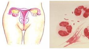
The transfer of images from retina to visual analyzer In the brain, there is a peculiar "cable" consisting of a variety of nerve fibers and packed in "insulation". The thickness of the optic nerve is not more than 2 mm, but it contains more than a million fibers. Each section of the image corresponds to a certain part of them, and when some of them cease to function, "Silent zones" (image violation) appear in the eye-perceived picture.
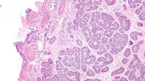
When moving the cells of nerve fibers, they gradually replace connective tissue Either the nerve auxiliary tissue (glia), which is normal to protect neurons.
Views
Depending on the causal factors, two types of atrophy of the optic nerve are distinguished:
- Primary. The disease is due to the affected X chromosome, therefore only men aged 15-25 years are sick. Pathology develops on recessive type and is inherited;
- Secondary. It occurs as a consequence of an eye or systemic disease associated with a violation of blood supply or a stagnation of an optic nerve. Such a pathological condition may appear at any age.
The classification is also conducted by lesion localization:

Also distinguish the following types of atrophy: initial, complete and incomplete; one-sided and bilateral; stationary and progressive; Congenital and acquired.
Causes of occurrence
The frequency of various pathological processes in a visual nerve is only 1-1.5%, and in 19-26% of them the disease ends with complete atrophy and incurable blindness.
The reason for the development of atrophy of the optic nerve can be any disease, the consequence of edema, squeezing, inflammation, damage to nerve fibers or damage to the vascular eye system:
- Eye pathologies: retinal pigment dystrophy, etc.;
- Glaucoma and increased VGD;
- Systemic diseases: hypertension, atherosclerosis, vessel spasms;
- Toxic impact: smoking, alcohol, quinine, drugs;
- Brain disease: abscess multiple sclerosis, arachnoiditis;
- Traumatic damage;
- Infectious diseases: meningitis, encephalitis, syfilitic lesion, tuberculosis, flu, measles, etc.
Is it possible to cure glaucoma read in.

Whatever the reason for the start of the atrophy of the optic nerve, the nerve fibers perish irretrievably, and the main thing is to quickly diagnose in order to slow down the process on time.
Symptoms
The main sign of the principle of pathology can serve a steadily progressive deterioration in view on one or both eyes, and it is not amenable to conventional methods of correction.
Gradually loss visual functions:

The manifestation of symptoms may leak depending on the acuteness of lesions of several days or months, but without timely response consistently leads to full blindness.
Possible complications
The diagnosis of "atrophy of the optic nerve" should be delivered as early as possible, otherwise the loss of vision (partial or complete) is inevitable. Sometimes the disease amazes only one eye - in this case the consequences are not so heavy.
Rational I. timely treatment Diseases that caused atrophy allows in some cases (not always) to preserve vision. If the diagnosis is made at the stage of the already developed illness, the forecast is most often unfavorable. 
If the disease began to develop in patients with indicators of view below 0.01, then medical events Most likely no result will be given.
Diagnostics
A targeted ophthalmological examination is the first mandatory step at suspected illness. In addition, it may be necessary to consult a neurosurgeon or neurologist.
To identify atrophy of the optic nerve, the following types of surveys can be carried out:
- Inspection of the eye dna (or biomicroscopy);
- - determination of violations spectatic perception (myopia, hyperopia, astigmatism);
- - study of the fields of view;
- Computer perimetry - allows you to determine the affected area of \u200b\u200bnervous tissue;
- Evaluation of color perception - determination of localization of lesions of nerve fibers;
- Video phthalmography - identifying the nature of damage;
- Craneography (X-ray skull) - the main object is the region of the Turkish saddle.
Read more about aK passes the inspection of the eye dna by .
To clarify the diagnosis and additional data, research is possible: CT, magnetic nuclear resonance, laser doppler.
Treatment
With partial damage to nerve fibers, treatment must be started quickly and intensively. First of all, the efforts of doctors are sent to eliminate the cause of the pathological condition in order to stop the progression of the disease.
Medical therapy
Since the restoration of dead nerve fibers is impossible, therapeutic measures are held, designed to stop the pathological process by all known means:
- Vasasculating: nicotinic acid, but-shp, dibazole, eufillin, complemin, papaverine, etc. The use of these means helps to stimulate blood circulation;
- Anticoagulants: heparin, ticklide. Preparations prevent blood thickening and thrombus formation;
- Biogenic stimulants: Vitreous body, Aloe extract, peat. Enhance metabolism in nervous tissues;

Heparin ointment is used in the treatment of optic nerve art
- Vitamins: Ascorutin, B1, B6, B2. Are catalysts of most biochemical reactions occurring in eye tissues, as well as amino acids and enzymes;
- Immunostimulators: Ginseng, Eleutherokokk. Necessary to stimulate the processes of regeneration and suppressing inflammation in infectious lesions;
- Hormonal agents: dexamethasone, prednisone. Used in the absence of contraindications to remove symptoms of inflammation;
- Improving the work of the central nervous system :, nootropyl, cavinton, cerebrolysin, flames.
Instruction D. examethazone for the eyes is located.

Dexamethasone is used in the treatment of optic nerve art
In each case, the treatment is assigned individually under the control of the attending physician.
In the absence of contraindications, an additional effect can be achieved by using needleflexotherapy, as well as physiotherapeutic treatment methods:
- Ultrasound;
- Electrophoresis;
- Electro- and laser diffraction of the optic nerve;
- Magnetic therapy.
Such procedures can have a positive effect with incompletely loss of nerve cells of its functionality.
Sururgically
Surgical methods are resorted to the threat of complete blindness, as well as other situations requiring operational intervention. For this, the following types of operations can be applied:

Various techniques for surgical treatment Success is practiced in clinics of Russia, Israel and Germany.
Folk remedies
The treatment of atrophy of the optic nerve should be carried out by drugs under the guidance of a qualified physician. However, often such therapy takes place for a long time, and in this case means folk recipes There may be invaluable help - because the action of most of them is aimed at stimulating metabolism and enhancing blood circulation:
- Dissolve in a glass of water 0.2 g mummy, drink before dinner on an empty stomach, as well as in the evening on a glass of means for 3 weeks (20 days);
- Make the infusion of crushed grass Astragala (2 art. L. Dried raw materials on 300 ml of water), insist 4 hours. For 2 months. Take 100 ml of infusion 3 r. in a day;
- Pepper mint is called eye herbs, it is useful to use it in food, and the eyes are injected with juice mixed with equal amounts of honey and water, in the morning and evening;
- Eliminate eye fatigue after long-term work on a computer can be using a row from indices of dill, chamomile, parsley, a blue and ordinary tea brewing cornflower;
- Unpretched pine cones Grind and 1 kg of raw materials cook 0.5 hours. After filtering, add 1 tbsp. Honey, stir and put in the refrigerator. Use 1 r. per day - in the morning before eating 1 tsp. ;
- Pour 1 tbsp. l. Parsley leaves 200 ml of boiling water, let it stand in a dark place 24 hours, after which take 1 tbsp. l. in a day.
Use in treatment folk remedies It follows only after consulting an ophthalmologist, since most of the plant components have an allergenic effect and can have an unexpected effect in the presence of some systemic pathologies.
Prevention
In order to avoid atrophy of the optic nerve, it is worth paying attention to measures of prevention not only eye, but also systemic diseases:
- In time to treat eye and systemic infectious diseases;
- Warn eye and cranial injuries;
- Make preventive inspections in the oncological clinic;
- Limit the use or exclude alcohol from your life;
- Take control of blood pressure.
Test on Dalton's online can be found.
Video
conclusions
Atrophy of the optic nerve is an almost incurable disease in the later stages, which threatens the patient with full blindness. but partial atrophy It can be suspended, and the main direction before developing medical tactics should be extensive diagnostics - it is it that will make it possible to establish the cause of the changes and try to stop them.
Therefore, try to pay reinforced attention not only to the health of the eyes, but also the whole organism. After all, it is all interconnected, and diseases of vessels or nerves can affect the quality of vision.
Also read about red spots under the eyes.
The optic nerve is a unique formation, a device and functionality of which differ from all other nerves in the body. In fact, these are intertwined nerve fibers. In the center of this interlacing is the arterial channel of the retina. Through it, the image is transmitted to the brain in the form of electronic impulses, which becomes impossible in the destruction of these fibers.
More than twenty percent of cases from the total number of blindness and weakness occur due to atrophy. Atrophy is the depletion of organs and tissues in the body or their reduction occurring during life. Atrophy of the optic nerve occurs when the components of its fibers begin to die, and the connective tissue is formed in their place. There are a lot of reasons for this, but in each case, it is possible to determine them accurately and choose treatment for only an ophthalmologist.
Heredity or congenital pathology are the root causes. In addition, it may arise due to the disease of the organs of vision, namely pathologies in visual nerve and retina. The reason for this ailment may also be a disease nervous system or diseases not related to organs of vision.
The main causes of atrophy of the optic nerve:
- Infectious diseases.
- Card and eye injuries and eye injuries.
- CNS diseases.
- Poisoning chemicals or alcoholic.
- Circulation failures of the organs of vision.
- Physical impact on organs of view, as a result of which the visual nerve is hung.
- Increased intracranial pressure.
There are several classifications of atrophy:
- the completed or complete and progressive depends on the degree of death of the visual nerve: when the patient first has a chance to restore vision, if the atrophy is finished, then the consequences of irreversible;
- hereditary and acquired;
- partial or complete;
- one-sided and bilateral.

Signs of the disease
The very first sign of atrophy is a violation of visual acuity. Wherein eyeball may not have pathologies, but electronic impulses from image transmission do not reach the brain.
Atrophy happens:
- primary, in which violated central vision And often the appearance of cattle, that is dark spots before your eyes,
- and secondary, affecting peripheral vision and resulting from various pathologies.
The patient experiences difficulties in reading, a color perception may be disturbed, a loss can begin in space. Signs of secondary atrophy of the optic nerve are caused by the reasons for its occurrence.
At the late stage of syphilis or paralysis, patient's vision drops gradually. If, for example, he has sclerosis, then a central field of view is possible.
With hypertension, lateral vision is affected. Also, this disease can be a consequence of abundant blood loss, then the lower boundaries of visibility are affected. If the visual nerve is composed, then the manifestations are probably different signs depending on the site that pressure is rendered.
Treatment is appointed depending on what reason it was formed. If there are signs of atrophy of the optic nerve, such as Amavrosis, that is, a sudden loss of vision, scotoma, mist in the eyes and blindness it is necessary to urgently examine at the oculist to avoid deposits.
Diagnosis and treatment
Before conducting an integrated ophthalmological examination, it turns out the presence of diseases in the patient leading to the atrophy of the optic nerve, information about the lifestyle, contacts with chemicals and alcohol are processed.

The main method of detection of atrophy - ophthalmoscopy, that is, the study inner structure eyes. It is carried out using an ophthalmoscope, in the process of the procedure in the patient's eye a ray of light is directed.
Distinguish several types of this diagnosis:
- With the opposite method, the Eye bottom is investigated in an inverted form.
- Direct ophthalmoscopy is possible if you pre-order a special vasodilatory solution into the eye, the study occurs when the image is increased to fifteen times.
In addition to ophthalmoscopy, perimetry is used to diagnose atrophy. It reveals the visible space available to the eye and its borders, thereby revealing the degree of violation of peripheral vision. The kinetic form of perimetry and statistical, that is, a computer.
The severity of atrophy may be different, a positive result in the treatment of this disease can be achieved only with partial fabric death. Adjust the course of treatment for a patient with an atrophy of the optic nerve is not an easy task for specialists, since the lost nerve fibers are practically not restored. There are chances for the treatment of nervous tissue, but provided that it is held on time.
As a rule, atrophy of the optic nerve does not arise in itself, but is a consequence of various pathologies of the eyes. For this reason, treatment is worth starting to eliminate these pathologies. If you have time to take care of the treatment since the beginning of the disease and for a couple of weeks, then the vision is possible to restore the entire one.
The treatment is carried out as follows: inflammation and swelling of nerve fibers are eliminated, the nutrition and blood circulation of the visual nerve is restored. Such a course of treatment occupies quite a long time and often does not bring the expected effect if it is not immediately immediately after the diagnosis is established. The main emphasis is on the treatment of the disease that caused the atrophy of the optic nerve. In parallel, a course of therapy is carried out to eliminate the consequences of this disease, which has given a complication for vision: drops, injections, and also medicinal products For receiving inside. This course, as a rule, consists of a number of events:
- Stimulation of blood circulation with the help of vasodilatory drugs.
- The use of biogenic stimulants accelerating the metabolism in tissues.
- Slowing inflammation with hormonal means.
- Activation of the nervous system with Emoxipin.
- In addition to physiotherapy, reflexotherapy is successfully used.
Some individual cases require surgical intervention.
It is worth noting that the treatment of atrophy of the optic nerve by folk remedies is not just inefficient, but also maliciously, in addition, takes advantage of the precious time in the patient. It is also impossible to ignore the signs of the disease that began. Operational appeal to verified medical institution Enhance the chances of recovery.
In the audience nerve there are many fibers that are intertwined with each other. In the center of this system there is a retinal channel, an image is transmitted through it that the man sees. During glaucoma, these nerve fibers are damaged, as a result of which you can completely lose sight.
Most cases of blindness occurs due to atrophy of the optic nerve, because it is impossible to restore damaged fibers. In any case, it is necessary to contact a good specialist, especially at the first manifestations of the disease.
According to the statistics of glaucoma, people are sick after 45 years of age, but there are cases and young age. This article will be discussed about atrophy of the visual nerves, the causes of the appearance, symptoms and methods of treating such a terrible disease.
Glaucoma
GlaucomaSource: MagicWorld.su.
Glaucoma is a common severe disease of the organ of vision. The cause of glaucoma is a violation of natural circulation intraocular fluid, raising intraocular pressure (WGD), which can lead to damage to the optical nerve transmitting information from the eye to the brain.
If the glaucoma is not treated, then the patient notes that he gradually loses the ability to see at the edges of the field of view (peripheral vision is lost). The progression of the disease leads to complete loss of vision.
Installed hereditary predisposition to glaucoma. If someone from parents have glaucoma, then children are in a group of increased risk.
Increased risk factors also include:
- myopia (myopia),
- age older than 60-65 years
- diabetes,
- hypotension,
- diseases thyroid gland and etc.
Glaucoma is the second most prevalence causes blindness in the world.
Speed \u200b\u200bnerve
 Source: brosaem.info.
 The optic nerve is a unique formation, a device and functionality of which differ from all other nerves in the body. In fact, these are intertwined nerve fibers.
In the center of this interlacing is the arterial channel of the retina. Through it, the image is transmitted to the brain in the form of electronic impulses, which becomes impossible in the destruction of these fibers.
More than twenty percent of cases from the total number of blindness and weakness occur due to atrophy. Atrophy is the depletion of organs and tissues in the body or their reduction occurring during life. Atrophy of the optic nerve occurs when the components of its fibers begin to die, and the connective tissue is formed in their place.
There are a lot of reasons for this, but in each case, it is possible to determine them accurately and choose treatment for only an ophthalmologist.
The main changes in glaucoma are associated with atrophy (dying) fibers of the optic nerve binding eyes with the brain. The optic nerve of the newborn contains 1.5 million conductive fibers. 500 thousand are constantly working, and 1 million - spare.
The intrauterine laying of these fibers can be compared with the gasket of the new telephone cable - there are always spare lines. Within 60-70 years of life, 1 million "spare" fibers gradually die from the moment of birth.
The remaining 500 thousand. Provide full-fledged sharpness and field of view. If, due to any circumstances, the process of "planned" shutdown of visual fibers is faster, it affects vision.
A special role in the "planned" atrophy of the optic nerve is played by the ratio of intraocular (WGD) and intracranial pressure (VHD). If it is greater than 2, then the process of atrophy of the optic nerve is complicated by the extrusion of a mechanically weak place in the eyeball - the lattice plate of the sclera.
In the glaucoma, the WGD is not necessarily elevated, and for the development of such excavation is sufficient (under reduced WGD) of the decline in VGF, which will increase their ratio than to 2. so develops the glaucoma of low (or normal) intraocular pressure. From the simple form of glaucoma, it is distinguished by an inconspicuous and hard flow.
With a simple form of glaucoma, the WGD is raised. It can be measured, somehow affect it, control. A decrease in the same State University does not care about anyone. Moreover, the decline in the HCH is also difficult to reveal, not to mention how to follow him.
Most often the reason for this is a decrease arterial pressurecaused by chronic heart or vascular failure. Treatment of such patients is carried out jointly with the therapist and neuropathologist.
Serious irreversible diseases of the organs of view are atrophy of the optic nerve. The disease is caused by the ignition of the nerve fibers, according to which the human brain receives nerve impulses and then converts them into the images.
The essence of the pathological process
The optic nerve is a weave of nerve fibers. In the center of such a weave is the arterial channel of the eye retina. Through this channel in the human brain, information is received in the form of nervous pulses that are converted into the picture.
With the defeat and death of neurons processes in their place, a connecting tissue is formed, and an atrophy of the optic nerve occurs. The brain ceases to receive information from the photosensitive cells of the eye retina and develops blindness. The disease affects the eyes partially or completely.
Stages of the disease
 Source: Operabelno.ru.
 - An increase in intraocular pressure indicators contributes to squeezing and damage to the tissue of nerve fibers.
- The clarity of view is reduced, dark circles appear before your eyes.
- The boundaries of view are narrowed and the functions of the optical system are disturbed.
- Embiring eye nerve occurs and the full loss of vision in the affected eye comes.
The disease amazes the same adults and children. Complete atrophy of the optic nerve is incurable.
Causes of the disease
Heredity or congenital pathology are the root causes of atrophy of the optic nerve. In addition, it may arise due to the disease of the organs of vision, namely pathologies in visual nerve and retina. The reason for this disease can also be a disease of the nervous system or illness not related to the organs of vision.
The main causes of atrophy of the optic nerve:
- Infectious diseases.
- Card and eye injuries and eye injuries.
- CNS diseases.
- Poisoning chemicals or alcoholic.
- Circulation failures of the organs of vision.
- Physical impact on organs of view, as a result of which the visual nerve is hung.
- Increased intracranial pressure.
There are several classifications of atrophy:
- the completed or complete and progressive depends on the degree of death of the visual nerve: when the patient first has a chance to restore vision, if the atrophy is finished, then the consequences of irreversible;
- hereditary and acquired;
- partial or complete;
- one-sided and bilateral.
There is an increase in the frequency and severity of the injury of the eyeball, which in most cases becomes the main cause of primary disability.
Atrophy of the optic nerve is not an independent disease. Its development is associated with complications and consequences of other diseases. The disease is often hereditary or congenital.
Signs
 Source: Bolezniglaz.ru.
 The very first sign of atrophy is a violation of visual acuity. At the same time, the eyeball may not have pathologies, but the electronic impulses from the transfer of images do not reach the brain.
Atrophy happens:
- primary, in which the central vision is disturbed and often the appearance of cattle, that is, dark spots before our eyes,
- and secondary, affecting peripheral vision and resulting from various pathologies.
The patient experiences difficulties in reading, a color perception may be disturbed, a loss can begin in space. Signs of secondary atrophy of the optic nerve are caused by the reasons for its occurrence.
At the late stage of syphilis or paralysis, patient's vision drops gradually. If, for example, he has sclerosis, then a central field of view is possible.
With hypertension, lateral vision is affected. Also, this disease can be a consequence of abundant blood loss, then the lower boundaries of visibility are affected. If the visual nerve is composed, then the manifestations are probably different signs depending on the site that pressure is rendered.
Treatment is appointed depending on what reason it was formed. If there are signs of atrophy of the optic nerve, such as Amavrosis, that is, a sudden loss of vision, scotoma, mist in the eyes and blindness it is necessary to urgently examine at the oculist to avoid deposits.
At the initial stage of development, the disease is asymptomatic. Symptoms of the disease depends on the level of nerve damage, the causes of the disease and the severity of its flow. The main symptoms of atrophy includes:
- loss of visual boundaries (anox);
- reduction of clarity and visual acuity (ambulopia);
- loss of view.
Penoplication is caused by a change in visual boundaries towards their narrowing. "Tunnel vision" develops, in which a person does not see lateral images. At the initial stage of amblyopia, a person does not see remote items, the image breaks down, but close items are visible clearly.
With the development of the disease, there is a damage to neurons, a person begins not to see objects located near. With complete fat, the nerve fibers comes blindness.
Diagnostics
 Source: nebolet.com.
 Before conducting an integrated ophthalmological examination, it turns out the presence of diseases in the patient leading to the atrophy of the optic nerve, information about the lifestyle, contacts with chemicals and alcohol are processed.
The main method of detection of atrophy - ophthalmoscopy, that is, the study of the internal structure of the eye. It is carried out using an ophthalmoscope, in the process of the procedure in the patient's eye a ray of light is directed.
Distinguish several types of this diagnosis:
- With the opposite method, the Eye bottom is investigated in an inverted form.
- Direct ophthalmoscopy is possible if you pre-order a special vasodilatory solution into the eye, the study occurs when the image is increased to fifteen times.
In addition to ophthalmoscopy, perimetry is used to diagnose atrophy. It reveals the visible space available to the eye and its borders, thereby revealing the degree of violation of peripheral vision. The kinetic form of perimetry and statistical, that is, a computer.
The severity of atrophy may be different, a positive result in the treatment of this disease can be achieved only with partial fabric death. Adjust the course of treatment for a patient with an atrophy of the optic nerve is not an easy task for specialists, since the lost nerve fibers are practically not restored.
There are chances for the treatment of nervous tissue, but provided that it is held on time.
- Physical examination - includes inspection and palpation of organs of view, the functioning of the visual system is determined, the level of color perception is measured intraocular pressure.
- Perimetry - the boundaries are determined by the visible area available for the eye.
- Ophthalmoscopy. With the help of electronic ophthalmoscope, the departments of the nervous barrel, eye iris, and the Eye Apple itself are examined.
- Fluorescent angiography. Color fluid is introduced into the vein. With a blood current, it moves along the eye vessels. The supply of blood vessels is evaluated through the device with the emission of light of different frequency, the level of tissue lesion and the structure of the eyeball.
- Laser tomography disk eye. The eye bottom and changes in the front of the nervous barrel are examined.
- Optical-coherent tomography. The condition of the tissues of neurons processes with infrared radiation is estimated.
Treatment of optic nerve at glaucoma
The treatment is carried out as follows: inflammation and swelling of nerve fibers are eliminated, the nutrition and blood circulation of the visual nerve is restored. Such a course of treatment occupies quite a long time and often does not bring the expected effect if it is not immediately immediately after the diagnosis is established.
The main emphasis is on the treatment of the disease that caused the atrophy of the optic nerve. In parallel, a course of therapy is carried out to eliminate the consequences of this disease, which has given a complication for vision: drops, injections, as well as drugs for intake. This course, as a rule, consists of a number of events:
- Stimulation of blood circulation with the help of vasodilatory drugs.
- The use of biogenic stimulants accelerating the metabolism in tissues.
- Slowing inflammation with hormonal means.
- Activation of the nervous system with Emoxipin.
- In addition to physiotherapy, reflexotherapy is successfully used.
Some individual cases require surgical intervention.
It is worth noting that the treatment of atrophy of the optic nerve by folk remedies is not just inefficient, but also maliciously, in addition, takes advantage of the precious time in the patient. It is also impossible to ignore the signs of the disease that began. Operational appeal to the proven medical institution will increase the chances of recovery.
The treatment of atrophy of the optic nerve is a rather complicated and laborious process for doctors, as it is necessary to remember that it is impossible to restore the deadly fibers of the optic nerve. In addition, the reasons for atrophy of the visual nerve set, so the diagnostic search may take a long time.
Treatment of the disease can be carried out in three directions:
- conservative treatment
- physiotherapeutic treatment
- surgery
The treatment of atrophy of the optic nerve is a very difficult task for doctors. It is necessary to know that the destroyed nerve fibers cannot be restored.
For some kind of treatment, it is possible to hope only when restoring the functioning of nerve fibers that are in the process of destruction, which still retained their livelihoods. If this moment is missing, then vision on the sore eye can be lost forever.
In the treatment of atrophy, it is necessary to keep in mind that this is often not an independent disease, but a consequence of other pathological processes affecting various parts of the visual path. Therefore, the treatment of atrophy of the optic nerve must be combined with the elimination of the reason that caused it.
In the case of timely elimination of the cause and, if atrophy has not yet managed to develop, during 2-3 weeks to 1-2 months, the film of the eye bottom and restoration of visual functions occurs.
Treatment is aimed at eliminating the enemy and inflammation in a visual nerve, to improve its blood circulation and trophy (nutrition), the restoration of conductivity is not to the end of the destroyed nerve fibers.
But it should be noted that the treatment of atrophy of the optic nerve is long, the effect of it is poorly expressed, and sometimes absent, especially in the launched cases. Therefore, it must be started as early as possible.
As mentioned above, the main thing is the treatment of the underlying disease, against which they spend comprehensive treatment Direct atrophy of the optic nerve. For this purpose various forms Preparations: eye drops, injections, both shared and local; Tablets, electrophoresis.
Conservative therapy
Conservative therapy implies the appointment of symptomatic treatment, including:
- vasodinating drugs;
- preparations that improve the blood supply to the optic nerve (Eutillin, Trental, but-shpa, nicotinic acid, papaverine)
- anticoagulants (ticklid, heparin)
- preparations that improve exchange processes In the nerve tissues (biogenic stimulants, enzymes, amino acids, immunostimulants)
- preparations whose action is aimed at stimulating metabolic processes and resorption of pathological processes (preoccupative), relief inflammatory process (hormones), improvement of the nervous system (Cavinton, Nootropyl, Emoxipin)
Electro-, magneto and laseristimulation of the optic nerve are prescribed as physiotherapy procedures.
Surgical treatment of atrophy of the optic nerve involves an operation to remove formations, squeezing it, dressing the temporal artery, which improve blood circulation and nerve vascularization.
If you eliminate pathology eye disease Within a few weeks from the beginning of the disease, the vision is completely restored.
Vioftans are ophthalmic agents, so-called animal bioregulators, as well as plant origin.
Preparations of medication therapy
Preparations of drug therapy:
- Preparations affecting eye vessels and accelerating their recovery: "Cavinton", "Pentoxifillain", "Agapurin", "Mexidol".
- Means for improving metabolic processes in the affected tissues: "Histochrom", Emoxipin, "Solkosaryl", "Aktovegin".
- Polyvitamin complexes: "Lutein Forte", "Ecomir"; Vitamins of group V.
Preparations must be taken to appoint a doctor after diagnosis. The doctor will select optimal treatment, given the concomitant diseases. In the absence of concomitant somatic pathology, it is possible to take a but-shlu, Papaverin, vitamin preparations, amino acids, emoxipin, nootropyl, flashes.
But there should be no self-medication with this serious pathology. Also used physiotherapeutic treatment, iggoreflexotherapy; Methods of magnetic, laser and electrical stimulation of the optic nerve have been developed. The course of treatment is repeated in a few months.
Food in atrophy of the optic nerve should be full, diverse and rich in vitamins. In food it is necessary to use as much as possible fresh vegetables and fruits, meat, liver, dairy products, porridge, etc.
The visually impaired and blind is appointed a rehabilitation course aimed at eliminating or compensating for the restrictions of vital activity arising from the loss of vision.
Treatment with folk remedies is dangerous by the fact that precious time is lost when curing atrophy and return the vision is still possible. It should be noted that with this disease, folk remedies are ineffective.
Operation
 Source: YZNAJ-KA.ru.
 Surgical treatment - the bandaging of the temporal artery and the implantation of various biogenic materials improve the vascularization and blood circulation of the nerve, and the operation of decompression of the optic nerve removes excessive compression of it.
It is impossible to restore damaged fibers, thus, the treatment of this ailment is concentrated on the preservation of available indicators of vision and stopping the progression of the disease.
Therapy, first of all, begins with eliminating the cause of atrophy, if it is not a hereditary factor. After a traditional treatment regimen is prescribed, consisting of vasodinating drugs, tonic blood circulation for the vitroless nerve, the operation and vitamins.
In addition, a magnetic, laser or electrical effect on the visual nerve is carried out. This contributes to the acceleration of tissue regeneration, activating metabolic processes and strengthening blood supply.
One of the newest methods for the treatment of this pathology is the implantation of the electrostimulator directly into the orbit of the eye. Despite high efficiency This method, it requires major cash investments, suggests a long period of rehabilitation, and the implant itself works only for several years.
Atrophy of the visual nerves is diagnosed based on the appropriate change in the fundus and violations of visual functions. In atrophy, there is always a pale of the optic nerve disk, the narrowing of the boundaries of the field of view and a decrease in visual acuity.
The severity of atrophy is determined not only by an ophthalmoscopic picture and the degree of violation of visual functions at the time of the study, but also whether the process is stable, complete or it is in progression stage.
Therefore, with the examination of patients with atrophy of optic nerves, it is necessary to take into account the data of dynamic observation, reflecting the nature of changes in the peripheral boundaries of the field of view and visual acuity.
The state of the disk of optic nerves, changes in the eye day and other symptoms depend on the etiology of atrophy. The etiology of the disease largely determines the severity of pathology, clinical and labor forecast.
Clinical diagnosis in atrophy of optic nerves should reflect the etiology of the disease, form clinical flow (progressive or stable, unpropressive), lesion localization (visual nerves, chiasm, visual tract), state of vision functions.
With a stable, unpressing form of atrophy of the visual nerves resulting from poisoning, bleeding and other (sometimes unknown) causes, patients, in particular, the work associated with the danger of intoxication is contraindicated, especially neurotropic poisons.
The neurosurgical surgery in atrophy of the optic nerve is carried out if the pathology is caused by the compression of this anatomical education. The factor that caused compression, while removing, normalizing the work of the nerve and preventing further development of atrophy.
IN postoperative period Patients prescribe a course medical treatment, physiotherapy, gentle eye mode.
In atrophy of the optic nerve, surgical methods are used, including vazejectory operations, the kateryization of the subtitonone space with the introduction of drugs into this area, the implantation of the electrodes to the disk of the optic nerve, as well as the transplant of various biomaterials.
The method of surgical correction of hemodynamics (separately or together with a conservative course of treatment) eyes. Such correction is carried out using local anesthesia.
The Ksenoplast collagen sponge is introduced into the subitanese space for the expansion of the vessels due to the developing aseptic inflammation in the microcirculatory beds surrounding its tissues. This is stimulated with newly formed vessels growth of connective tissue.
After 1-2 months after the operation at the place of administration of the sponge, the formation of granulation tissue occurs. After 2-3 months, the sponge is completely absorbed, and the degree of vascularization of newly formed episcileral tissues remains rather high.
Improving blood flow in the vascular shell, which participates in the blood supply to the retina and the disk of the optic nerve will become a factor that leads to an increase in sharpness by 61.4%, also to expansion by 75.3%, and visual expansion. The operation can be carried out repeatedly, but not more often than after the expiration of 2 months after the previous one.
Operation: Implant from collagen (width - 6 mm, length - 20 mm) is impregnated with an antioxidant or vasodilatory drug and through a sectional in conjunctiva is introduced into the subitanese space (Limba or Nizhneus-Search quadrant, 8 mm from the limb) without superimposing seams.
Indications:
- Visual acuity with correction to 0.4
- in atrophy (glaucomatous) optic nerve with stabilized intraocular pressure;
- with the rear and front ischemic neuropathy of non-inflammatory genesis;
Contraindications will be:
- Age over 75 years;
- Vision, if its sharpness is less than 0.02 d;
- Heavy noncompensated somatic diseases (Collagenoses, GB III Art. Oncological, etc.);
It is impossible to completely restore visual sharpness, but without treatment, the disease will lead to blindness. The main method of therapy will depend on what is the cause of atrophy of the optic nerve.
The drugs that are used in treatment are tools for improving the blood supply of the nerve, improving metabolism, vasodilators, polyvitamins, biostimulants.
These funds reduce swelling, inflammation in the area of \u200b\u200bthe optic nerve disk, improve its nutrition, blood supply, stimulate the activity of the remaining nerve fibers.
Partial form of optic nerve when glaucoma
- Atrophy caused by a chronic or acute circulatory disorder. The use of vasoactive drugs (Tanakan, Cavinton, Sermion) and Antioxidant (Mildronat, Mexide, Emoxipin) is shown.
- Atrophy on the background of the pathologies of the central nervous system. Requires aggressive nootropic therapy (SopaCyril, Nootropyl, Actovegin) and Fermenzyme (FPogenzym, Vobenzyme).
- Downward atrophy. The bioregulating therapy is prescribed by peptide preparations (Etitalamine, Cortexin).
- Toxic atrophy. Disinfection measures, vasoactive, nootropic and peptide preparations are shown.
- Atrophy of the volatile, innate and post-traumatic genesis. Requires the use of cytomotines (Retinalamine, Cortexin) and laser, magnetic and light exposure courses.
Full shape
As a rule, complete atrophy of the optic nerve is not amenable to correction. However, in the case of the continuing degenerative process, the vision is possible. Therapy for atrophic diseases is carried out in several directions:
- Anticoagulants (heparin, aspirin in small doses). This pharmacological group It helps to improve the rheological properties of blood (improves its fluidity), prevents the formation of blood clots, contributes to a better supply of oxygen tissues and nutrients.
- Vasodilators (Trental, Actovegin, Pentoxifillin). Temporarily expand the clearance of blood vessels, increasing the amount of blood flowing through them per unit of time. In this way, it is possible to achieve an anti-ischemic effect and improve tissue blood supply.
- Stimulants of metabolism (vitamins of group B, aloe, ginseng). Preparations contribute to strengthening regenerative processes in the field of affected nerve and in the body as a whole.
- Corticosteroids (prednisone, hydrocortisone, dexamethasone). It has a pronounced anti-inflammatory and anti-ethnic effect, inflammation is removed, including in a paraproorbital region.
- Nootropic tools (piracetam, cerakson, cerebro). Contribute to improving the activities of the central and peripheral nervous system.
How effective are the methods of treatment of atrophy of the optic nerve?
The dead nerve cells and the fibers of the optic nerve cannot be restored. The treatment aims to increase the resistance to the effects of adverse factors are not yet damaged nerve cells and restore the functions of those that have not yet died completely, but are in the depressed state.
Thus, treatment allows you to delay or stop further impairment, and in part of patients with glaucoma even improve visual functions.
Prescribed medicines that enhance blood circulation and metabolic processes in the visual nerve and retina, drugs with antisclerotic action, vitamins.
- means acting on the vessels of the eye: Pentoxifillain (Trental, Hehst, Germany; Agapurin, Biotika, Czech Republic), Vinpocetin (Cavinton, Richter, Hungary);
- preparations that improve exchange processes in the tissues of the eye: Retinalamin (Herofarm, Russia), Histochrom (Russia), Emoxipin (Russia), deproteinated hemoderiovats (Solfossyril, Solko, Switzerland, Actovegin, Hafslund Nikomed, Austria);
- complex vitamin preparations with microelements (especially with selenium): multivitamin complexes, such as Anthocian Forte, Lutein Forte (Ecomir, Russia), Okwit (Baush & Lomb. USA).
The useful effect is noted from the inhalation of a mixture of oxygen (95%) and carbon dioxide (5%) - carbogen therapy. When inhaling this mixture carbon dioxide Expands brain and eye vessels, and oxygen stimulates energy processes in the same structures.
Carbogenicherapy contributes to the expansion of the field of view and an increase in visual acuity in glaucoma patients.
Recently, such physiotherapeutic methods of treatment as electrostimulation and magnetotherapy have spreads.
One of the new techniques medicinal treatment Atrophy of the optic nerve is to sum up to the rear section of the eye (that is, closer to the retina and visual nerve) surgically by a special system.
Surgical methods of treatment of atrophy of the optic nerve include operations on blood vessels To redistribute blood circulation in the artery of the optic nerve, as well as the dissection of a dense gloil ring around the nerve outlet location from the eye to reduce the pressure on the nerve fibers.
Forecast for recovery
In the absence of timely and effective treatment The pathology of the disease is rapidly developing and changes in the tissues of the optic nerve takes irreversible. Atrophied nerve fibers are not restored, therefore a positive result in the fight against illness is achieved only with partial die into the tissue of nerve fibers.
When determining signs of illness to early stages And eliminating all causes of pathology can be partially or fully restored visual functions.
What mode of labor and life should be adhere to the patient glaucoma?

Atrophy of the optic nerve is called the pathological process, in which the nerve fibers are partially or completely destroyed, replaced by a connective tissue. As a result, there is a violation of the functions of nervous tissue. Most often atrophy is a complication of any other eye disease.
When progressing the process, there is a gradual death of neurons, as a result of which the information coming from the retina information enters the brain in a distorted form. With the development of the disease, the cells perishes more and more, the entire nervous trunk is affected.
In this case, it becomes almost impossible to restore the visual function. Therefore, treatment should be started at the earliest stage, when the first signs of the disease appears.
How is the atrophy of the optic nerve, the symptoms of the manifestation of this eye disease? We will talk about all this today on this page "Popular about health" with you. But let's start our conversation with characteristic signs This pathology:
Symptoms of atrophy of the nerve eye
It all begins with reducing vision. This process can occur gradually or rapidly, suddenly. It all depends on the dislocation of nerve damage, on which cut cut it develops. Depending on the severity of the pathological process, the reduction of vision is divided into degrees:
Uniform decrease. It is characterized by a uniform deterioration of the ability to see objects, distinguish between colors.
Loss of side fields. A person clearly distinguishes objects that are in front of him, but he sees badly, or does not see what is on the side.
Spot of spots. Normal vision prevents the stain in front of the eye that may have different sizes. In its limits, a person does not see anything, beyond the vision is normal.
In severe cases, complete atrophy, the ability to see is lost completely.
Treatment of atrophy of the optic nerve
As we already know, this pathological process is often complicated by another eye disease. Therefore, after the reason for the detection, the complex treatment of the underlying disease is prescribed and take measures to prevent the further development of eye nerve atrophy.
In the event that the pathological process has just begun and has not yet managed to develop, cure the nerve usually succeeds and visual functions are restored on time from two weeks to several months.
If, by the time of the beginning of the treatment, atrophy managed to develop enough, it is completely impossible to cure the visual nerve, since destroyed nerve fibers in our time can not be restored. If the damage is partial, it is still possible to carry out rehabilitation in order to improve vision. But, at a severe stage, complete damage to cure atrophy and restore visual functions so far.
The treatment of atrophy of the eye is to use medicinal preparations, drops, injections (general and local), the action of which is aimed at improving blood circulation in a visual nerve, a decrease in inflammation, as well as to restore those nervous fibers that are not completely destroyed. Additionally apply physiotherapy methods.
Preparations used in the treatment:
To improve the blood circulation of the eye nerve, vasodilators are used: Nicotinic acid, But-Shpu, Papaverin and Dibazole. Also, patients are prescribed complemin, Eufillin, Trental. As well as Galidor and Sermion. For the same purpose, anticoagulant drugs are used: ticklide and heparin.
In order to restore exchange and regenerative processes in the tissues of the affected nerve, patients prescribe biogenic stimulants, in particular, the vitreous body, peat and aloe preparations. Vitamins, amino acids, enzymes and immunostimulants are also prescribed.
To relieve, reduce the inflammatory process often use hormone therapy with prednisone and dexamethasone.
In addition, complex treatment includes drugs aimed at normalizing the work of the central nervous system: cerebrolysin, flashes, as well as an Emoxipin, nootropyl and cavinton.
All the above and other medicines, the doctor appoints individually, after clarifying the causes of the pathological process and the diagnosis of the underlying disease. It takes into account the degree of defeat of the optic nerve, the age of the patient, its general state and the presence of concomitant diseases.
In addition to drugs, physiotherapy techniques, iggoreflexotherapy, are actively used. Apply the methods of magnetic, laser and electrical stimulation of the stem of the optic nerve. According to the testimony, the patient may be recommended surgical treatment.
Complex therapy is appointed by courses that repeat every few months.
In conclusion of our conversation it should be noted that atrophy of the optic nerve cannot be cured by unconventional means. You will only lose time. The pathological process will progress, increasingly reducing the chances of successful treatment and restoration of vision.
Therefore, in the presence of the described above, or other symptoms, indicating the development of pathology, do not lose precious time and sign up for reception to an experienced ophthalmologist. With a timely manner, the chances of restoring vision are significantly increasing. Be healthy!
Atrophy of any organ is characterized by a decrease in its size and loss of functions due to lack of nutrition. Atrophic processes are irreversible and talking about the severe form of any disease. Atrophy of the optic nerve is a complex pathological condition that is almost not treatable and often ends with loss of vision.
In this article
Functions of the optic nerve
The optic nerve is white substance Big brain, as if rendered on the periphery and the brain associated. This substance conducts visual images from the mesh shell on which light rays fall into the bark of the brain, where the final image is formed, which sees a man. In other words, the visual nerve serves as a supplier of messages in the brain and is the most important component of the entire transformation process obtained by the eyes of lighting information.
Atrophy of the optic nerve: general description
In atrophy of the optic nerve, its fibers are completely or partially destroyed. They are subsequently replaced by a connective tissue. Decommissioning fibers leads to the fact that the light signals obtained by the retina are converted into electrical signals that are transmitted to the brain. For the brain and eye, this process is pathological and very dangerous. In his background, various violations are developing, including a decrease in visual acuity and the narrowing of its fields. Atrophy of the optic nerve is found in practice quite rarely, although even the most minor eye injuries can provoke its beginning. However, approximately 26% of cases of disease ends with the fact that the patient completely loses his eyesight on one eye.
Causes of atrophy of the optic nerve
Atrophy of the optic nerve is one of the symptoms of various eye diseases or the stage of development of any illness. The reasons that can lead to this pathology are very much. Among the ophthalmological diseases that are able to provoke atrophic changes in the optic nerve, the following ailments:
- glaucoma;
- retinal pigment dystrophy;
- myopia;
- uveitis;
- retinit;
- neurrit of the optic nerve,
- damage to the central artery of the mesh shell.
Also atrophy may be associated with tumors and eye diseases: Glioma optic nerve, neurinoma, orbit cancer, meningoma, osteosarcoma and others.
All sorts of diseases of the brain and the central nervous system in some cases lead to atrophic processes in the eyes, affecting, above all, visual nerves. These diseases include:
- multiple sclerosis;
- pituitary tumors;
- meningitis;
- brain abscess;
- encephalitis;
- card and brain injuries;
- damage to the skeleton of the face marked in the eye nerve.
Types and forms of atrophy of the optic nerve
This pathological condition is congenital and acquired. Acquired atrophy is divided into descending and ascending. In the first case, the fibers of the direct nerve are affected. In the second, the cells of the mesh shell fall under the blow.
In accordance with another classification, acquired atrophy may be:
- Primary. She also is called a simple form of atrophy, in which the disk of the optic nerve is pale, but it has clear boundaries. The vessels in the retina at this type of pathology are narrowed.
- Secondary, which develops due to inflammation of the visual nerve or its stagnation. The disk boundaries become fuzzy.
- Glaucomatous, accompanied by elevated intraocular pressure.
On the scale of the lesion of the fibers of the optic nerve atrophy is divided into partial and complete. Partial (initial) form manifests itself in a strong impairment of vision that fails to adjust contact lenses and glasses. At this stage, you can preserve the remaining visual functions, but the color perception will be very violated. Full atrophy is the defeat of the entire visual nerve, in which a person does not see anything with a sick eye. Atrophy of the optic nerve is manifested in a stationary form (not developing, but remains on the same level) and progressive. With stationary atrophy, visual functions remain in a stable state. The progressive form is accompanied fast decline Acute view. Another classification shares atrophy to one-sided and bilateral, that is, with damage to one or both of the organs of vision.
Symptomatic atrophy of optic nerve
The first and main symptom, which manifests itself in any form of atrophy of the optic nerve, is a deterioration of vision. At the same time, it is not amenable to correction. This is a sign of which an atrophic process can be distinguished from AMETROPIA - changes in the ability of the human eye correctly refractive light rays. Vision can deteriorate gradually and rapidly. It depends on the form in which atrophic changes occur. In some cases, visual functions are reduced within 3-4 months, sometimes a person is completely blind to one or both eyes in a few days. In addition to the overall reduction of visual acuity, its fields narrow. 
The patient almost completely loses his lateral vision, which leads to the development of the so-called "tunnel" type of perception of the surrounding reality, when a person seems like a pipe. In other words, it is seen only what is directly in front of a person, and not on the side of it.
Another frequent sign of atrophy of the optic nerve is the appearance of cattle - dark or blind sites arising in sight. By location, the cattle can be determined, the fibers of which area of \u200b\u200bthe nerve or retinal are damaged most. If the stains occur right in front of the eyes, the nerve fibers are affected, located closer to the central selection of the mesh shell or directly in it. Coloring disorder becomes another problem with which a person is facing atrophy. Most often, the perception of green and red shades is disturbed, a rarely blue-yellow spectrum.
All these symptoms are signs of primary shape, that is, it initial stage. They can notice the patient himself. Symptoms of secondary atrophy is visible only during the examination.
Symptoms of secondary atrophy of the optic nerve
As soon as a person appeals to the doctor with such symptoms, as a decrease in visual acuity and the narrowing of its fields, the doctor conducts a survey. One of the main methods is ophthalmoscopy - inspection of the eye bottom with the help of special tools and appliances. In the course of ophthalmoscopy detected the following signs Atrophy of the optic nerve: 
- narrowing of vessels;
- expansion of veins;
- disc pale;
- reducing the reaction of the pupil into light.
Diagnostics
As already described above, the first method used to identify pathology is ophthalmoscopy. However, the symptoms that can be detected using this study do not allow you to accurately diagnose. The deterioration of vision, the absence of the reaction of the pupil into the light, the narrowing of the vascular of the eye is the signs of many eye ailments, for example, the peripheral form of cataracts. In this regard, many different methods apply to the diagnosis of atrophy:

Laboratory studies are also held. The patient gives blood and urine on the analysis. Tests are prescribed on syphilis, borreliosis and to determine other diseases of Neophthalmological nature.
How is the atrophy of the optic nerve?
Restore fibers that have already been destroyed is impossible. The treatment helps to stop atrophy and save those fibers that still function. There are three ways to combat this pathology:
- conservative;
- therapeutic;
- surgical.
For conservative treatment The patient is prescribed vaconishing drugs and medicines whose actions are aimed at normalizing the blood supply to the optic nerve. The doctor also prescribes anticoagulants that oppress the activity of blood clotting. 
Help to stop the fibers of the drugs, stimulating metabolism, and drugs that take inflammation, including hormonal.
Physiotherapeutic impact involves the appointment:

The surgical treatment method is focused on the removal of formations, which are pressed on the eye nerve. During the operation, the surgeon may implant the patient with biogenic materials that will help improve blood circulation in the eye and in an atrophied nerve, in particular. Transferred pathology in most cases leads to the fact that a person is assigned to a person. Blind patients or visually impaired are sent to rehabilitation.
Prevention
To prevent the atrophy of the optic nerve, ophthalmic diseases should be initiated in a timely manner. 
At the first signs of reduction of visual acuity, it is necessary to immediately make an appointment with an ophthalmologist. With the beginning of atrophy, it is impossible to lose a minute. If at the initial stage it is still possible to maintain most of the visual functions, then as a result of further atrophic changes, a person can become disabled.