The norm of the thickness of the cornea of the eye for vision correction. Influence of the central thickness of the cornea on intraocular pressure is normal. Structure and function of the cornea
For citation: Egorov E.A., Vasina M.V. Effect of corneal thickness on level intraocular pressure among different groups of patients // BC. Clinical ophthalmology. 2006. No. 1. S. 16
Influence of corneal thickness on IOP level in different groups of patients
in different groups of patients
E.A. Egorov, M.V. Vasina
Department of eye diseases of medical faculty of RGMU
Ophthalmological center “Dr. Visus".
Purpose: To make a comparative analysis of corneal thickness and IOP level of healthy subjects, patients with POAG and after excimer laser treatment.
Materials and methods: The study lasted 2 years. Main group included 269 patients (418 eyes), 109 males and 160 females. Main group composed of subjects healthy, patients with POAG and patients after excimer laser treatment. All patients detection of postoperative visual acuity with correction, computer perimetry, pachymetry, biomicroscopy and ophthalmoscopy. In the group of patients with POAG gonioscopy was also performed, and in the group of patients after refractive operation - keratotopography.
results:
First group included 62 healthy subjects (110 eyes). Average data of corneal thickness was the following: center 548.01±31.13 mcm, top - 594.43±38.36 mcm, lower part - 571.02±35.52 mcm, internal part - 580.36±37 .22 mcm, external - 575.87±37.94 mcm. IOP (P0) was 17.52 ±3.33 mm Hg in average. In the POAG group with central corneal thickness (CCT)<520 mcm (34 patients; 55 eyes) Р0 was18,7±1,64 mm Hg and CCT 500,09±20,71 mcm in average.
In the POAG group with central corneal thickness (CCT) 521-580 mcm (70 patients; 96 eyes) P0 was 19.26±1.68 mm Hg and CCT 548.61±15.41 mcm in average. In the POAG group with central corneal thickness (CCT) >581 mcm (25 patients; 39 eyes) P0 was 20.36±1.20 mm Hg and CCT 600.34±17.71 mcm in average.
Conclusion:
Average CCT is 548 mcm, which correlates with IOP level - 17.5 mm Hg. Each 10 mcm of CCT changing leads to IOP level changing by 0, 63 mm Hg.
Refractive anomalies don't affect CCC and IOP level. Patients with CCT<520 mcm should be at the risk group of glaucoma.
The problem of glaucoma occupies one of the important places in modern ophthalmology. The frequency of blindness from glaucoma in the world over the past 30 years has remained approximately at the level of 14-15% of the total number of all cases. Such a high percentage of adverse outcomes is associated with both the late diagnosis of glaucoma and the incorrect assessment of the eye hydrodynamic data obtained during the examination of the patient.
An important role in the early diagnosis and monitoring of patients with open-angle glaucoma has recently become an assessment of the correlative relationships between the strength characteristics of the eye (rigidity, thickness of the cornea), the level of ophthalmotonus, and the stages of the disease. (Brucini P. et al., 2005; Ogbuehi K.C., Imubrad T.M., 2005; Sullivan-Mee M. et al., 2005; Yagci R. et al., 2005).
The results of the study of IOP can be considered correct if it is taken into account that they are influenced by such a factor as the thickness of the cornea. There are options for both overdiagnosis (when receiving elevated IOP) and underestimation of the ophthalmotonus data obtained by measuring.
In the last decade, excimer laser refractive surgery on the cornea has become widespread. As a result of this intervention, there is a decrease in the thickness of the cornea, and along with this, not only the refraction of the eye changes, but also the parameters of the measured IOP (Cennato G., Rosa N., La Rana A., Bianco S., Sebastiani A., 1997; Ogbuehi KC , ImubradTM, 2005). In this regard, in the future, it is necessary to learn how to correctly assess the measured IOP in patients who underwent refractive surgery.
Purpose of the study
To conduct a comparative analysis of corneal thickness and measured IOP data among patients in a healthy population, with primary open-angle glaucoma and in patients undergoing excimer laser refractive surgery.
Materials and methods
This study was carried out over 2 years. The study group included 269 patients (418 eyes). Among them, 109 men and 160 women aged 16 to 84 years. All patients were divided into three main groups: healthy patients, patients with primary open-angle glaucoma (POAG), and patients after refractive excimer laser surgery.
All patients underwent the determination of visual acuity with correction, computer perimetry, tonometry, pachymetry, biomicroscopic and ophthalmoscopic examination. Patients with glaucoma - gonioscopy, and refractive patients - keratotopography. The measurement of ophthalmotonus was carried out on a non-contact pneumotonometer "NIDEK NT-1000". Determination of the thickness of the cornea - on the ultrasonic pachymeter "NIDEK UP-1000". After instillation of a local anesthetic (oxybuprocaine), the thickness of the cornea was determined at 5 points (in the center and 4 along the periphery: top, bottom, inside, outside). At each point, a threefold value was obtained, after which the average was calculated. The pachymeter probe was held perpendicular to the cornea, with the patient in the "lying and looking up" position. Patients from the refractive group underwent LASIK (laser in situ keratomileusis) surgery using the NIDEK EC-5000 excimer laser.
Patients with contact lenses, injuries and diseases of the cornea, who underwent any eye laser or surgical operations, were excluded from the study group.
The exception was 78 patients (118 eyes) from the group who underwent refractive excimer laser surgery (eye parameters were assessed before and after laser correction). Of these, 33 men and 45 women aged 16 to 59 years.
In the healthy group - 62 people (110 eyes) - visual acuity with correction was not lower than 0.7, and the refractive error (for myopia and hypermetropia) did not exceed 3 diopters, astigmatism did not exceed 1 diopter. The mean age was 40.8±17.1 years (from 17 to 81 years). This group also did not include patients suffering from somatic diseases such as diabetes mellitus, bronchial asthma, rheumatoid arthritis, and some others.
In the group with POAG - 129 patients (190 eyes) - patients were selected regardless of the stage of the glaucoma process, but with normalized ophthalmotonus (P0 to 21 mm Hg). The age of the patients ranged from 17 to 86 years, 59 men and 70 women. All patients received medical treatment with drugs from various pharmacological groups.
results
According to the literature data (Doghty M. J., Zaman M. L., 2000, Stodtmeister R., 1998, Whitacre M. M., Stein R. A., Hassanein K., 1993), the average central thickness of the cornea is 548.01 ± 31.13 µm.
Based on this, patients from the first (healthy) and second (with POAG) groups were divided by us into subgroups according to the thickness of the cornea: a)<520 мкм, б) 521-580 мкм, в) >581 µm. The third group of patients (refractive patients) was divided according to the degree of myopia and hypermetropia (weak, moderate, high).
The group of healthy patients included 62 people (110 eyes). The average data for this group according to the thickness of the cornea were distributed as follows: center 548.01±31.13 µm, top 594.43±38.36 µm, bottom 571.02±35.52 µm, inside 580.36±37.22 µm, outside 575.87±37.94 µm. IOP (P0) averaged 17.52±3.33 mm Hg. Art. After obtaining these data, subgroups were identified (Table 1).
An analysis was made of changes in IOP (P0) with an increase in the thickness of the cornea in the center in the respective groups (Fig. 1).
As a result of the study, an analysis of patients in different age groups was carried out (Table 2).
In the second group, 129 patients (190 eyes) with POAG were examined. The patients were also divided into groups depending on the obtained data on the CTR:
1) <520 мкм обследовано 34 пациента (55 глаз). Измеренное ВГД (Ро) составило 18,7±1,64 мм рт. ст., а среднее значение ЦТР 500,09±20,71 мкм. Распределение по стадиям глаукомы выглядело следующим образом: с 1-й - 13 глаз (23%), со 2-й 18 глаз (32%), с 3-й - 22 глаза (38%), с 4-й - 4 глаза (7%) (рис. 2);
2) from 521 to 580 microns. This group included 70 patients (96 eyes). The mean IOP was fixed at 19.26±1.68 mm Hg. Art. The CTR values were 548.61±15.41 µm. The first stage of glaucoma was respectively in 34 eyes (35%), the second - in 40 eyes (42%), the third in 18 eyes (19%) and the fourth - in 4 eyes (4%) (Fig. 3);
3) >581 µm. 25 patients (39 eyes) were examined. IOP indicators were 20.36±1.20 mm Hg. Art., and the average CTR is 600.34±17.71 microns. The first stage of glaucoma was recorded in 26 eyes (66%), the second in 10 eyes (26%), the third in 2 eyes (5%), and the fourth in 1 eye (3%) (Fig. 4).
The third group consisted of refractive patients who underwent excimer laser surgery. A total of 78 patients (118 eyes) were examined. All measurements were recorded before and after refractive surgery (Table 3).
Discussion
In the diagnosis and monitoring of patients, measurements of IOP are important, as well as data on the CTR. Significant changes in corneal thickness were thought to occur only in patients with keratoconus, keratoplasty, scarring, and corneal disease. Johnson M. et all. (1978) noted a case with a CTR of 900 µm and an IOP of 30 to 40 mmHg measured with a Goldman applicator tonometer, while IOP measured with a water pressure gauge was 11 mmHg. Art. . During our study, there was only one patient with a maximum CTR of 701 µm in the right eye and 696 µm in the left. IOP data obtained by measuring on a non-contact tonometer were 27 and 26 mm Hg. Ehlers N., Bramsen T., Sperling S. (1975) took CTR = 520 µm as the norm and obtained the results of IOP measurements on a Goldman applicator tonometer, at which the CTR value was accurate. At the same time, they found that the deviation from the value of CTR=520 µm in 10 µm leads to a deviation of IOP measured on the applicator tonometer by 0.7 mm Hg. Art. . According to the study of Whitacre M. M., Stein R. A., Hassanein K. (1993), a change in CTR by 10 μm leads to a change in the obtained IOP from 0.18 to 0.23 mm Hg. . Doughty M. J., Zaman M. L. (2000) analyzed 80 ultrasound pachymetric studies and found that the normal CTR=544 μm. They concluded that every 10 μm deviation in CTR resulted in a deviation in IOP of 0.5 mmHg. Art. .
In our study, 110 pachymetries were analyzed in a group of healthy patients. The CTR values averaged 548 μm, and the measured IOP was 17.5 mm Hg. Art. We concluded that every 10 μm deviation in the CTR results in a 0.63 mm Hg change in IOP. Art.
After processing the data, we got the following formula:
X= 0.063 x Y - 17.0 where
X is the current IOP (P0) for this patient;
0.063 - IOP deviation for every 1 micron from the CTR;
Y - CTR of the given patient;
17.0 - constant (constant value).
After analyzing 269 patients (418 eyes) from different age groups, we came to the conclusion that the corneal thickness is more common in the range from 520 to 580 microns. We saw confirmation of this both in patients with glaucoma and in the group of refractive patients. The change in refraction from high myopia to high hyperopia did not affect the CTR values, which corresponded to the data obtained in these groups (549.1 and 551.5 μm, respectively).
Having obtained data from patients from this group before and after excimer laser surgery on the cornea, we concluded that a decrease in CTR for every 10 µm led to a decrease in IOP by 0.83 mm Hg. Art.
In the group of patients with POAG, we selected patients with, as it seemed to us, normalized ophthalmotonus (P0 did not exceed 21 mm Hg). However, we obtained data that in the group with thin cornea (<520мкм) частота встречаемости далекозашедших стадий намного больше, чем в 2-х других группах с большими показателями ЦТР.
In other words, when measuring ophthalmotonus, the thin cornea, which easily bends under the weight of the plunger, made it possible to obtain low or normal IOP values that did not correspond to the true, higher pressure level. Accordingly, the ophthalmologist chose the tactics of a light version of antihypertensive therapy, which led to the rapid progression of the glaucomatous process and the transition of the disease to advanced stages.
conclusions
1. The average thickness of the cornea in the center is 548 microns, which corresponds to an IOP of 17.5 mmHg. The deviation of the CTR value for every 10 microns leads to a change in IOP by 0.63 mm Hg. Art.
2. Anomalies of refraction (myopia, hypermetropia, astigmatism) do not affect the CTR and the indicators of the received IOP.
3. The relationship between corneal thickness and measured IOP does not change significantly over the course of life in a healthy population.
4. The obtained data of elevated IOP must be correlated with the data on the CTR, as this can lead to overdiagnosis and unreasonable prescription of treatment. In turn, the underestimated effective IOP with a thin cornea leads to late detection of glaucoma and incorrect medical management of the patient.
5. Patients with CRTD< 520мкм должны находиться в группе риска по появлению глаукомы.
6. The frequency of occurrence of advanced stages of glaucoma with a thin cornea confirms the fact that there is an underestimation of IOP and further uncontrolled progression of the glaucoma process.
7. The presence of a larger percentage of patients with the initial stage of glaucoma in the group with a thick cornea can be explained by the fact that when receiving increased IOP (largely associated with a thicker and more rigid cornea at applanation), with preserved visual functions, an earlier referral to laser or surgical treatment.
8. When examining a patient with glaucoma, we recommend taking into account the ratio of corneal thickness and ophthalmotonus. It is necessary to reduce IOP to a tolerable level, focusing on the data on the level of ophthalmotonus and CTR obtained in groups of healthy patients.
9. It is necessary to introduce the measurement of CTR into the practice of an ophthalmologist, which will largely contribute to a more accurate and early diagnosis and further monitoring of patients, especially from the group with glaucoma and suspicion of it.


Literature
1. Stodtmeister R. “Applanation tonometry and correction according to corneal thickness”. Acta Ophthalmol Scand 1998; 76:319-24.
2. Cennamo G., Rosa N., La Rana A., Bianco S., Sebastiani A. “Non-contact tonometry in patients that underwent photorefractive keratectomy”. Ophthalmica 1997; 211:341-3.
3. Chatterjee A., Shah S., Bessant D. A., Naroo S. A., Doyle S. J. “Reduction in intraocular pressure after excimer laser photorefractive keratectomy”. Ophthalmology 1997; 104:355-9.
4. Zadok D., Tran D. B., Twa M., Carpenter M., Schanzlin D. J. “Pneumotonometry versus Goldmann tonometry after laser in situ keratomileusis for myopia”. J Cataract Refract Surg 1999; 25:1344-8.
5. Ehlers N., Bramsen T., Sperling S. “Applanation tonometry and central corneal thickness”. Acta Ophthalmol Copenh 1975; 53:34-43.
6. Whitacre M. M., Stein R. A., Hassanein K. “The effect of corneal thickness on applanation tonometry”. Am J Ophthalmol 1993; 115:592-6.
7. Johnson M., Kass M. A., Moses R., Grodzki W. J. “Increased corneal thickness simulating elevated intraocular pressure”. Arch Ophthalmol 1978; 96:664-5.
8. Doughty M. J., Zaman M. L. “Human corneal thickness and its impact on intraocular pressure measures: a review: a meta-analysis approach.” Surv Ophthalmol 2000; 44:367-408.
9. Damji K. F., Muni R. H., Munger R. M. “Influence of corneal variables on accuracy of intraocular pressure measurement”. J Glaucoma 2003; 12:69-80.
10. Brucini P., Tosoni C., Parisi L., Rizzi L. European Journal of Ophthalmology 2005; 15:550-555.
11. Ogbuehi K.C., Imubrad T.M. “Repeatability of centralcorneal thickness measurements measured with the Topcon SP2000P specular microscope”. Graefe's Archive for Clinical and Experimental Ophthalmology 2005; 243:798-802.
12. Sullivan-Mee M., Halverson K.D., Saxon M.C., Shafer K.M., Sterling J.A., Sterling M.J., Qualls C. “The relationship between central corneal thickness-adjusted intraocular pressure and glaucomatous visual-field loss”. Optometry 2005; 76:228-238.
13. Yagci R., Eksioglu U., Mildillioglu I., Yalvac I., Altiparmak E., Duman S. “Central corneal thickness in primary open glaucoma, pseudoexfoliative glaucoma, ocular hypertension, and normal population”. European Journal of Ophthalmology 2005; 15:324-328.
Pachymetry is a diagnostic method for measuring the thickness of the cornea. Together with biomicroscopy, this study is used to obtain more detailed information about the condition of the patient's eyes. In ophthalmologists, pachymetry is important both for dynamic observation and for preparation for surgical treatment. Knowledge of the thickness of the cornea helps in the diagnosis of increased intraocular pressure and other eye diseases.
Note! "Before you start reading the article, find out how Albina Gurieva was able to overcome vision problems using ...
Tasks
Among the practical tasks of the method are noted:
- Determination of edema of the stratum corneum in case of pathologies of the endothelium;
- Diagnosis of the degree of thinning of the cornea of the eye;
- Examination of surgical patients after corneal transplantation;
- Thinking through the tactics of keratotomy or laser correction.
Indications
The basis for the appointment of the survey are:
- glaucoma;
- corneal edema;
- keratoconus, keratoglobus;
- endothelial corneal dystrophy (ICD-10 code H18.5).
Development of pachymetry
The beginning of measurements of the cornea falls on the beginning of the 50s of the XX century. At this time, doctors Maurice and Giardini described a method using split lenses.
Thirty years later, the ultrasonic method appeared. Due to its accuracy, ease of use, it has become widespread in medical practice.
Despite the obvious advantages of ultrasound, it still has its drawbacks. Let's take a look at both.
Advantages:
- security;
- high speed of examination;
- obtaining information about the thickness at a given point.
- relative subjectivity (the result partly depends on the doctor of ultrasound diagnostics and the device used);
- dependence on the location of the sensor;
- the inability to obtain data on how deep the point of interest is from the researcher.
With the advent of the tomograph, pachymetry received volumetric images of the anterior and posterior surfaces of the cornea and pachymetric maps.
Multifunctional devices have been developed for studying various parameters of the eye, the tasks of which include measuring the thickness of the cornea. These devices include refractometers that allow for auto refractometry, keratometry, pachymetry in non-contact mode.

2 measurement methods
There are two ways in which corneal pachymetry can be performed.
Optical non-contact measurement
The optical method consists in using a slit lamp and a nozzle made of two glass plates. They are fixed in such a way that one plastic remains motionless, the second one rotates vertically. The patient is located opposite the slit lamp, rests his chin on the stand. The doctor directs the light of the slit lamp to the patient's eye and uses the nozzle to take measurements.
The optical method also includes coherent pachymetry of the cornea using a tomograph. The device directs rays to the eye, which, reflected from the media of the eyeball, form an image. This method allows you to get an idea of the thickness of the cornea over the entire surface. The study is non-contact, does not require special preparation and takes no more than 15 minutes.

Contact diagnostics
Ultrasound is the second way to obtain data on the condition of the cornea. This is a contact procedure, performed using a special sensor. To eliminate discomfort, the patient is instilled with painkillers. During ultrasound diagnostics, the patient lies on the couch. The doctor's task is not to act too hard on the eye with the sensor in order to exclude false results.
Cornea parameters in healthy people
The cornea is not the same thickness in different parts. The norm for the central region is from 490 to 560 microns. At the same time, for the limbic zone, the normal value is higher: from 700 to 900 microns. As a rule, women have thicker corneas. In the fair sex, its thickness averages 551 microns, in men - 542 microns. The thickness of the cornea changes throughout the day. Changes do not exceed 600 microns.
Contraindications
It is necessary to refuse diagnostics if:
- The patient was taking drugs or alcohol before the study;
- The patient shows signs of emotional arousal, psychosis, in the presence of mental pathologies;
- There is damage to the cornea (in this case, non-contact pachymetry is prescribed);
- There are inflammations of the eyes, especially with the release of pus (a contraindication for ultrasound).
Examination cost
The price of a service for measuring the thickness of the cornea starts from 500 rubles. and can reach 1500 rubles.
The outer shell of the eyeball is spherical in shape. Five-sixths of it is the sclera - a dense tendon tissue that performs a skeletal function.
The cornea or cornea, occupies the anterior 1/6 of the fibrous membrane of the eyeball and performs the function of the main optical refractive medium, its optical power is on average 44 diopters. This is possible due to the peculiarities of its structure - a transparent and avascular tissue with an ordered structure and a strictly defined water content.
Normally, the cornea is a transparent, shiny, smooth, spherical tissue with high sensitivity.
The structure of the cornea
The corneal diameter averages 11.5 mm vertically and 12 mm horizontally, the thickness varies from 500 microns in the center to 1 mm at the periphery.

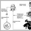
The cornea consists of 5 layers: anterior epithelium, Bowman's membrane, stroma, Descemet's membrane, endothelium.
- The anterior epithelial layer is a stratified squamous non-keratinized epithelium that performs a protective function. Resistant to mechanical stress, if damaged, it quickly recovers within a few days. Due to the extremely high ability of the epithelium to regenerate, scars do not form in it.
- Bowman's membrane is a cell-free layer of the surface of the stroma. When it is damaged, scars form.
- Corneal stroma - Occupies up to 90% of its thickness. Consists of properly oriented collagen fibers. The intercellular space is filled with the main substance - chondroitin sulfate and keratan sulfate.
- Descemet's membrane - the basement membrane of the corneal endothelium, consists of a network of thin collagen fibers. It is a reliable barrier to the spread of infection.
- Endothelium is a monolayer of hexagonal cells. It plays an important role in the nutrition and maintenance of the cornea, prevents its swelling under the influence of IOP. Does not have the ability to regenerate. With age, the number of endothelial cells gradually decreases.
The innervation of the cornea is carried out by the endings of the first branch of the trigeminal nerve.
The cornea is nourished by the surrounding vasculature, corneal nerves, anterior chamber moisture, and the tear film.
Protective function of the cornea and corneal reflex
Remaining the outer protective shell of the eye, the cornea is exposed to harmful environmental influences - mechanical particles suspended in the air, chemicals, air movement, temperature effects, and so on.

The high sensitivity of the cornea determines its protective function. The slightest irritation of the surface of the cornea, for example, a speck of dust, causes an unconditioned reflex in a person - closing of the eyelids, increased lacrimation and photophobia. Thus, the cornea protects itself from possible damage. When the eyelids are closed, the eye simultaneously rolls up and copious release of tears, which washes away small mechanical particles or chemical agents from the surface of the eye.
Symptoms of corneal diseases
Changes in the shape and refractive power of the cornea
- With myopia, the cornea may have a steeper shape than normal, which causes a greater refractive power.
- With farsightedness, the opposite situation is observed, when the cornea is flattened and its optical power is reduced.
- Astigmatism is manifested when the cornea is irregularly shaped in different planes.
- There are congenital changes in the shape of the cornea - megalocornea and microcornea.

Superficial damage to the corneal epithelium:
- Point erosions are small defects in the epithelium stained with fluorescein. This is a non-specific symptom of corneal diseases, which, depending on the localization, can occur with spring catarrh, poor selection of contact lenses, dry eye syndrome, lagophthalmos, keratitis, and toxic effects of eye drops.
- Edema of the corneal epithelium indicates damage to the endothelial layer or a rapid and significant rise in IOP.
- Pinpoint epithelial keratitis is common in viral infections of the eyeball. Granular swollen epithelial cells are found.
- Threads - thin mucous threads in the form of a comma, connected on one side with the surface of the cornea. They occur with keratoconjunctivitis, dry eye syndrome, recurrent corneal erosion.
Corneal stroma damage:
- Infiltrates are areas of an active inflammatory process in the cornea, which are both non-infectious - wearing contact lenses, and infectious in nature - viral, bacterial, fungal keratitis.
- Edema of the stroma - an increase in the thickness of the cornea and a decrease in its transparency. It occurs with keratitis, keratoconus, Fuchs' dystrophy, endothelial damage after eye surgery.
- Ingrown vessels or vascularization - manifests itself as an outcome of the transferred inflammatory diseases of the cornea.

Damage to the Descemet's membrane
- Tears - with trauma to the cornea, also occur with keratoconus.
- Folds - are caused as a result of surgical trauma.
Methods for examining the cornea
- Biomicroscopy of the cornea - examination of the cornea using a microscope with an illuminator, allows you to identify almost the entire range of changes in the cornea in its diseases.
- Pachymetry - measurement of the thickness of the cornea using an ultrasound probe.
- Specular microscopy is a photographic study of the endothelial layer of the cornea by counting the number of cells per 1 mm2 and analyzing the shape. The cell density is normal - 3000 per 1 mm2.
- Keratometry - measurement of the curvature of the anterior surface of the cornea.
- Topography of the cornea is a computer study of the entire surface of the cornea with an accurate analysis of the shape and refractive power.
- In microbiological studies, scrapings from the surface of the cornea are used under local drip anesthesia. A corneal biopsy is performed when the results of scrapings and crops are indicative.

Principles of treatment of diseases of the cornea
Changes in the shape and refractive power of the cornea, such as myopia, hyperopia, astigmatism, are corrected with glasses, contact lenses or refractive surgery.
With persistent opacities, corneal leukomas, it is possible to perform keratoplasty, corneal endothelial transplantation.
Antibacterial, antiviral and antifungal drugs are used for corneal infections, depending on the etiology of the process. Local glucocorticoids suppress the inflammatory response and limit scarring. Drugs that accelerate regeneration are widely used for superficial damage to the cornea. Moisturizing and tear replacement drugs are used for tear film disorders.
Thanks
The site provides reference information for informational purposes only. Diagnosis and treatment of diseases should be carried out under the supervision of a specialist. All drugs have contraindications. Expert advice is required!
What is corneal pachymetry?
 pachymetry is a research method ophthalmology (a science concerned with the study, diagnosis, treatment and prevention of eye diseases), with which the doctor evaluates the thickness of the cornea ( cornea). This allows you to identify a number of diseases accompanied by its thinning or thickening. In addition, pachymetry can be used when planning or performing various surgical operations on the cornea, as well as to monitor the effectiveness of such operations. The procedure is relatively safe and absolutely painless, and therefore it can be prescribed to almost all patients, regardless of gender, age, the presence of concomitant diseases and other factors.
 Pachymetry technique
To understand when and why this study is needed, as well as how it is carried out, certain knowledge from the field of anatomy of the eyeball is necessary.The cornea belongs to the outer shell of the eyeball and is located in its anterior part, having a slightly convex ( outside) shape. Under normal conditions, the cornea is transparent, as a result of which light rays pass freely through it, entering the inside of the eyeball and then reaching the retina, where images are formed. The cornea belongs to the so-called refractive system of the eye ( it also includes the lens and some other structures of the eyeball). Due to a certain curvature and thickness of the cornea passing through it ( and then through the lens) light rays are refracted and focused at a certain point in the eyeball ( namely on its back wall, right on the retina), which ensures the formation of a clear image of the objects that a person looks at. Violation of the curvature of the cornea, as well as a change in the thickness of the entire cornea or certain sections of it, will be accompanied by a violation of its refractive power, which can cause a violation ( decrease) visual acuity. Measurement of the thickness of the cornea in its various departments allows you to identify the existing pathology and choose the most optimal treatment, as well as evaluate its effectiveness.
How is pachymetry done?
 To measure the thickness of the cornea, you need to use special equipment ( pachymeters) and technology.
 Preparation for pachymetry
No special preparation for the study is required. On the appointed day or right during the first visit to an ophthalmologist - a doctor specializing in the diagnosis and treatment of eye diseases) the patient can undergo a pachymetry procedure, after which he can immediately go about his business. If the patient wears contact lenses, they will be asked to remove them immediately prior to the examination.Apparatus for conducting and types of pachymetry
To date, there are several studies that measure the thickness of the cornea of the eye. They differ from each other both in terms of execution technique and information content.To study the thickness of the cornea, use:
- optical pachymetry. For the study, a special slit lamp is used, which can direct a beam of light into the patient's eye in the form of a strip, the length and width of which the doctor can adjust. Using a slit lamp and special lenses allows you to most accurately determine the thickness of the cornea.
- Ultrasonic pachymetry. To conduct this study, ultrasound devices are used that allow you to study the structure and thickness of various tissues of the eyeball.
- computerized pachymetry. For the study, a special apparatus is used ( tomograph), which "sees through" the eye structures, allowing you to get images of the eyeball, cornea and other tissues.
Optical pachymetry
This technique was first used to study the thickness of the cornea more than 50 years ago, however, due to its simplicity and information content, it is still relevant today. As already mentioned, the essence of the method is to use a slit lamp and special lenses.The slit lamp is a kind of "microscope". The lamp allows you to direct a strip of light at the patient's eye, and then study the structures visible in it under high magnification. For pachymetry, two additional lenses are installed on the lamp.
The procedure goes as follows. The patient comes to the ophthalmologist's office and sits down at the table on which the slit lamp is located ( it is quite voluminous and usually tightly fixed to the table). Then he puts his chin on a special stand, and presses his forehead against the fixing arc. The doctor asks him to remain still and not blink, while he adjusts the optical system of the lamp so that it is directly opposite the eye being examined.
After the slit lamp is placed, a beam of light is directed into the patient's eye. The thickness of the cornea is measured using special lenses mounted on a lamp and arranged parallel to each other. One lens is fixed while the other is movable. Slowly rotating a special handle, the doctor changes the angle of the movable lens, as a result of which the nature of the light rays passing through the cornea changes. Based on this, the specialist measures its thickness in various areas.
Ultrasound pachymetry
This technique is also called contact pachymetry, since during the study there is a direct contact of the ultrasound probe with the patient's cornea ( with the optical method of research, there is no such contact).Before starting the study, anesthesia of the cornea should be performed. The fact is that during the procedure, the working part of the sensor will come into contact with the outer surface of the cornea, which is rich in sensitive nerve endings. Any, even the most insignificant touch to its surface causes a blinking reflex, as a result of which the patient's eyelids involuntarily close. It also stimulates increased lacrimation ( as a protective reaction to corneal irritation). It will be impossible to carry out research in such conditions.
Anesthesia solves these problems. Its essence is as follows. 3 - 6 minutes before the start of the study, a few drops of local anesthetic are instilled into the patient's eyes. This drug penetrates the cornea and temporarily "turns off" the nerve endings located there, as a result of which the patient ceases to feel touching the surface of the cornea.
After performing anesthesia, the doctor proceeds directly to pachymetry. For this, the patient must lie down or sit on the couch and keep his eyes open. Having picked up an ultrasonic sensor, the doctor lightly touches the surface of the cornea of the eye with the working part of the device. Within a few seconds, the device takes measurements, after which the built-in display shows the thickness of the cornea in the examined area.
The essence of the ultrasound method ultrasound) is as follows. Ultrasonic waves generated by a special emitter can propagate in various tissues of the body that they meet on their way. At the border between tissues, the composition of which differs, sound waves are partially reflected and recorded by a sensor located inside the apparatus. The analysis of reflected waves makes it possible to determine the thickness of the examined tissue, as well as to evaluate its structure.
As mentioned earlier, the cornea is the front part of the shell of the eyeball. Behind it is the so-called anterior chamber of the eye, filled with intraocular fluid ( aqueous humor). When the probe is applied to the anterior surface of the cornea, ultrasonic waves begin to propagate along it, however, reaching its posterior border, they are partially reflected from the aqueous humor. Evaluation of the nature of the reflected waves and the time of their reflection and allows you to determine the thickness of the cornea. All this takes about 1-3 seconds from the device. Using this technique, within a few minutes, the doctor can examine the thickness of the cornea along its entire length.
If after the end of the study the patient feels any discomfort in the eyes, he can rinse them with warm clean water. At the same time, it is worth noting that usually the examination is absolutely painless, without causing any inconvenience to the patient. The sensitivity of the cornea is restored after a few minutes or tens of minutes ( depending on the anesthetic used and its dose). In this case, the patient can go about his business immediately after the end of the procedure.
Computed pachymetry
A computerized evaluation of the thickness of the cornea can be carried out during the so-called optical coherence tomography. The essence of the study lies in the fact that the human eye is "translucent", "scanned" by infrared radiation. Special sensors register the nature of the reflection of infrared rays from various structures of the eyeball, and after computer processing, the doctor receives an accurate, detailed image of the area under study.The procedure goes as follows. The patient comes to the ophthalmologist's office and sits in front of the apparatus ( tomograph). He applies his chin and forehead to special fixators ( as in a slit lamp examination), which ensures the immobility of the head throughout the procedure. Next, the doctor brings the working part of the device closer to the examined eye and scans the cornea and ( if necessary) other structures of the eye.
The duration of the procedure usually does not exceed 3-10 minutes, after which the patient can immediately go home, having received the results of the study.
Interpretation of pachymetry results ( norm and pathology)
After the examination, the doctor gives the patient a conclusion in his hands, which indicates the thickness of the cornea, measured in its various parts. Although this indicator can vary widely ( depending on the age, race and other characteristics of the patient), researchers have established certain average limits for corneal thickness.The normal thickness of the cornea is:
- in central departments– 490 – 620 micrometers ( 0.49 - 0.62 mm).
- In the peripheral areas (along the edges) – up to 1200 micrometers ( 1.2 mm).
Indications for pachymetry
 Indications for the appointment of this study may be diseases that are characterized by thickening, thinning or curvature of the cornea. As a rule, the clinical signs of such diseases are detected by an ophthalmologist during the examination of the patient, evaluation of his complaints and evaluation of the results of simpler studies. If after that it is not possible to make a definitive diagnosis, the patient may be assigned pachymetry.
 Indications for pachymetry are:
- Edema of the cornea. With corneal edema, its tissue is affected by the pathological process, thickens and deforms. The cause of this pathology may be inflammation of the cornea or other structures of the eye, allergy, foreign body entering the cornea, eye injury, poor hygiene when wearing contact lenses, and so on. The patient may complain of the appearance of fog before the eyes, increased tearing, redness of the eyes, pain in the eyes. When conducting pachymetry, it is possible to detect widespread thickening of the cornea, as well as the appearance of individual "folds" and other deformations in its various parts.
- Corneal ulcers. An ulcer is called a defect ( deepening) in corneal tissue. The cause of the development of an ulcer can be trauma, inflammatory or infectious lesions of the cornea of \u200b\u200bthe eye and other damage to it. With ulceration of the cornea, its thickness in the affected area decreases, resulting in a violation of its refractive power. Patients may complain of pain and burning in the area of the affected eye, increased lacrimation. Pachymetry allows you to determine the depth of the ulcer, as well as evaluate the effectiveness ( or inefficiency) of the treatment.
- Dystrophic diseases of the cornea. Corneal dystrophies are a number of hereditary diseases that are characterized by a violation of the processes of renewal of the corneal tissue. These disorders can be manifested by excessive formation of corneal tissue and its thickening, clouding of the cornea, metabolic disorders and ulceration ( partial or complete) cornea and so on. Pachymetry allows to detect pathological changes at an early stage of the disease. At the same time, the treatment of these pathologies is not always effective, since in most cases they are caused by disorders in the human genetic apparatus ( i.e. considered incurable). The only effective treatment for these pathologies can be considered a corneal transplant from a donor.
- Preparation for operations on the cornea. Before transplanting the cornea, it is important for the doctor to know the thickness of the cornea at the transplant site, its structure and other features of its structure. Pachymetry can help in resolving these issues. In addition, this study can be prescribed before operations on other structures of the eye ( for example, when replacing the lens).
- Assessment of the state of the cornea in the postoperative period. After corneal transplantation, pachymetry allows you to assess whether the donor tissue has taken root, whether corneal edema or other complications develop.
Keratoconus
This pathology is characterized by a cone-shaped outward protrusion of the cornea. At the same time, its thickness is significantly reduced. A change in the shape and thickness of the cornea disrupts its refractive power, as a result of which patients begin to complain of blurred images, double vision ( if only one eyeball is affected by keratoconus), increased lacrimation, photophobia, and so on.Diagnosis can usually be made by examining the patient's eyeball ( especially in advanced stages, when the bulge of the cornea becomes extremely pronounced). Pachymetry can be used to determine corneal thickness before surgical treatment of keratoconus. The essence of the operation is that the surgeon performs several incisions on the cornea, which is accompanied by a change in its shape. However, with severe thinning of the cornea ( what is characteristic of keratoconus) the doctor risks piercing it through and through. Pachymetry allows you to determine the exact thickness of the tissue and calculate the required incision depth.
Glaucoma
Glaucoma is an eye disease characterized by an acute or chronic, slow increase in intraocular pressure ( IOP). This happens due to the accelerated formation or impaired removal of intraocular fluid. An increase in IOP can lead to damage to the nerve structures of the eye ( optic nerve), which can lead to complete blindness.To determine if a patient has glaucoma, intraocular pressure should be measured. The essence of this procedure is that a special weight with a known mass is placed on the cornea of the patient lying on his back. The lower part of the weight is pre-coated with a special paint. Under its weight, the cornea bends, as a result of which part of the paint is washed off from the surface of the weight, which is directly adjacent to the cornea. The lower the intraocular pressure, the more the cornea will bend and, conversely, the higher the IOP, the less the cornea will bend and the less paint will be washed off the weight. At the final stage of the study, the weight is applied to special paper and the diameter of the ring formed as a result of washing off the paint is determined. This allows you to evaluate the IOP.
The problem of the study is that the measurement does not always take into account the thickness of the cornea. At the same time, it was experimentally established that the IOP parameters measured by the method described above depend on the thickness of the cornea of the eye. The fact is that the corneal tissue has a certain elasticity, the thicker it is, the weaker it will sag under the pressure of the weight and, conversely, the thinner the cornea, the more it will sag. So, for example, an increase in the thickness of the cornea by 100 micrometers ( 0.1 mm) can increase intraocular pressure by 3 mmHg. This can lead to a false diagnosis of glaucoma and unreasonable prescribing of treatment that the patient does not need. At the same time, thinning of the cornea may be accompanied by too low IOP, as a result of which the patient's glaucoma may go unnoticed.
Today, in all modern clinics, the measurement of intraocular pressure should be accompanied by pachymetry. After determining the thickness of the cornea, an appropriate correction is made, which allows you to determine the intraocular pressure as accurately as possible.
Contraindications and adverse reactions for pachymetry
 The list of contraindications to the study is small, due to its simplicity and safety.
 Pachymetry is contraindicated:
- Patients in an inadequate condition. These can be both mentally ill people and patients who are in a state of alcohol and / or drug poisoning. In this state, the patient will not be able to sit still during the entire procedure ( 3 – 15 minutes), as well as look straight ahead, which is a prerequisite for pachymetry.
- With perforation of the cornea. In this case, contact pachymetry using an ultrasound probe, which must be applied directly to the cornea, is contraindicated. The fact is that when performing a study, an infection can penetrate through a defect in the cornea of \u200b\u200bthe eye, which can be accompanied by the development of formidable complications, up to complete blindness.
- With purulent-inflammatory diseases of the eye. In this case, contact pachymetry is also contraindicated, since the procedure can provoke the spread of infection and increase the severity of the inflammatory process.
- If you are allergic to local anesthetics. In this case, the patient is also contraindicated in contact ultrasonic pachymetry, during which anesthetics are used. The fact is that instillation of such a drug into the eyes of a patient who is allergic to it can lead to the rapid development of allergic reactions ( from redness and swelling of the eye to anaphylactic shock and death of the patient). Complete patient interview and test execution ( test) for allergies allows you to almost completely eliminate the risk of developing this complication.
Where to do pachymetry?
 Pachymetry can be done in any major hospital or clinic where an ophthalmologist sees, as well as in ophthalmological rooms and clinics equipped with the necessary equipment. Depending on the type of research, its price can range from 250 to 3000 rubles.
 Sign up for pachymetry
To make an appointment with a doctor or diagnostics, you just need to call a single phone number
+7 495 488-20-52 in Moscow
+7 812 416-38-96 in St. Petersburg
The operator will listen to you and redirect the call to the right clinic, or take an order for an appointment with the specialist you need.
In Moscow
In St. Petersburg
The address | Phone |
|
Medical Center MEDEM | st. Marata, house 6. | 7 (812 ) 336-33-36 |
All-Russian Center for Emergency and Radiation Medicine. A.M. Nikiforovich EMERCOM of Russia | st. Academician Lebedeva, house 4/2. | 7 (812 ) 607-59-00 |
Military Medical Academy. CM. Kirov | st. Academician Lebedev, house 6. | 7 (812 ) 573-99-04 |
Hospital for war veterans | st. Narodnaya, house 21, building 2. | 7 (812 ) 446-17-91 |
Ophthalmological center "Vision" | st. Ryukhina, house 12. | 7 (812 ) 900-85-42 |
In Ekaterinburg
In Krasnoyarsk
In Krasnodar
In Novosibirsk
In Vladivostok
In Rostov-on-Don
In Voronezh
In Perm
In Chelyabinsk
Name of medical institution |
LASIK (L aser- A sistedin Si tu K eratomileusis - "laser keratomileusis") - today is the most common method for correcting refractive errors, such as: myopia, hypermetropia, astigmatism.
Most excimer laser interventions are performed to correct myopia. High efficiency, painlessness and fast recovery are the main criteria for choosing Lasik for patients who want to have high visual acuity without additional correction tools.
The suitability of a Lasik procedure for highly myopic patients is determined by the thickness of the cornea. The optimal thickness of the cornea for excimer laser correction in mild and moderate myopia is from 450 microns. In keratoablation, the excimer laser vaporizes 13-14 microns per diopter. The minimum residual corneal thickness after keratoablation is 280 microns under the flap. But with a high degree of myopia and a cornea thickness of less than 500 microns, there is a danger of thinning of the cornea below a critical level. Since the excimer laser should evaporate 104-140 microns.
The task of the refractive surgeon is the correct calculation of the thickness of the evaporated and residual cornea, as well as the assessment of possible intra- and postoperative complications.
Purpose of the study. Study of the features of calculating the residual thickness of the cornea and evaluating the effectiveness of excimer laser correction of high myopia with a cornea thickness of less than 500 microns.
Material and methods. We performed excimer laser correction of high myopia in 6 patients (12 eyes) with corneal thickness less than 500 microns. At admission, all patients underwent visometry (with/without correction), visometry against the background of cycloplegia, tonometry, autokeratorefractometry, keratotopography, corneal OCT, and biomicroscopy.
Laser correction was carried out on the VISXStarS4 (AMO) excimer laser system:
- Wavelength 193 nm
- Beam diameter 0.65-9.5 mm
- Variable patch ablation
- Ablation speed from 2 s/d
- 3-D pupil tracking system with automatic centering
- Strobe-saving algorithms
- Short operation time
The formation of the corneal flap was performed using an automatic microkeratome ML-7 (USA). The thickness of the flap is 80 microns.
The calculation of the residual thickness of the cornea was carried out according to the formula:
TR - 80 - (14×D)
where TP is the total thickness of the cornea;
80 – flap thickness (micron);
14 - the number of microns evaporated by the laser per 1 diopter
D - diopters that need to be adjusted
Results. The mean uncorrected visual acuity before LASIK was 0.079±0.05; the mean corrected visual acuity was 0.83±0.2; the average value of spherical refraction was -9.0 diopters.
The average thickness of the cornea is 492.8 microns.
The result was evaluated 1 month after stabilization of visual acuity and refraction.
The mean value of uncorrected visual acuity after the LASIK procedure was 0.93±2.0; the average value of spherical refraction was -0.29±0.3 diopters.
Re-epithelialization of the corneal flap was completed by the second day of observation. The cornea remained transparent, smooth, shiny. Subepithelial opacities were observed in one patient, which resolved within 2 weeks.
All patients were prescribed drops of Dexatobropt according to the scheme: 1st day, 1 drop every 1.5 hours; 4 days 1 drop 4 times a day; 4 days 1 drop 3 times a day. In case of dry eye syndrome after the LASIK procedure, patients were prescribed Vizmed gel 1 drop 2-3 times a day.
Conclusions.
After analyzing the results of the correction of high myopia by the Lasik method in patients with a cornea thickness of less than 500 microns, high visual acuity results were obtained. The correct calculation of the residual thickness of the cornea in patients with high myopia made it possible to safely perform excimer laser ablation with the preservation of the residual corneal stroma in the range from 280 to 300 microns.


