Visual hygiene analyzer. Organ view. How is the perception and transmission of visual information
1. What are the analyzers? What links do it consist of? 2. Who first introduced this term? What is the difference between the concept of the analyzer from the concept of the sense body? 3. What is the most significant analyzer for a person and why? What is his structure? 4. What place in this chain occupy your eyes? Explain the words of William Blake: "Through the eye, not an eye to look at the world knows how the mind can ..." Answer questions:

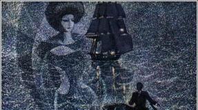
Her eyes are like two fog, semi-coulting, half-fake, her eyes - as two deceptions covered with a mix of failure. The connection is two riddles. Half-resistant, semi fright, insane tenderness of the seafood, anticipating mortal flour. When the dotmon comes and the thunderstorm approaches, from the bottom of my soul, my beautiful eyes flicker. N. Zabolotsky. F. Rokotov "Portrait of the Store"

Today in the lesson we have to: consider the structure of the eye as an optical system and identify the connection of the structure with the function of the eyes. Determine the causes and types of violations of vision. Find out the rules of hygiene view, because It is necessary to preserve the health of our eyes.


If the tear fluid does not stand out, then: retinal cells die? The corneal cells will die? Crystalik will change the curvature? Pupil narrows? In every century 80 eyelashes. How many eyelashes have a person? Daily: A man blinks once our lacrimar glands produce 3 Thors of the tears you know ...


Close the left eye, place the drawing at a distance of 20 cm from the right eye and look at the green circle depicted on the left. Slowly bring the drawing to the eye, the moment will certainly come when the red circle will disappear. How to explain this phenomenon? "Detection of the Blind Spot".


Detect the narrowing and expansion of the pupil. Look in your eyes your neighbor in the desk and mark the magnitude of the pupil. Close your eyes and depart their palm. Count up to 60 and open your eyes. Watch the change in the magnitude of the pupils. How to explain this phenomenon?

Questions to class: What body of the eye is called a live lens? What shell focus on the rays? What happens in retina receptors? How are nerve impulses transmit? Where are the nerve impulses transmit? Is it true that the eye looks, and does the brain sees? How do babies see? What violations of view said in a video clogging?

In congenital myopia, the eyeball has an extended form. Therefore, a clear image of items located far from the eyes occurs not on the retina, but as if in front of it. Acquired myopia develops due to an increase in crust curvature, which may occur with improper metabolism or impaired hygiene. Mostic people see remote objects vague. Glasses with bonotherapy lenses help ensure that distinct images of objects occur accurately on the retina. Violations. The most common impairment of vision is myopia and hyperopia. The presence of these violations establishes a doctor when measuring visual acuity using special tables. Myopia is congenital and acquired.

Acquired hyperopia arises due to a decrease in crust scavenge and most characteristic of elderly people. Falnotherbore people see close objects vague, cannot read text. Glasses with double-way lenses help the image of a close object exactly on the retina. Violations. Falnarity also happens congenital and acquired. For congenital distancelessness Eye apple shortened. Therefore, a clear image of items located close to the eyes arise as if behind the retina.


Repetition: Test 1. Who introduced the concept of analyzers? 1.I.P.Pavlov. 2.I.m.Sechenov. 3.N.y.pirogov. 4.I.I.Thennik. ** Test 2. What parts are different in the analyzers? 1. Organ sense. 2. Receptors (peripheral link). 3. Nervous pathways (conductor), for which the excitement is carried out to the central link. 4. Centers in the cerebral cortex processing information. 5. Nervous paths (conductor), for which the excitement is carried out from the central link. Test 3. Where are the highest sections of the visual analyzer? 1. In temporal fractions. 2. In frontal fractions. 3. In parcels. 4. In the occipital shares.

Repetition: Test 4. How many muscle pairs are responsible for the movement of the eye? 1. One pair. 2. Two pairs. 3. Three pairs. 4. Four pairs. Test 5. What is the name of the front transparent part of the outer shell of the eye? 1.Clare. 2.Reight. 3. Voltage. 4.Connet. Test 6. What is the name of the middle shell of the eye and its front part, in the center of which is the pupil? 1.Conscious. 2.Scar. 3. Voltage. 4. Schedule.

** Test 7. What changes in the structures of the eye arise with the acquired myopia? 1. The eyeball is shortened. 2. The eyeball is lengthened. 3. The crystal becomes more flat. 4. The crystal becomes more convex. Test 8. What is the eyeball with congenital hypodiness? 1.Ort. 2.). Test 9. What changes in the structures of the eye occur when acquired hyperopias? 1. The eyeball is shortened. 2. The eyeball is lengthened. 3. The crystal becomes more flat. 4. The crystal becomes more convex. Reiteration:

Test 10. Where is the layer of ferrous pigment cells? 1. On the outer surface of the retina. 2. On the inner surface of the vascular shell. 3. On the inner surface of the protein shell, sclera. 4. On the inner surface of the iris. What is indicated in the figure figures 1 - 14?

The learning process passes through the recess in the material studied,
Then through the deepening in itself.
I.F. Herbart
Objectives:
Educational goal: Socialization of students in an educational situation, the development of a feeling of tolerance to each other and self-esteem.
Developing goal: the formation of elements of the natural science worldview by students with knowledge of the basics of anatomy and physiology, the development of communicative skills through the formation of skills to work in mini-groups and the ability to analyze their activities
Complex learning (Didactic) Purpose (cds): - mastering the content of the "Analyzers" theme. Formation of students' understanding between the structure and functions of the constructs of organs and the body on the example of analyzers.
Private didactic objectives (CDC):
- Development of skills to recognizing eye structures.
- Formation of readiness to use the knowledge and skills obtained in the lesson.
- Expansion of students' presentings on functional structural links of the visual analyzer.
Students should know: Terminology on the topic " Spectator analyzer", The main structures of the eye and their work.
Students should be able to:
- Find on the proposed didactic material of the structure of the visual analyzer,
- Describe anatomy and physiology of analyzers.
- Justify the need for a valeological approach to yourself and the surrounding people.
- Have a skill of healthy behavior.
The formulated area of \u200b\u200bunderstanding is a structural and functional analysis of the eye and the visual analyzer at the propagation level.
Pedagogical strategy: "In order to digest knowledge, it is necessary to absorb them with appetite" (Anatol Franz)
Pedagogical tactics: individualization of frontal learning by means of knowledge differentiation at the stage of explanation of the new material.
Leading forms of u. roca:euristic conversation, work with a digital microscope, analysis of the materials presentation materials, reflection as part of team activities.
Pedagogical technologists: personal-oriented learning.
Equipment lesson: multimedia projector, digital microscope QX3 + CM, preparations of dried bull eyes.
Monitoring forms: self-control, interconnection and expert control.
Summary lesson
Part 1. Setting the problem: the value of the visual analyzer (slides number 1-2)
To solve the problems of this lesson, the formation of children's understanding of the leadership role of the visual analyzer. Therefore, the teachings are proposed for working with a running polylinghouse string. Students create their own list of words and expressions about vision and eyes. The functional contribution of this part of the lesson can be characterized as an emotional-intellectual immersion of children in the theme.
Part 2. Explanation and consolidation of a new material: the structure of the eye. (Slides number 3, 4, 5, 6)
Propaede study of the structure of the eye is carried out in 6-7 classes. Therefore, the main complexity in presenting the topic in the 8th grade is the "alluring" of children, which can be avoided by the appeal to the analysis of "household knowledge" with the repetition and deepening of the studied earlier. Combining heuristic conversation with teamwork in intellectual pairs, the teacher brings students to demonstration laboratory work.
Part 3. Demonstration laboratory work: the structure of the mammal eye. (Slide number 3)
The most dynamic and therefore the memorable form of comparative analysis of structures is microscopation . The training situations are:
a) The presentation of the student-demonstrators of a highly specialized task in the form of individual drugs.
b) a consistent discussion in the teams of "pictures" of digital microscopation.
Part 4. Explanation and consolidation of a new material: the main refractive mediums of the eye and the eye bottom. (Slides number 7, 8, 9, 10, 11, 12)
In this part, the continued Intrigue of the lesson: the collision of various domestic observations and turn them into scientific knowledge. In the same part of the lesson introduced new complex conceptsForming children understanding the features of the color and light perception of a person. Therefore, 3 slides from 6 is devoted to the discussion of information.
Part 5. Explanation and consolidation of new material: image perception. (slides number 13-15)
The complex part is determined by its integitivity. Discussion of the unexpected consequences of the brain asymmetry for the perception of the picture of the world by trace method allows children to clearly appreciate the degree of mastering the material, and the degree of reproductiveness and the creativity of the responses can be expressed both in shorten track trails and in a change in the color of the step.
Demonstration laboratory work lasts 10 minutes. Students-demonstrators and observers discuss drugs. A - appearance of the eye, in - internal structure eyes, with - retina
Part 2 (continued). Explanation and consolidation of new material: the structure of the eye. (Slides number 5, 6)
Slide number 13. Creating a visual image It occurs in the occipital fraction of the cortex of the brain. It is very important how the image is transmitted to the brain, because the brain is asymmetric. Remember the chicken. It will not connect information from two halves of the brain, so chicken sees autonomously every eye. In person, the right side of the retina each eye transmits the image into the left analytical hemisphere, and the left part of the retina transmits the image into the right shaped hemisphere.
Slide number 14. Features of the eyes of a woman
In the female eye more sticks. Therefore:
- Peripheral vision is better developed.
- It is better to see in the dark.
- Perceive information more than men at every moment of time
- Instantly fix any movement.
- Sticks work on the right, specifically-shaped hemisphere.
Slide number 15. Features of the eye of a man
In the men's eye more colums.
On columns, there is a focus of the eye lens. Therefore:
- Better perceive colors.
- Clever see a picture.
- Concentrate on one aspect of the image, reducing the entire field of view to the tunnel.
- Columns work on the left, abstract hemisphere.
Part 6. Reflection (slides number 16, 17). These slides did not enter the presentation presented at the festival
A) students introduces students with a fragment of the educational and research project "Functional dependence of the condition of the eye from the student of the schoolboy's day."
Hygiene Eyes is mainly in compliance with the day, night rest (night sleep is at least 8 hours), work at a computer (students of 8th grade can work at a computer for about 3 hours a day). It is necessary to systematically do eye exercises.
- Write a nose.
- See through.
- Move eyebrows.
B) Students record the main thing, in their opinion, the thought thought in the day's diary diary, thereby summarizing its own schedule of sleep and daily employment chart.
Homework: on the textbook N.I.Sonin, M.R. Sapin biology. Human. M.Drof.
- Reproductive task
High School N8.
« Human visual analyzer
Student 9A class.
Sherstyukova A.B.
obninsk
Introduction
I. Eyework and Functions
1. Feling
2. Auxiliary systems
2.1. Overall muscle
2.4. Temaful apparatus
3. Shells, their structure and functions
3.1. Outer shell
3.2. Medium (vascular) shell
3.3. Inner sheath (retina)
4. Transparent intraocular environments
5. Perception of light stimuli (light-crossing system)
II. Speed \u200b\u200bnerve
III. Mozgian center
IV. Hygiene view
Conclusion
Introduction
Human eye is an amazing gift of nature. He is able to distinguish the finest shades and the smallest dimensions, well to see the day and not bad at night. And compared to the eyes of animals, has great opportunities. For example, the pigeon sees very far away, but only during the day. Owls and bats are well seen at night, but during the day they are blind. Many animals do not distinguish between a separate color.
Some scientists say that 70% of all information from the world around us we get through the eyes, others are called even a large figure - 90%.
Works of art, literature, unique monuments of architecture became possible thanks to the eye. In the development of space, the organ of vision belongs to a special role. More cosmonaut A. Neoonov noted that in conditions of weightlessness, not a single sense body, in addition, does not give proper information to the perception of a spatial situation.
The emergence and development of the organ of vision is due to the variety of environmental conditions and the inner environment of the body. The light was a stimulus, which led to the emergence of an organ of vision in the animal world.
Vision is ensured by the work of the visual analyzer, which consists of a perceive part - eyeball (With its auxiliary apparatus), conducted by the path, which is perceived by the eye is transmitted at the beginning of the subcortex centers, and then in the bark of a large brain (occipital shares), where the highest visual centers are located.
I. The structure and function of the eye
1. Feling
The eyeball is located in the bone container - a red-having width and a depth of about 4 cm; In shape, it resembles a pyramid of four faces and has four walls. In the depths of the eyelid, there are upper and lower and nourish-eye cracks, a visual channel, through them nerves, artery, veins. The eyeball is located in the front of the orbit, separated from the rear section of the connective membrane - the vagina of the eyeball. In the backyard it is located speed \u200b\u200bnerve, muscles, vessels, fiber.
2. Extractive systems
2.1. Overall muscle.
In the movement, the eyeball leads four straight (top, bottom, medial and lateral) and two oblique (upper and lower) muscles (Fig. 1).
Fig.1. Overall muscles: 1 - medial straight line; 2 - upper straight; 3 - upper oblique; 4 - lateral straight line; 5 - Lower straight; 6 - lower oblique.
Medial straight muscle (reducing) turns the eye of the bed, the lateral - knutrice, the upper straight moves upwards and knutrice, the upper oblique is the book and the duck and the lower oblique - up and the duck. Eye movements are provided by the innervation (excitation) of these muscles with eye, block-shaped and discharge nerves.
2.2. Eyebrows
Eyebrows are designed to protect the eyes from droplets of sweat or rain flowing from the forehead.
2.3. Century
These are movable dampers covering in front of the eyes and protect them from external influences. The skin of the age is thin, under it there is a loose subcutaneous tissue, as well as the circular muscle of the eye, providing the clamp of the eyelids with a dream, blinking, and grinding. In the thuscale of the century there is a connecting plate - cartilage, giving them the form. In the edges of the eyelids grow eyelashes. In centuries are located sebaceous glandsThanks to the secret of which it creates a sealing bag seal when closing the eye. (Conjukiwa is a thin connecting sheath that widespread the back surface of the eyelids and the front surface of the eyeball to the cornea. With closed eyelids, the conjunctiva forms a conjuncture bag). It warns the eye clogging and drying the cornea during sleep.
2.4. Temaful apparatus
The tear is formed in the tear gland located in the upper gender corner of the orbit. The tear gland's output ducts falls into the conjunctival bag, protects, nourishes, moisturizes the cornea and conjunctival. Then, on the lacrimal routes, it enters the nasal cavity through the nasal duct. With a constant blowing age on the cornea, a tear is distributed, which supports its humidity and flushes small foreign bodies. The secret of the tear glands acts as a disinfectant liquid.
3. Shells, their structure and functions
The eyeball is the first important component of the visual analyzer (Fig. 2).
The eyeball is not quite the right spherical shape. It consists of three shells: external (fibrous) capsule, consisting of a cornea and sclera; medium (vascular) shell; internal ( retina, or retina). The shells surround the inner cavities (cameras) filled with transparent water melted moisture ( intraocular fluid), and internal transparent refractive media (crystal and vitreous body).
Fig.2. Eyeball: 1 - cornea; 2 - front camera eye; 3 - crystal; 4 - sclera; 5 - vascular shell; 6 - retina; 7 - optic nerve.
3.1. Outdoor shell
This is a fibrous capsule, which causes the form, the tour (tone) of the eye, protects its contents from external influences and serves as a place of attachment of the muscles. It consists of a transparent cornea and an opaque sclera.
The cornea is a refractive medium when light rays come into the eye. There is a lot of nervous endings in it, so the hit of even small suture on the cornea causes pain. The cornea is dense enough, but has good insight. Normally, it does not contain blood vessels, it is covered with epithelium.
The scler is an opaque part of the fibrous eye capsule having a bluish or white color. Overhead muscles are attached to it, the vessels and nerves of the eye are passing through it.
3.2. Medium (vascular) shell.
Vascular provides meals with an eye, it consists of three departments: iris, ciliary (ciliary) body and a vascular shell itself.
Rainbow - The most forefront of the vascular shell. It is located behind the cornea so that there remains free space between them - the front chamber of the eye filled with transparent water moisture. Through the cornea and this moisture, the iris is clearly visible, its color determines the color of the eyes.
In the center of the iris, there is a round hole - the pupil, the dimensions of which vary and regulate the amount of light falling inside the eye. If there are a lot of light, the pupil is narrowing if it is not enough - expands.
The ciliary body is the middle part of the vascular shell, the continuation of the iris, it has a direct impact on the lens, thanks to the bundles of its composition. With the help of ligaments, a lens capsule is stretched or relaxed, which changes its shape and refractive force. From the refractive strength of the lens, the ability of the eye to see near or away. The clarity body is like an iron of the internal secretion, since it takes place from the blood of a transparent water heater, which enters the eye and feeds all its internal structures.
Actually vascular shell - This is the back of the middle shell, it is located between the scleria and the retina, consists of the vessels of different diameters and blood supply to the retina.
3.3. Inner sheath (retina)
The retina is a specialized brain tissue made on the periphery. With the help of retina vision is carried out. The retina is a thin transparent shell adjacent to the vascular shell at all over it up to the pupil.
4. Transparent intraocular environments.
These environments are designed to transmit light rays to the retina and their refraction. Light rays, having loved in roghoric pass through the front chamber filled with transparent water moisture. The front camera is located between the cornea and iris. A place where the cornea goes into the scler, and the iris into the ciliary body is called rainbound corner (The angle of the anterior chamber), through which waterproof moisture is exposed (Fig. 3).
Fig.3. Rainbow corneal angle: 1 - conjuctiv; 2 - sclera; 3 - venous sinus sclera; 4 - cornea; 5 - Rainbow Corneal Angle; 6 - iris; 7 - crystal; eyelash belt; 9-ciliary body; 10 - front camera eye; 11 - Rear eye camera.
The next refractive medium of the eye is crystalik . This is an intraocular lens, which can change its refractive force depending on the tension of the capsule due to the work of the ciliary muscle. Such an adaptation is called accommodation. There are violations of vision - myopia and hyperopia. Myopia is developing due to an increase in lens curvature, which may occur with improper metabolism or impaired hygiene of view. Falcastness arises due to a decrease in crustacea. Crystalik does not have vessels, nerves. It does not develop inflammatory processes. It has many proteins that can sometimes lose their transparency.
Vitreous body - Lighting medium of the eye located between the lens and the eye. This is a viscous gel that supports the shape of the eye.
5. Perception of light stimuli (light-crossing system)
Light causes irritation of light-sensitive retinal elements. In the retina there are photosensitive visual cells that have the form of sticks and colodes. The sticks contain the so-called visual purple or Rhodopsin, thanks to which the sticks are excited by very quickly weak twilight light, but can not perceive the color.
Vitamin A is involved in the formation of Rhodopsin, with its lack of "chicken blindness" develops.
Columns do not contain a visual purple. Therefore, they are slowly excited and only bright light. They are able to perceive the color.
In the retina there are three types of colums. Some perceive the red color, others - the green, third - blue, depending on the degree of excitement of the colodes and the combination of irritation perceived various other colors and their shades.
In the eye of a person there are about 130 million chopsticks and 7 million colodes.
Right-opposite pupil in the retina is rounded yellow spot - Stain of the retina with a hole in the center, in which a large number of colums are concentrated. This segment of the retina is the area of \u200b\u200bthe best visual perception and determines the eye sharpness, all other sections of the retina - a field of view. From the photosensitive elements of the eye (sticks and colodes), nerve fibers are deployed, which, connecting, form a visual nerve.
The place of exit from the retina of the optic nerve is called disk of the optic nerve.
In the area of \u200b\u200bthe disk of the optic nerve of the photosensitive elements. Therefore, this place does not give a visual sensation and is called blind spot.
6.Binular vision.
To obtain one image in both eyes of the line of view, it is converged at one point. Therefore, depending on the location of the subject, these lines when looking at distant items diverge, and on the close - converge. Such an adaptation (convergence) is carried out by arbitrary muscles of the eyeball (straight and oblique). This leads to a single stereoscopic image, to the embossed vision of the world. Binocular vision makes it possible to also determine the mutual location of items in space, visually judge their remoteness. When looking with one eye, i.e. With monocular vision, it is also possible to judge the remoteness of objects, but less exactly than with binocular vision.
II. Speed \u200b\u200bnerve
The visual nerve is the second important component of the visual analyzer, it is a conductor of light irritation from the eye to the auditorium and contains sensitive fibers. Figure 4 shows the conducting paths of the visual analyzer. Out of the rear pole of the eyeball, the optic nerve comes out of the eye and, entering the cavity of the skull, through the visual channel, along with the same nerve of the other side, forms the cross (chiam). Between both retina there is a link through a nervous beam going through the front angle of the cross.
After crossing, the visual nerves continue in visual tracts. The optic nerve is like a brainstanty, rendered on the periphery and associated with the nuclei of the intermediate brain, and through them with the crust of large hemispheres.
Fig.4. Ways of the visual analyzer: 1 - field of view (nasal and temporal halves); 2 - eyeball; 3 - optic nerve; 4 - visual cross; 5 - a visual tract; 6 - subcortical visual assembly; 7 - visual radiation; 8 - visual core centers; 9 - eyelary corner.
III. Mozgian center
The auditorium is the third important part of the visual analyzer.
According to I.P. Pavlov, the center is the brain end of the analyzer. Analyzer is a nervous mechanism, the function of which is to decompose the entire complexity of the external and inner world into separate elements, i.e. carry out analysis. From the point of view, I.P.Pavlova, the brainstall, or the cortical end of the analyzer, has no strictly outlined boundaries, but consists of a nuclear and diffused part. The "kernel" presents a detailed and accurate projection in the crust of all elements of the peripheral receptor and is necessary for the implementation of higher analysis and synthesis. "Scattered elements" are on the periphery of the nucleus and can be scattered away from it. They carry out simpler and elementary analysis and synthesis. Under the damage to the nuclear part, the scattered elements can up to a certain extent to compensate for the feed function of the nucleus, which is of great importance for the restoration of this function in humans.
Currently, the entire brain crust is considered as a solid perceiving surface. The bark is a set of circular ends of analyzers. Nerve impulses From the external environment of the body enter the cortical ends of the analyzers of the external world. An auditorium analyzer belongs to the analyzers of the outside world.
The core of the visual analyzer is in the occipital share - fields 1, 2 and 3 in fig. 5. On the inner surface of the occipital share in the field 1 ends the visual path. The retina of the eye is designed here, and the visual analyzer of each hemisphere is associated with the retina of both eyes. When defeating the core of the visual analyzer, blindness occurs. Above the fields 1 (in Fig. 5), the field 2 is located, with the defeat of which the vision is saved and only the visual memory is lost. Even above - field 3, with the defeat of which the orientation is lost in an unusual setting.
IV. Hygiene view
For normal operation, it is necessary to protect them from different mechanical influences, read in a well-lit room, holding a book at a certain distance (up to 33-35 cm from the eyes). The light should fall on the left. It is impossible to be close to the book, since the lens in this position is long in convex condition, which can lead to the development of myopia. Too bright lighting is harmful, destroys light-crossing cells. Therefore, for example, Stalalem. Welders and persons of other similar professions are advised to wear dark protective glasses during operation.
You can not read in moving transport. Due to the instability of the position of the book, the focal length is changing all the time. This leads to a change in crystal curvature, a decrease in its elasticity, as a result of which the ciliary muscle is weakened. When we read lying, the position of the book in your hand towards my eyes is also constantly changing, the habit of reading is harmful.
Vision disorder may also arise due to lack of vitamin A.
Stay in nature, where a large horizon is provided - a wonderful vacation for the eyes.
Conclusion
Thus, the visual analyzer is a complex and very important tool in human vital activity. No wonder, the science of eyes, called ophthalmology, stood out into an independent discipline as due to the importance of the functions of the organ of vision and due to the characteristics of the methods of its survey.
Our eyes provide perception of the magnitude, shape and color of objects, their mutual location and the distance between them. Information about the changing outer world man gets the most over the visual analyzer. In addition, the eyes still decorate the face of a person, no wonder they are called the "soul mirror".
The visual analyzer is very significant for a person, and the problem of preserving good vision is very relevant for a person. Comprehensive technical progress, universal computerization of our life is an additional and hard load on our eyes. Therefore, it is so important to observe hygiene of view, which, in essence, is not as difficult: not to read in uncomfortable for the eyes of the conditions, take care of the eye in production through protective glasses, work on a computer with interruptions, do not play games that can lead to eye injury etc.
Due to the vision, we perceive the world as it is.
Literature
1. Big Soviet Encyclopedia.
GL. A.M. Prokhorov., Ed.3-E.S. " Soviet Encyclopedia", M., 1970.
2. Dubovskaya L.A.
Eye diseases. Ed. "Medicine", M., 1986.
3. Grees M.G. Lysenkov N.K. Bushkovich V.I.
Human anatomy. Ed.5. Ed. "Medicine", 1985.
4. Rabkin E.B. Sokolova E.G.
Color around us. Ed. "Knowledge", M.1964.
Organ of sight - One of the main senses, it plays a significant role in the process of environmental perception. In the diverse human activity, performed by many of the most subtle works, the body of vision is paramount. Having achieved perfection in a person, the organ of view catches the light stream, directs it to special photosensitive cells, perceives the black and white and color image, sees the object in the volume and at various distances. The organization is located in the eye and consists of an eye and auxiliary Fig. 144. Eye structure (scheme) 1 - scler; 2 - vascular sheath; 3 - retina; 4 - Central Snack; 5 - blind spot; 6 - optic nerve; 7 conjunctive; 8- Ciliary bunch; 9-cornea; 10 pupil; eleven, 18- optical axis; 12 - front camera; 13 - crystal; 14 - iris; 15 - rear chamber; 16 - cilic Muscle; 17- Vitreous body
Eye (Oculus) consists of an eyeball and optic nerve with his shells. The eyeball has a rounded shape, front and rear poles. The first corresponds to the most protruding part of the outer fibrous shell (cornea), and the second - the most protruding part, which is located the lateral yield of the optic nerve from the eyeball. The line connecting these points is called the outer axis of the eyeball, and the line connecting the point on the inner surface of the cornea with a point on the retina, received the name of the inner axis of the eyeball. Changes in the ratios of these lines cause violations of focusing images of objects on the retina, the appearance of myopia (myopia) or hyperopia (hypermetropium). Eyeball consists of fibrous and vascular shells, retina and eye cores (aqueous moisture of the front and rear cameras, lens, vitreous body). Fibrous shell - The outer dense shell, which performs protective and light-conducting functions. Its front part is called a cornea, the back - scler. Cornea - This is a transparent part of the shell, which has no vessels, and in shape resembles an hour glass. The diameter of the cornea is 12 mm, the thickness is about 1 mm.
Sclera It consists of dense fibrous connective tissue, a thickness of about 1 mm. On the border with a cornea in the thickness of the sclera is a narrow channel - venous sinus sclera. Opel muscles are attached to the sclera. Vascular shell Contains a large number of blood vessels and pigment. It consists of three parts: its own vascular shell, ciliary body and iris. The vascular shell itself forms most of the vascular shell and lifts the back of the sclera, it grips loosen with the outer sheath; Between them is an eye-seeking space in the form of a narrow slit. Ciliary body Reminds the average pulp of the vascular shell, which lies between its own vascular shell and iris. The basis of the ciliary body is a loose connective tissue, rich in vessels and smooth muscular cells. The front department has about 70 radially located cilia processes that make up a cereal crown. Radially located fibers of the ciliary belt are attached to the latter, which then go to the front and rear surface of the lens capsule. The posterior department of the ciliary body is a cruise circle - resembles thickened circular strips, which are moving into a vascular shell. The ciliac muscle consists of complex beams of smooth muscle cells. With their reduction, the crystal curvature and adaptation to a clear vision of the subject (accommodation) occur. Rainbow - The most front part of the vascular shell, has a disk form with a hole (pupil) in the center. It consists of connective tissue with vessels, pigment cells that determine the color of the eyes, and muscle fibers located radially and circularly. Internal (sensitive) eye apple shell - retina - Tightly adjacent to vascular. The retina has a large rear visual part and a smaller front "blind" part that combines the ciliary and iris part of the retina. The visual part consists of inner pigment and inner nervous parts. The latter has up to 10 layers of nerve cells. In the inner part of the retina includes cells with converts in the form of colodes and chopsticks, which are photosensitive elements of the eyeball. Columns perceive light rays in bright (day) light and are both color receptors, and sticks Function during twilight lighting and play the role of twilight light receptors. The remaining nerve cells perform a binding role; Axes of these cells, connecting into a beam, form the nerve that comes out of the retina.
IN core eye The front and rear cameras filled with water-melted moisture, a lens and a vitreous body are included. The front camera of the eye is the space between the cornea in front and the front surface of the iris back. Crystalik. - This is a bicon-like lens, which is located behind the chambers of the eye and has a light-rapid ability. It distinguishes the front and rear surfaces and the equator. The substance of the lens is colorless, transparent, dense, does not have vessels and nerves. Internal part of it - core - Much dense peripheral part. Outside, the lens is covered with a thin transparent elastic capsule, to which the clarification belt is attached (Zinnov a bunch). When cutting the ciliac muscle, the dimensions of the lens and its refracting ability are changed. Vitreous body - This is a jelly transparent mass that has no vessels and nerves and is covered with a membrane. It is located in the vitreous chamber of the eyeball, behind the lens and fits tightly to the retina. From the side of the lens in the vitreous body there is a deepening, called a vitreous fossa. Refractive ability fiscame body Close to such a watery moisture that fills the chambers of the eye. In addition, the vitreous body performs a reference and protective function.
Auxiliary bodies of the eye. An eyeball muscles include the muscles of the eyeball (Fig. 145), the fasciaries, eyelids, eyebrows, tear, fat body, conjunctival, eyeball vagina. Eye apple:
A - view from the lateral side: 1 - upper straight muscle; 2 - muscle raising the upper eyelids; 3 - lower oblique muscle; 4 - Lower straight muscle; 5 - lateral straight muscle; B - top view: 1 - block; 2 - the vagina of the tendon of the upper oblique muscle; 3 - top oblique muscle; 4- medial straight muscle; 5 - Lower straight muscle; 6 - upper straight muscle; 7 - lateral straight muscle; 8 - muscle raising upper eyelids
Eye proprietary machine is represented by six muscles.
Eyeless In which the eyeball is located, consists of an edema's periosteum, which in the region of the visual channel and the top of the orphanage is growing with a solid cerebral sheath. The eyeball is covered with a shell (or a tone capsule), which loosen is connected to the scler and forms episcileral space. There is a fat body of the orphanage between the vagina and an enemy's venue, which serves as an elastic pillow for the eyeball.
Century (Top and Lower) They are the formations that lie in front of the eyeball and cover it from above and below, and when closed, it is completely closed. Eyelids have anterior and rear surface and free edges. The latter, connecting with spikes, form medial and lateral corners of the eye. In the medial corner there are a tear lake and a tear meat. On the free edge of the upper and lower eyelid near the medial angle, a slight elevation is visible - tear nipples with a hole on the top, which is the beginning of the lacrimal canal. The space between the edges of the age is called eye slit . Eyelashes are located along the front edge of the eyelids. The basis of the century is cartilage, which is covered with skin from above, and from the inside - the conjunctive century, which then goes into the conjunctival of the eyeball. The deepening that is formed during the transition of the occasion of the eyelid on the eyeball is called a conjunctival bag. Essentials, besides the protective function, reduce or overlap light flux access. On the border of the forehead and upper century located eyebrow, Presenting a roller covered with hair and performing a protective function.
Temaful apparatus Consists of tear glands with output ducts and tear paths. The tear gland is located in the vertex of the same name in the lateral corner, at the top wall of the orbit and is covered with a thin coupling capsule. The output ducts (about 15) of the lacrimal gland are opened in the conjunctival bag. The tear ishes the eyeball and constantly moisturizes the cornea. The movement of tears contributes to the blinking of the eyelids. Then the tear on the capillary slit near the edge of the eyelids flows into the tear lake. In this place, the lacrimal canals are beginning to be started in a lacrimal bag. The latter is located in the same vertex in the lower society of the orcap. The book he goes into a rather wide nose-colored channel, along which the tear liquid enters the nasal cavity
This is a set of rules, conditions and requirements to be carried out to create optimal conditions for the activity of the visual analyzer.
1. Compliance with the norms of natural and artificial illumination.
2. Proper selection of furniture, taking into account the growth of the child (the distance from the eyes to the table is 30-35 cm).
3. Compliance with the rules and requirements for viewing television gear.
4. The correct dosage of visual loads (the font for each age is not to read lying, in the moving transport - observe the distances, comply with the norms of the continuity of the letter: for students 6-7 years 5-7 min, 7-10 years 10 minutes, 11-12 years 15 min, 13-15 years 20 minutes, 16-18 years 25-30 min. Continuing reading: 6-7 years 5-10 min, 8-10 years 15-20 min, 11-15 years 25-30 min, 16 -18 years 35-45 min, in the intervals should be given to the eyes of the eyes for about 10 minutes).
Hearing analyzer
The auditory analyzer is the second most important analyzer in ensuring adaptive reactions and cognitive human activity, his special role in a person is related to a self-parting speech.
Hearing perception is the basis of a self-parting speech. The child who lost his rumor in early childhood, loses both speech ability, although his entire articular apparatus remains impatient.
The auditory analyzer perceives hearing waves, distinguishing in height, frequency and inner ear. Sound waves come into an outdoor ear consisting of ear shells and an auditory passage goes into the middle ear consisting of drumpatch and 3-hearing bones-hammer, anvil, rapidly, then come in interior Ear, comprising a labyrinth that consists of three parts: in the center of the run, in front of it there is a snail consisting of 2.5 turns, from behind - semicircular channels. In the center of the Snail there are a hearing analyzer receptors - sound-by-spiral, or cortiyev organ, which is an auditory hairstyle, hitting which sound wave is converted into an electrical pulse transmitted to auditory nervewhich enters the auditory center.
The auditory analyzer includes a vestibular apparatus, providing the hold of the body in space.
Age features of the auditory analyzer
The smaller the child:
1. The less the thresholds of hearing, the smallest magnitude of the thresholds of hearingness, i.e. The greatest hearing acuity is characteristic of adolescents and young men (14-19 years old)
2. More lower hearing acuity.
3. The weight of the auditory analyzer is developing faster.
Human analyzer hygiene
Human analyzer hygiene is a set of rules, conditions and requirements aimed at protecting hearing, the creation of optimal conditions for the activity of the auditory analyzer that promotes its normal development and operation.
1. For hearing children, excessively strong sounds are harmful. This can lead to a resistant reduction in hearing and even full deafness.
2.Filact "School Noise".
3. The teacher should be alive rich in various intonations, words should be pronounced clear.
4. Right dosage of auditory loads.
5.Gygenic hearing dictates the size of the classroom.
Lecture 6. Iron internal secretion and musculoskeletal system
Plan
1. The effect of endocrine glands.
2. Relief of the activities of the domestic secretion.
3. Performance glands of the internal secretion.
4. Zheleza internal secretion
5. The value of the musculoskeletal system.
6. The functions of the musculoskeletal system.
7. Skeleton is a structural base of the body.
8. Growth and development of bones.
9. Parts of the skeleton and their development
10.Sheuma system.
11. Treatment features of the musculoskeletal system.
12.gigienne bone muscular system.
Keywords
Iron internal secretion, endocrine system, pituitary gland, epiphysis, thyroid gland, pancreas, adrenal glands, thymus, hormones, movement, skeleton, muscle, bone-muscular system, Lordoz, kyphosis, scoliosis, flatfoot.
Literature
- Kharkova A. G., Antropova M. V., Farber D. A. Age physiology and school hygiene: allowance for students ped. In-Tov - M.: Enlightenment, 1990. - 319 p.
- Irgashev A. S. Age physiology. Tashkent, 1989.
- Farber D. A., Kornienko, Sonykin V. D. Schoolboy Physiology. - M.: Pedagogy, 1990. - 64 p.
- Sonin N. I., Sapin M. R. Biology 8 CL. Man: studies. For general education. studies. establishments. 2nd ed, rear. - M.: Drop, 2000. 216 p.
- Sapin M. R., Bryxina Z. G. Anatomy and physiology of children and adolescents: studies. Manual for stud ped. universities. - M.: Ed. Center "Academy", 2004. - 456
- First aid for damage and accidents. / Ed. V. A. Poleva. - M.: Melicin, 1990 - 120 s.
Questions to the practical lesson
1. Name the features of the internal secretion glands.
2. What is the difference between the glands of the internal secretion from the gland of the external secretion?
3. What is a hormone?
4. ROLE thyroid gland.
5. The main functions of the musculoskeletal system.
6. Muscular system value.
7. What disorders of the musculoskeletal system do you know?
8. Hygienic requirements for school furniture.
9. Age features of the musculoskeletal system.
Endocrine glands.
Endocrine system
Endocrine glands. In the regulation of the body functions, an important role belongs to the endocrine system. The organs of this system - the gland of the internal secretion are special substances that have a significant and specialized effect on the metabolism, structure and function of organs and tissues.
The glands of the internal secretion are carried out with the nervous nervous system humoral regulation The activities of organs and systems aimed at maintaining homeostasis (constancy) of the inner environment of the body.
The glands of the internal secretion are carried out by humoral regulation of reflexively, by excretion in blood during their excitation of hormones - highly active biological substances affecting growth and development, metabolism in the body and to preserve the constancy of the inner medium. The glands of internal secretion include:
Pituitary,
Pancreas,
Thyroid,
Adrenal glands
Parathyroid or pancake glands,
Watchfish (soda) iron, thymus, sex glands (male and female).
The inner secretion glands differ from other glands having output ducts (external secretion glands), in that the substances produced directly into the blood produced by them. Therefore, they are called endocrine glands (Greek-Endon - inside, Krinein - allocate).
As mentioned above, the gland of the inner secretion or endocrine glands allocate their hormones directly into the blood, unlike them, the gland of the external secretion allocate their secret either to either into the cavity (sweat, greasy, tear, gastric, intestinal, salivary). There are mixed glands, which part of the secret are distinguished outside, and part in the form of hormones in blood. These include: pancreas, partially intestinal and sex glands. Pancreas and sex glands are mixed, since part of their cells performs an excessive function, the other part is intracerecretory. Separate glands produce not only sex hormones, but also sex cells (egg and spermatozoa). The pancreatic cells produces hormone insulin and glucagon, the other cells produce digestive and pancreas.
Human endocrine glands are small in size, have a very small mass (from a fraction of grams to several grams), richly equipped blood vessels, Blood brings them the necessary building material and takes chemically active secrets. An extensive network of nerve fibers is suitable for endocrine glands, their activities constantly controls the nervous system.
The inner secretion glands are functionally closely related to each other, and the defeat of one gland causes the function of the function of other glands.
Hormones. Specific active substances produced by the glands of internal secretion are called Ormones (from Greek. Herman - excite). Hormones have high biological activity and are relatively rapidly destroyed by tissues, so to ensure long action It is necessary to constantly release into blood. Only in this case it is possible to maintain the constant concentration of hormones in the blood.
Hormones have relative species specificity, which is important, as it allows the lack of one or another hormone in the human body to compensate for the introduction hormonal drugsderived from the corresponding glands of animals.
Hormones act on metabolism, regulate cellular activity, contribute to the penetration of metabolic products through cell membranes. Hormones affect breathing, blood circulation, digestion, selection; The reproduction feature is associated with hormones.
The growth and development of the body, the change of various age periods is associated with the activities of the internal secretion glands.
Depending on the amount of hormone released, the normal, reduced (hypofunction) is distinguished, increased (hyperfunction) of a function of one or another gland.
For example, with a small highlighting of the hormone growth of the pituitary gland, the pituitary dwarf develops, with large pituitary giants.
The feature of the glands of the internal secretion is:
High specificity of hormones, that is, the thyroid gland allocates thyroxine only;
Multiplicity of functions (emotions and other functions); - High interdependence and interconnectedness. The inner secretion glands refer to the category of those organs that are very small in size, creating truly great things, as it stands out in blood and dealing throughout the body, they have an impact on the functions of all organs and systems.
The king of all hormones is pituitarySanding on the Turkish saddle. The pituitary is a small formation of oval shape, it is iron weighing up to 0.5 g. In an adult and significantly smaller in children. With its microscopic study, the adult distinguishes three lobes: the front, rear and intermediate.
The intracerene effect of the pituitary gland is distinguished by versatility, which is associated with the presence of a set of hormones isolated by iron into the blood and the spinal fluid.
The pituitary - affects the functions of almost all the glands of the internal secretion, as well as the growth rates and child development. This iron allocates the following hormones:
1) Somatotropin, or growth hormone, determines the growth of bones in length, speeds up the metabolic processes, which leads to increased growth, increase body weight. The disadvantage of this hormone is manifested in malvority (growth below 130 cm), sexual delays; The proportions of the body are saved.
2) adrenocorticotropic hormone (ACTH) prevents hyperfunction of adrenal cortex, leading to on the mating of metabolism, increase the amount of blood sugar.
3) Lactogen hormone (milk isolation at the birth of a child).
4) Luntsintropic hormone (regulates the formation of a yellow body in the uterus).
5) Oxytocin stimulates a smooth muscles of the uterus during childbirth, and also has a stimulating effect on the release of milk from the mammary glands. Several hormones of the front lobe of the pituitary gland affect the function of the sex glands. These are gonadotropic hormones. Some of them stimulate the growth and ripening of follicles in the ovaries (foliculotropin), activate spermatogenesis. Under the influence of luteintopine in women there is ovulation and the formation of a yellow body; In men, he stimulates the development of Tomosterone.
Epiphiz, or prycoid iron. This gland is called another top brain appendage. In children of iron relatively large sizes than in adults. Epiphiz participates in the exchange of substances and maintains the concept of "childishness", operates mainly up to 3 years, after 3 years swims with fat.
Thyroid. It is located on the larynx and the trachea. It distinguishes the right and left lobes and experiencing between them. Iron is rich in blood vessels. Contains many sympathetic and parasympathetic nerve fibers. The thyroid gland has a regional peculiarity for us.
Thyroxin thyroid hormone contains up to 65% iodine. Thyroxine is a powerful metabolic stimulator in the body; It accelerates the exchange of proteins, fats and carbohydrates, activates oxidative processes in mitochondria, which leads to increased energy exchange. The role of the hormone in the development of the fetus is especially important, in the processes of growth and tissue differentiation.
Thyroid hormones have a stimulating effect on the central nervous system. Insufficient flow of hormone in blood or its absence in the first years of the child's life leads to a sharply pronounced mental developmental delay.
The failure of the function of the thyroid gland in childhood leads to crealth. At the same time, the growth is delayed and the proportions of the body are disturbed, the sexual development is delayed, mental development is lagging behind.
The thyroid gland also distinguishes the hormone triiodothyronine, which regulates the content of iodine A, with a large secretion of which the Basedov disease develops, with hypofunction - myxedema (entire body edema). With the hyperfunction of the thyroid gland, the lack of content and absorption of iodine is compensated, which causes the gland to actively produce a hormone, which leads to an increase in the mass, size of the gland, due to either the body, or two appendages, is the endemic goiter. The clinical signs of the Basned Disease are: the increase in heartbeat (tachycardia); an increase in thyroid sizes; Pucheglasie; An increase or intensification of metabolism, leading to an increase in the excitability of the nervous system.
The parathyroid gland parachitoid or pancake glands are located along the rear surface of the thyroid gland. Therefore, we carry such a name. These are small glands, in the amount of 4 pieces, weighing up to 0.4 g.
The parachitoid gland is released by parachonon (parathyroid), which regulates calcium exchange, increasing its amount in the blood and reduces the excitability of the nervous system. When hypofunction, the excitability of the nervous system increases sharply.
Pancreas. Behind the stomach, next to duodenalistLies pancreas. This iron mixed function. The pancreas highlights insulin into the blood, which contributes to the disposal of blood glucose. When hypofunction develops diabetes.
Insulin acts mainly on coal water exchangeHaving influenced him opposite to adrenaline. If adrenaline contributes to the fastest spending in the liver of carbohydrate stocks, then insulin saves, replenishes these reserves.
In case of diseases of the pancreas, leading to a decrease in insulin production, most of the carbohydrates entering the body is not delayed in it, and is derived from the urine in the form of glucose. This leads to sugar diabetes (diabetes). Most characteristic signs Diabetes is a permanent hunger, the abundance of thirst, abundant allocation of urine and increasing alliance.
Due to the interaction of adrenaline and insulin influence, a certain level of blood sugar is maintained, necessary for the normal state of the body.
Adrenal. Adrenal glands - a pair of organ; They are located in the form of a small taurus over the kidneys. Mass of each of them 8-10 g. Each adrenal gland consists of two layers having a different origin, miscellaneous structure. Distinguish: externally cortical and internal - brain layers. The cortical layer allocates corticosteroids, or corticoids affecting the exchange of water and salts. The activity of this gland is particularly important in hot climate conditions, for which the sharp intensification of water-salt metabolism is characteristic. Three main groups of hormones of the cortex layer of adrenal glands are distinguished:
1) glucocorticoids - hormones acting on metabolism, especially on the exchange of carbohydrates. This includes hydrocortisone, cortisone and corticosterone. The ability of glucocorticoids to suppress the formation of immune bodies is noted, which gave the basis to apply them when transplanting organs (heart, kidney). Glucocorticoids have an anti-inflammatory effect, reduce increased sensitivity to some substances.
2) Mineralocorticoids, they regulate mainly mineral and water exchange. The hormone of this group is aldosterone.
3) Androgens and estrogens are analogues of male and female sex hormones. These hormones are less active than the hormones of the sex glands are produced in a slight amount.
The main cerebral hormone is adrenaline, it is approximately 80% of hormones synthesized in this adrenal part. Adrenaline is known as one of the fastest hormones. It accelerates the blood circuit, strengthens and participates heartfrections; improves pulmonary breathing, expanding bronchi; Increases the decay of glycogen in the liver, the yield of sugar into the blood; Enhances the reduction of muscles, reduces their fatigue, etc. All these influences of adrenaline are leading to one general result - mobilization of all the forces of the body to perform hard work. Elevated adrenaline secretion is one of the most important mechanisms for restructuring in the functioning of the body in extreme situations, with emotional stress, sudden physical LoadsWhen cooling. Adrenaline as a rule forms human emotions. In hyperfunction, adrenaline causes an increase in blood pressure level, leading to pertonic disease.
The inner brain layer also distinguishes norepinephrine, which, on the contrary, reduces blood pressure levels before leading to hypotension (sharp decrease in blood pressure level).
Milk iron (Timus) - participates in ensuring the development of a child and maintaining immune system, delays the child at the level of the second childhood.
Read also ...
- The work of "Alice in Wonderland" in a brief retelling
- That transformation. "Transformation. Attitude towards the hero from the sister
- Tragedy Shakespeare "King Lear": the plot and the history of the creation
- Gargantua and Pantagruel (Gargantua et Pantagruel) Francois Rabl Gargantua and Pantagruel Brief
