ECG alone and after loading norm. Methods for conducting an ECG with exercise. Technique of the ECG with exercise: how do, normal indicators, interpretation
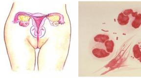
Different heart pathology is diagnosed and monitored using e-cardiography. Infarcations, heart thresholds, thromboembolism and much more is very noticeably changing the work of the heart and vessels, which is clearly visible on the cardiogram.
One way to find out the presence of certain heart disease is to spend an ECG with a load. Many medical centers Provide such an opportunity.
Essence of ECG with load
Electrocardiography has been applied for about a hundred years, proven by the introduction of medicine in the sphere, and so far there were no similar diagnostic methods. Many heart disease is fixed precisely thanks to her.
But unfortunately, there are some pathologies that are more difficult to find out even with ECG. Patients are treated at the moment of remission of the manifestation of pathology, and symptoms gives only a small hint of a likely diagnosis. Therefore, it was invented to carry out electrocardiography during the physical activity of a person.
What is the advantage of the ECG under load? Diagnostics ECG It is carried out at the moment of high physical exertion on the patient. For this, there are both several options for old and proven ways to simulate everyday load and newer medical developments.
ECG with load can be carried out in the following ways:
- functional samples;
- diagnostics on the cyergometer;
- the method of American scientists - Tredamil;
- with the help of Halter monitoring.
The above-mentioned exercises during the removal of ECG data have several subspecies. Consider them in more detail.
Functional tests
This method is used to evaluate the work of the heart during the passage of professionality of some professions - athletes, pilots, military. With the help of functional samples, you can define hidden pathologies, heart endurance to some loads.
It is also permissible to apply the methodology for assessing the health of children before entering the sports section.
Sample technique has several options:
- metartine method is 20 squats performed within 30 seconds. Readings are removed before physical activity, immediately after it and three minutes later;
- running samples - similar to the first sample, only with running instead of squats;
- step test - has more than 20 types of tests, each of them has its own rules of indicators;
- wedigoTostatic - procedure for children. The child is fixed by the necessary equipment, after which they remove the testimony in the lying and standing position. Normal indicators - an increase in heart abbreviations in the range of 20-40%.
BeltEergometer
In the role of loading, a vehicle similar to the exercise bike - a bicycle game - it is offered by many private medical centers. During the occupation, ledge is well fixed on the cardiogram change in the heart of the heart, which can be invisible without load. As a rule, this method is effectively determined by ischemic disease, a variety of disorders in the normal rhythm of the heart, other disorders, and simply diagnosed the body's reaction rate on physical activity.
One of effective methods Diagnostics with load - cyergometry
Before cycle ergometry, you need to prepare:
- for the appointment of a doctor for a few days before the start of the diagnosis, drugs (beta-blockers, nitrates) are canceled;
- it is impossible to diagnose after stress or exercise, the patient must be calm both physically and emotionally;
- before the study, you need to change clothes into light comfortable clothes;
- during diagnostics, electrodes are attached to the patient's body, so men having a chest hair Pokrov, it is necessary to get rid of it;
- for three hours, it is impossible to eat and drink any liquid before conducting the procedure.
The test belt is worn on the chest, a special belt or a multiple electrodes. The first data in the patient is removed alone, then a diagnostic diagnostic step occurs, which is forcibly increased after certain periods. The increase is stopped at the moment when the patient begins to complain about unpleasant symptoms - pain, fatigue, dizziness, other signs. After this, the testimony of physical changes is removed for another ten minutes, until the body returns the normal state of rest.
Note!The subject may require the termination of the procedure at any time.
It is recommended to conduct an inspection in the morning, after 2-3 hours after breakfast. Such a survey is used mainly for adults, but sometimes acceptable in pediatrics to assess the work of the heart in adolescents, the weight of which exceeds 40 kg.
For research there are some contraindications:
- period of aggravation of heart attack;
- heart disease infectious nature;
- aggravation of the disease in the blood circulation of the brain;
- complex heart pathology;
- heart failure at 2-4 stages;
- arrhythmia, conduction blockade;
- arteries 3 level hypertension;
- different kinds thrombosis;
- mental diseases;
- other reasons.
You can only pass the survey after discussion with the doctor of all sorts of contraindications.
Simulated method of cyergometry, but a treadmill with a changing angle of inclination of the plane is used as a simulator - imitating running hill. The diagnostic method is acceptable for children - the simulator does not have restrictions on the growth and weight of the subject, unlike the exercise bike.
 Tredamil - a similar method of diagnosing a bicycle game, invented by American scientists
The changes in the changes are removed during the class and after the load, after the analysis of the norms is carried out.
Halter monitoring
The method is considered to be conditionally load: the patient carries the electrodes during the day, and the special device Halter displays the indicators. A person lives in the usual rhythm, but every physical activity, emotional stress he lies into the diary.
During such a study, one of the above samples is allowed.
At the end of the study, a cardiologist analyzes changes in the body in various conditions, makes deciphering the cardiogram, and at this stage the informativeness of the diary plays an important role.
The above-described method of diagnosis has no contraindications, since targeted loads on the patient's body does not turn out. Exception can only be mental disorder.
Indications for ECG with Forced Load
Indications for the use of ECG with load perform the following signs:
- painful syndromes in the field of hearts that are not diagnosed under the usual examination;
- small changes in the results on the electrocardiogram, which are not accompanied by signs of angina;
- changes in the lipid balance without signs of IBS;
- people with an increased risk of EBS appearance;
- the probability of habering myocardial ischemia.
 ECG with forced load is prescribed only by a cardiologist
After the examination, the patient receives a full result of the body's reaction on physical activity, changes in blood pressure, indications restorative process After the end of physical exertion, and, of course, the recommended permissible level of load.
The ECG Examination Method under load is a safe method of medical research. But, given the load on the heart during testing, risk side Effects, and in very rare cases - complications that can lead to a heart attack. Therefore, the survey should be carried out by qualified specialists in order to provide medical care if necessary.
The study of changes in the electrical activity of the heart under the influence of muscle work allows you to identify the nature of the dependence of heart rate from the load capacity, establish disorders heart Rhythm, conductivity, excitability, functional state of myocardium. A high diagnostic value of such studies in identifying ischemic heart disease (IBS) is established, clarifying the degree of severity of coronary insufficiency.
The sample is based on the following common patterns. With exercise, the blood supply to the working organs and tissues increases sharply by incorporating a number of compensatory mechanisms and, in particular, a significant increase in the work of the heart. Under these conditions, the discrepancy between the actual blood supply to myocardium and the needs in it can cause the ECG ischemic changes in myocardial, which will allow to judge the state of coronary blood supply. The value of the sample is underlined by a certain parallelism of its results with a degree and number of affected coronary arteries according to coronaryogiography, the forecast of IBS.
When studying the electrical activity of the heart under muscle conditions, the following types of loads can be used:
- rise and descent on special steps (step test);
- pedaling on the cyergometer;
- Running and walking on Tredban;
- Work on a manual ergometer.
Indications for conducting a test with exercise:
1. For diagnostic purposes: a) identification of ECG signs of myocardial ischemia associated with coronary failure; b) identifying hidden heart rate and conductivity disorders.
2. Differential diagnosis of ECG changes due to disruption of coronary blood circulation, from changes related to diseases of non-coronaic nature.
3. Definition of the load tolerance, i.e., the level of physical activity (and the values \u200b\u200bof physiological indicators with this load - the frequencies of heart abbreviations, blood pressure, etc.), in which subjective or objective signs of coronary failure appear in patients with IHD.
4. Conducting control over the effectiveness of rehabilitation activities (drug, physiotherapeutic, surgical treatment, physical training, etc.).
5. Evaluation functional state Hearts, the nature of his adaptation to physical exertion from beginners to engage in healthy physical education, in persons, whose professional activity is related to the performance of severe physical exertion and so on.
Contraindications for testing with exercise:
Absolute:
Pronounced blood circulation deficiency (heavier than IIA).
Acute period of myocardial infarction.
Quickly progressive or unstable angina.
Hypertension II - III stage.
Aneurysm of vessels.
Pronounced aortic stenosis.
Pronounced heart rate disorders (tachycardia over 100 -110 in 1 min, political extrasystoles).
Acute thrombophlebitis.
Pronounced respiratory failure.
Acute infectious diseases.
Relative:
Frequent suctivativericular and ventricular extrasystoles, flickering arrhythmia.
Indications as a history of serious violations of the heart rhythm, a tendency to sudden loss of consciousness.
Aneurysm heart.
Moderate aortic stenosis
Endocrine diseases ( diabetes, goiter endemic and diffuse toxic, mixedma).
Significant increase in the heart.
States requiring special attention and (or) precautions:
Conductivity disorder (full, atrioventricular blockade, blockade of the left feet of a beam of Gis, WPW syndrome).
The presence of an implanted driver of the heart rhythm with a fixed frequency.
Violation of cardiac rhythm.
Violation of the electrolyte balance.
The use of certain drugs (drugs of instretzing, etc.).
Pronounced hypertension (diastolic pressure of over 120 mm Hg. Art.).
Stenzardia and other manifestations of coronary failure.
Pronounced anemia.
Pronounced obesity.
Renal, hepatic and other types of metabolic failure.
Explicit psychoneurotic disorders.
Diseases of the joints, nervous and neuromuscular systems that interfering with samples.
Research procedure. The sample with physical exertion should be carried out by an experimental physician, familiar with electrocardiography and with load tests that own resuscitation methods. In the room where the sample is carried out is, it is necessary to have a set of medicines and technical means to provide medical care (defibrillator, an apparatus for artificial ventilation, syringes, adrenaline, nitroglycerin, ammonia alcohol, Promedol, etc.).
For continuous monitoring of changes in ECG during sample, you must have a computer. To control the tolerability of physical exertion, the heart rate is regularly determined regularly, the size of the blood pressure is monitored, followed by the appearance and well-being of the patient.
On the day of research, physical activity should be reduced. Beginning the procedure follows no earlier than 1.5 - 2 hours after meals and preliminary rest duration of at least 30 - 60 min.
The trial with cyergometric loads, as a rule, is carried out in the patient's position sitting with a frequency of pedaling 40 - 80 revolutions per minute. Before starting the study, it is necessary to adjust the height of the steering wheel and the seat of the cyergometer, depending on the growth of the surveyed. The saddle is set in such a way that the sitting on it could have almost completely straightened leg to get to the bottom pedal.
The type of load and the nature of its execution is determined by the doctor before the procedure starts. At the same time, the gender, age, physical development, training and health status of the surveyed are taken into account. To eliminate possible psycho-emotional reactions, the patient must be adapted to the conditions of pedaling on the cyergometer (or walking along the steps), for which it is previously not less than the I-2 h to the main study, it is necessary to familiarize it with the upcoming work directly on the device at low load power.
The recording of the original ECG is carried out immediately before the start of muscle work in the conditions of rest in the horizontal position of the patient, and then necessarily in the position in which physical exertion will be performed. This makes it possible to eliminate ECG changes associated with orthostatic effects.
In the process of muscular work, the ECG is registered at the end of each minute of the sample, immediately after its end, as well as in the recovery period on the 2nd, 3rd, 5th, 10th minute of rest, and if necessary - more often (every minute) And in the later periods of restitution. Every minute of load and in the restorative period measure arterial pressure.
In some cases due to movements chest and arising from this displacements of the electrodes, which ultimately reflected on the quality of the ECG, it is difficult to establish the exact position of the isoelectric line and in connection with this measure the amplitude of the teeth, the degree of displacement of the ST segment. With a "floating" record, the recording should be made on a long tape, in some cases - with a short respiratory delay.
To prevent development trimming state (As a result of a sharp decrease in the venous return of blood to the heart, due to the termination of the work of the "muscle pump") after the end of the cycle ergometric loads (especially relatively large in power), pedaling should be continued, but already at the lowest load capacity for about 1 min.
Considering the daily fluctuations of various cardiac indicators and, in particular, the possible dependence of the degree of shift of the ST segment under the influence of muscle work on the time of day (the degree of shift of the ST segment during exercise, as a rule, the smallest in the first half of the day, and the maximum - between 20.00 and 23.00), it is desirable, especially with dynamic observations, conduct examinations with exercise at the same time.
Hyperveptulation can cause changes in the electrocardiogram, in many respects similar to those observed with muscle work in patients with coronary heart disease. At the same time, the physical activity itself leads to a significant increase in pulmonary ventilation. Therefore, to eliminate the "false-positive" data during cyergometric loads, it is recommended preliminary (better a day before the survey) to test hyperventilation.
Criteria for termination of the sample with physical activity
Pulse reaction. One of the main criteria for stopping the test with the physical activity on the WHO recommendation is to increase the pulse to the submaximal value (Table 2), which is approximately 75% of the maximum possible for the given person (the maximum pulse value is determined by the formula: 220 minus age in years). The informativeness of the sample increases with a further increase in load power. However, the risk of acute pathological conditions limits the widespread use of the limit or relatives to them.
Table 2.
The values \u200b\u200bof the maximum and submaximal pulse in persons of different ages
Electrocardiographic changes.
1. Horizontal or arcuate (sickle, habitual) shift of the ST segment down relative to the isoelectric line by 0.2 mV and more.
2. The shift of the ST segment up to 0.2 mV and more, accompanied by the displacement of the ST segment in opposite leads.
3. Significant heart rate disorders - registration of frequent (4:40) extrasystole, group, political or early extrasystoles, paroxysmal tachycardia, fluttering or atrial fibrillation.
4. pronounced disorders of atrioventricular or intraventricular conductivity.
When examining patients with coronary insufficiency, more stringent criteria for the cessation of physical exertion are adhere to. For example, when determining the load tolerance, the sample should be completed already when the ST segment is shifted according to ischemic type by 0.1 mV (as well as with increasing this segment to the same value), depression by type IS - T, exceeding 0.2 MV (with ratio Q - X / Q - T is greater than 50%), inversion or reversion of T. T.
Changes in blood pressure (AD).
1. Increased blood pressure up to 220/120 mm RT. Art.
2. Lack of increment or drop of blood pressure while increasing the load capacity.
Other signs.
1 use of the attack of angina.
2. Excessive shortness of breath or suffocation
3. A sharply changed color of the face.
4. Dizziness or condition close to a fainted.
5. Total pronounced fatigue, weakness.
6. Feeling pain or sense of fatigue in the muscles of the legs.
7. Patient refusal to continue the study.
Sample believes positive When the following two signs appear together either each of them separately: the attack of angina; Electrocardiographic signs of myocardial ischemia.
Negative The sample is considered in the event that there are no pathological changes on the ECG with loads that cause an increase in the pulse at least 75% of the maximum possible for a particular person.
The negative results of the sample (especially with a certain clinical symptoms) do not exclude completely ischemic heart disease, but indicate only the absence of sharply pronounced coronary failure.
2.3.11. Double (Holter) ECG monitoring
Holter monitoring is continuous registration of ECG over the day. The study is performed both in hospital and outpatient. Since arrhythmia in many patients occurs periodically, make an ECG and burn the work of the heart during the attack of arrhythmia at the doctor's reception can be quite difficult. Daily monitoring ECG just allows you to do this. It is shown to patients with heart rate complaints and interruptions in the work of the heart - to identify violations of the rhythm and conductivity of the heart, with obscure fainting, and also partially for registration of "mute" (non-combing) myocardial ischemia, to assess some parameters of the electrocardiotimulator. ECG record is carried out using a small portable device that the patient carries on the belt under clothing. During research, the patient leads a normal lifestyle, noting the time and circumstances of the occurrence of unpleasant sensations in the field of heart, as well as the time of loads in a special diary, as well as load time (walking, climbing the stairs), meals, sleep. When evaluating the ECG record during the day, you can get the following information:
information on rhythm violations: extrasystoles of supervaluation and ventricular (indicating quantity, morphology and other features), paroxysms of arrhythmias;
information about rhythm pauses;
information about changes in the PQ and Qt intervals if these changes occurred, information about the changes in the morphology of the QRS complex due to disorders of intraventricular conductivity;
information about changes in the final part of the ventricular complex (ST) and the links of these changes with the physical activity of the patient and its sensations in the diary;
information about the work of an artificial rhythm driver - if it is.
The identified features or pathology should be illustrated by ECG printouts for the corresponding monitoring period.
this is the removal ECG heart For a long time, and is intended to evaluate the activities of the heart in the process of ordinary daily activities. There are several types of Holter monitoring ECG.
Continuous daily monitoring of ECG. The most common type is the continuous recording of the heart of the heart for 24-72 hours. Unlike the usual ECG procedure, which fixes about 40-50 heart blows for a short period, such an ECG Halter can record about 100,000 heart blows in 24 hours,
Periodic monitoring ECG.With periodic daily monitoring ECG record is not constantly, but with interruptions. This is convenient if the symptoms of heart rate disorders occur very often. The ECG Halter Monitoring Device will record only when the patient presses a special button.
Electrocardiography (ECG) with exercise - a diagnostic procedure showing how the heart of your child works well during activity and how it adapts to different levels of physical exertion. If an exercise bike is used as a load, then electrocardiography with a load is often called cyergometry. Also, a treadmill can be used to create physical exertion on the child's body. This diagnostic procedure is carried out for children who can walk and run along the treadmill, or rotate the bike pedals, and quite mature to understand what they are asked about. Usually an ECG with a load is performed for children aged 5 years and older.
Physical bases of electrocardiographic study with a load.
In the process of operation, the electrical activity of the heart is constantly and rhythmically varies in accordance with the stages of compression or relaxation (systole and diastole). The electrocardiograph registers the difference between the electrical potentials between the two parts of the heart projection on the surface of the child's body and gives the results obtained in the form of a characteristic schedule - cardiograms on a paper strip or computer screen. The number of waves per minute on the chart shows the heart rate, the distance between the same cardiogram elements is the heart rate. Wave forms show how electrical heart pulses are formed and how well separate parts of the heart work together. In the form and size of the cardiogram elements, the doctor can judge the quality of the child's heart and identify the signs of possible diseases.
During exercise, the body needs additional oxygen. The heart and lungs should work more than usual to ensure the supply of the body with an additional amount of oxygen. When performing physical exercises, the heart begins to work with a larger load: it is reduced more often and stronger to increase the release of blood pumped blood and maintain the blood supply to the muscles and other devices involved on adequate load level. At the same time, the heart muscle itself begins to consume more oxygen supplied with blood on heart blood vessels.
The record of electrical activity from the body surface is made through small metal plates (electrodes), which are placed in certain places on the chest, hands and feet of the child. Electrodes are connected to the cardiograph with wires through which information is recorded. During the exercise of the physical activity, the electrocardiograph will register changes in the heart rate, heart rate, blood pressure and other symptoms while your child exercises - first slow paced, and then faster and faster until it reaches the highest level. All the obtained data of the electrocardiogram will be measured, analyzed by a computer and a doctor who will form a conclusion according to the results of the study.

Preparation for research
ECG with load is usually held in the morning when your child rested well and full of strength. Your child should have a light breakfast, and drink clean water at least 2 hours before joining the study. 12 hours before the study is forbidden to drink coffee, tea and energy drinks. It is necessary to take with you sportswear for the season and sneakers, towel and replacement clothes for dressing up dry after the study. If the child uses breathing inhaler - you need to take it with you. If you control the blood glucose level in the child's blood - bring with you a glucometer to control before and after the study. If the level of glucose before the study is low - immediately inform the doctor. Medications for controlling the level of glucose ("sugar") in the blood can only be taken after the completion of the procedure.
If the child takes "hearty" medicines - check in advance from your attending physician and a doctor who will conduct an ECG with a load, which of them and how can be taken on the study day. Perhaps the doctor will recommend in advance to stop the reception of such drugs such as cardiac glycosoids, diuretics, calcium antagonists, prolonged nitrates.
Children are usually less afraid of and feel more confident during research when they know what to expect. Tell your child in advance how the study passes, focusing on the fact that the child will see, hear and will feel. Tell your child that the study does not hurt his health, and answer in detail all his questions.
What is the ECG with the load?
- screening (prophylactic) research
- evaluation of the functional state of the cardiovascular system
- identification of diseases of coronary arteries - blood vessels ( ischemic disease hearts)
- identification of heart rhythm disorders
- assessment of the results of the treatment
- risk assessment in front of surgical intervention
- definition of disability
How is the study?
Electrocardiography with load is performed in a separate room, where the electrocardiograph and the simulator are installed. Electrodes (small plastic plasters with metal plates) will be attached to your child's chest, and one electrode will be attached to each hand and leg. Electrodes will be connected to the electrocardiograph with wires. The cuff apparatus for measuring blood pressure will be imposed on your child's hand. When the blood pressure is measured during the study, the cuff will inflate the air tightly to crimp the child's hand. During ECG recording, the child will not feel anything unpleasant. No current will be skipped through the electrodes - it's just sensors for information.
Initially, a cardiogram is recorded in a state of rest (without load), and then the child is asked to start performing a physical exercise in a certain rhythm, during which the ECG is also registered. At the end of your child will be asked to perform an exercise at the maximum effort. When your child is tired or an ECG will be marked by abnormal changes, or the level of blood pressure will increase significantly, the study will be discontinued.

Attention! The child should know that immediately should be informed about the doctor about the appearance during the study of pain or discomfort in the chest, in the heart, in hand, in jaw, dizziness and other unusual sensations.
After performing exercises with the load, the ECG will again be registered in the state of rest to see how quickly the indicators of the child's cardiovascular system to normal levels are returned.
Since the child will have to do an exercise on high level physical activity, one should expect some shortness of breath and muscle fatigue, which is a normal physiological response. The doctor will closely monitor the condition of your child throughout the procedure to make sure that the child and his heart should normally cope with the increasing load. The most important thing is that parents can make is to encourage their child to perform exercises at the maximum level of opportunity, explaining the child that exercises are not harmful or painful.
The execution time of physical activity usually does not exceed 15 minutes, and all the research takes about 45 minutes.
Contraindications for conducting an ECG with a load
Since during the study, the load on the heart of the child increases, there are a number of absolute contraindications for the procedure. In children, they are rare, but, nevertheless, you need to know about them:
- acute myocardial infarction (within 48 hours)
- unstable shape of angina, uncontrolled heart rate disorders,
- acute endocarditis
- myocarditis or pericarditis
- aortic stenosis of severe
- decompensated heart failure
- acute pulmonary embolism or lung infarction
- thrombosis of deep veins
Also conducting research is contraindicated with infectious diseases, renal failure, thyrotoxicosis. Caution should be approached with an ECG with a load with hypertrophic cardiomyopathy, stenosis of coronary arteries, tahiartimia or bradyarithmia, hypertensive disease, hyperthodosis, stroke (less than 1 month), violation of the conductive heart system.

Decoding results
RESULTS RESEARCH The results contain a conclusion about the presence of either the absence of signs of violation of the blood supply to the heart muscle during exercise, on the reaction to the load level of blood pressure and the rate of recovery of the initial indicators after the load. On the basis of the study, recommendations may be given permissible level physical exertion for your child. In some cases, a re-electrocardiographic study may be required or a special type of ECG, accompanied by other types of research. Ultrasound of the heart, doppler Sushes).
Miklukho-Maclay Russia, Moscow +7 495 735 88 99 +7 495 134 25 26
Leninsky Prospect Russia, Moscow +7 495 735 88 77 +7 495 134 25 26
2017-03-09
Electrocardiography - a method that allows you to estimate the state of the cardiovascular system. Such a study is included in the mandatory dispensarization program. For the first time it is carried out by the kids back in the hospital. Thereafter healthy child You must attend cardiologist once a year.
ECG with load - test with functional breakdown. With it, the work of the heart muscle after exercise is checked. The child's procedure is recommended to pass before entering the sports section. This will allow you to evaluate the reaction of the cardiovascular system in sports.
Note: this procedure It is forbidden to children with heart or vessel pathology, as well as increased blood pressure. The functional test is also not recommended if there are complaints of chest pain, dizziness or fainting.
Preparation and holding
The study includes several stages. The first is the reception of a cardiologist and therapist. Doctors will determine the admissibility of the appointment of the ECG with the load, and will also select physical exercises.
Anxiety and excitement kid can distort the test results. Before visiting the clinic, discuss with him the procedure, tell us that it is absolutely not hurt. On the ECG Day, try to limit physical activity and mobile games. It is advisable to come to the examination in advance so that the little patient can sit a little and calm down.
There are various techniques electrocardiography with load sample, including new ones. All of them have their advantages and disadvantages. In Germany, a bicycle ergometry is used, performed in the patient's position sitting. And in the United States, for example, a running track (Tredamil) is widely used to perform a test with exercise.
In practice, when performing a load sample is often used the following ECG registration programs:
Leads I, II, III, V2 (or V1), as well as V4 and V6
Leads I, II, III, V4, V5 and V6
ECG registration In addition, V1 is preferable compared with the V2 assignment, as in this lead, it is more convenient to judge the teeth of the rhythm and the overload of the LP (deep and broad negative part of the left-subnedental teeth of P).
Before doing load test You should register at rest to know the source data.
If using load It is necessary to identify coronary failure, then it is necessary to try to set a short and rapidly increasing load so that in a small patient to achieve submaximal heart rate. A patient subjected to cyergometry is needed a load equal to 80% of the maximum load for this age group. The implementation of the submaximal load is judged by the achievement of the CSS, the values \u200b\u200bdetermined by the formula below:
Chersubmas \u003d 200 - age.
For maximum load definitions Use the following formula:
CHSMAKS \u003d 220 - Age.
With help bicycle Roomergomera You can accurately dispense physical exertion; It is desirable that it makes up approximately 150-200 W for men. For men, the sample begins with a load of 50 W, and for women and the elderly - from 25 watts. The load every 2 minutes is raised by 25 W. Persons of young age reaches a submaximal frequency with a load, approximately equal to 150 W, the elderly - already at 100 W.
The most important criteria for termination of the ECG sample with exercise are the oversized pain and severe ventricular rhythm disorders, as well as a significant increase in blood pressure.
For performing physical exertion Exercise ECG monitoring, every minute recording short fragments and measuring every 2 minutes of hell. The feed rate in the electrocardiograph is 50 mm / s. In the restorative period (which lasts about 3-6 minutes) the ECG is also recorded after short time intervals.