Batalov duct in newborns is. Symptoms and methods of treating open arterial duct for what features can be suspected that the duct remained open
Botals duct was first described in 1564. With intrauterine blood circulation, it plays a big role, since it takes most of the blood from the pulmonary artery directly in the aorta. It departs from the place of lung artery division into 2 branches, sometimes from her left branch. The duct flows into the aorte below the so-called isge into its downward part 2-3 mm below and opposite the mouth of the left connector artery. Duch length, according to Kushev, in newborns and infants 6.9-6.2 mm, diameter is 4.3-3 mm. The duct differs from large vessels with a predominance of muscle elements with a weak development of elastic tissue.
After birth, the closure first occurs, and later the infection of Botall duct. In this case, the increase in blood pressure in the aorta, as well as the movement of the breasts. Following the physiological closure, the anatomical tightness of the duct begins, which ends during the first 6 weeks, but sometimes tightened to 3-4 months. By the end time of the obliteration process, the duct turns into Lig. ARTERIOSUM MAGNUM. If the injecting duct turns out to be incomplete or not at all, there is a malformation. The absence of a Botallian duct may be the only heart defect, sometimes it is combined with other defects, for example by stenosis and atresia of pulmonary arteries, stenosis of the mouth of the aorta, its isthmus, narrowing of the left venous atrioventricular mouth, etc. In cases of combinations with other blobes of Botalan, the duct makes a compensatory role. Of the 1000 patients with early congenital heart defects, the open Botallov duct was found in 242. The width of its lumen is different - from 4 to 12 mm, an average of 7 mm, and it can further expand depending on blood pressure. Through it can be thrown into the pulmonary arteries a large amount of blood coming into the aorta. Diagnostics of open Botallian duct in many cases is simple, affordable and based on well-studied clinical signs. But it should be remembered that the occasionally observed the incoming cases of the Botallian duct, which are not manifested during life and only by chance-opening people who died from other diseases. Indimacy clinical picture Not always depends on the width of the lumen.
In a small part of cases, there is a light cyanotic staining of the skin or transient cyanosis in early ageassociated with physical stress. In most cases, cyanoticism is absent and the skin seems normally painted or even unnecessary pale.
In this regard, children with an open Botallah dummy never have fingers in the form of drum sticks, nails in the form of watchcases. In many cases, there is an easy-to-appear shortness of breath and fast fatigue. Often there is a tendency to disease respiratory tract. Quite often there is a lag in physical development. But many children adapt to the environment and lives, visit the normal school.
When examining the patient, there is sometimes clear ripple in the stubborn yam. When palpation of the heart area, it is sometimes possible to note the presence of systolic jitter in the second intercontalone left. The heart boundaries at percussion are often slightly expanded both to the left and right. Part of the children (in 20%) it is possible to determine the lintel-like dullness of the percussion sound to the left of the sternum in the first, second and third intercostal, observed mainly in older children. This dullness, first noted by Hergardt, partially corresponds to the expanded Botallah of the protuch, partially expanded pulmonary artery.
The most characteristic are auscultative data. On the basis of the heart, a distinct loud rough noise is heard in the second interval. The noise is long, continuous, resembling the operation of the machine or the noise of the mill wheel. This noise is well carried out throughout the heart of the heart, it is heard in the plug-in area and in the left half of the chest. In the neck vessels, it is usually not held, but sometimes heard. On the back, the noise in the inter-opumen space is well listened. It fills most of the systole and diastole and disappears only at the end of the diastole. In a lying position, it is pronounced stronger. The noise is perceived as systolic-diastolic, is vortex. Sometimes up to 3 years can only be heard over the coarse systolic noise, which is sometimes enhanced during the inhalation and decreases when exhaling. Sometimes the maximum noise is listened to the right from the sternum or on the back. Listened rough noise to the right of the sternum can sometimes be a manifestation of relative aorta stenosis or subaartal stenosis.
Along with the noise, there is a significant increase in the second tone on the pulmonary artery, but this is not always observed.
As a result of increased blood flow, the amount of blood flowing through the pulmonary veins in the left atrium is increasing, and in the future and the left ventricle. But on the other hand, it is easy to imagine that at the same time due to the influx of blood from the aorta in powerful artery Obstacles are created for emptying and right ventricle.
According to the clinic, the maximum arterial pressure With an open Botallah, it turns out to be normal, minimal - reduced, and with a wide protocol can reach zero. By virtue of this, the amplitude of the pulse pressure increases, i.e. the difference between the maximum and minimum pressure.
In the study of hemodynamics with an open Botallah duct played great importance Heart probing. The more the difference between the pressure in the aorta and the pulmonary artery, the more blood will be held through the dashing from the aorta in the lungs and the more clearly there will be noise. With the same diastolic pressure in both vessels, only the blood flow from the aorta into the pulmonary artery during systole can occur. With an open Botallah duct, both the oxygen container and the content of O2 and CO2 in arterial and venous blood are almost no differ from the norm and blood saturation reaches 95-96%.
Sometimes they managed to notice the presence of a significant increase in pressure in a small circle circle. At the same time, patients find some features of the clinical picture. They usually do not have a diastolic component of noise, they poorly carry their vice, in the sample with the load, it is noticeable. arterial blood Oxygen, reducing the coefficient of use of oxygen, they are more easily occurring the phenomena of cyanosis.
On the basis of the above, an increase in pressure in the pulmonary artery and a higher content of oxygen in it is characteristic of the open Botallov and a higher oxygen in it than in the right ventricle, due to the impurity of arterial blood from the aorta.
From other, less characteristic and less permanent symptoms, with an open Botallah duct, it is possible to indicate the notifier of the pulse on the hands, tacking the stronger pulse on the right. Occasionally, the pulse takes a paradoxical character, the disappearance of pulse oscillations can be observed with deep breath. In isolated cases, it is possible to observe the phenomena of Afony due to the compression of the left return nerve. Systolic pressure with an open Botallah duct is normal, diastolic is reduced, and, by virtue of this, the amplitude of the pulse pressure increases (above 40-50 mm Hg. Art.). Accordingly, it is often observed by Pulsus Celer Et Altus as in the lack of aortic valves.
The electrocardiogram with an open Botallah protocol does not have natural and characteristic changes. It is often noted the right type, at an older age, the leftogram. More often deviation of the axis to the vertical, violation of vascularity, the elongation of p-q and q-t.
X-ray study usually confirms the presence of a heart expansion to the left, less often to the right. The increase in the messenger artery cone is striking, which gives the left heart contour a typical form. It is characterized by the amplification of the vascular pattern and the strong systolic pulsation of the pulmonary artery arc, the Hilus and the aortic arcs. Translucent is better to produce in the anterorable and left oblique position. On the X-ray-kimogram of the tea, the presence of an intermediate diastolic teeth of the pulmonary artery arc.
With the help of the probing method, it is sometimes possible to establish the presence of a passable Botallian duct. From the upper floor of the vein, the probe can be held in the right atria, the right ventricle and the pulmonary artery, and through the preserved duct - and in the aorta, from where the turn from the outside is sent inside vertically down in the abdominal aorta. But this method is very difficult, you need to have great patience to bring the probe to the right place, and often it is not possible at all. Therefore, the diagnosis is most often confirmed on the basis of the study of blood saturation with oxygen in the cavities of the heart. Increase oxygen content in a. Pulmonalis, compared to venous blood In the right ventricle, it says about the presence of communication between the aorta and the pulmonary artery, that is, on the existence of the Botallian duct.
Valuable data also gives an angiocardiographic research method. The contrast agent is introduced through the elbow vein and the top hollow vein in the right atrium. Tracking the contrast moves on seconds, it is possible to install features characteristic for open Botallov. First of all, the expansion of the pulmonary artery and especially its left branch. After filling the left sections of the heart on the angiocardiogram, there is long contrasting of the vessels of the lungs, left atrium, left ventricle and aortic.
Gotz offered a new diagnostic sign. When the contrast passes through the pulmonary artery, you can note the defect on the lung artery arc circuit. This defect is created due to the dilution of the contrast of a mass of blood coming from the aorta through Botallov duct into the pulmonary artery.
Sometimes to solve the issue you have to use the aortography, with which you can see the flow of contrast from the aorta to the pulmonary artery.
The described picture is peculiar to clean forms of open Botallian duct. The picture changes in the case of a combination of this vice with another, for example, with the stenosis of the pulmonary artery, stenosis of aortic and other defects. It is always necessary to differentiate this vice from the narrowing of the mouth of the pulmonary artery, since the systolic noise in the second intercostal left is also listened to the last interchange. Therefore, it is necessary to remember that with the narrowing of the mouth of the pulmonary artery II, the tone of the pulmonary artery is usually weakened, and sometimes not even listened.
The ability of the Botallian duct in general is a non-heavy defect and gives a relatively favorable forecast. Children can lead a normal lifestyle, attend school. But it should be remembered that at the same time there is a predisposition to stagnation In the lungs, and this in turn leads to a more frequent development of pneumonia. In the 2/3 of our patients in history there were repeated pneumonia. Of any kind infectious diseases Such children are worse. You can always be afraid of the development of endocarditis, the attachment of rheumatic infection and, which is particularly important, the sclerosis of the vessels of the lungs with subsequent hypertension in the pulmonary artery system. According to Shapiro and Case, 40% of patients die from subacute endocarditis, some from the gap of the duct or pulmonary artery.
Treatment of Botalls of duct is possible only by the operational way and consists in ligation of the duct or its intersection. Children transfer the operation relatively easily, after surgery they disappear auction phenomena, noise ceases to listen or is being done weaker. The performance of patients increases sharply.
Risk from operation less than the risk from possible complications In later life. In case of suspicion of complication, endocarditis must be pre-conducted by antibiotic treatment. According to domestic scientists, mortality in operations over the Botallian duct is 0.5-2%. In children, surgical intervention is rational even in the absence of any symptoms.
Open arterial duct (OAP) - a disease due to violation of the normal development of the heart and trunk vessels in the intrauterine and postnatal period. Congenital usually formed in the first months of fetal development as a result of an atypical formation of intracardiac formations. Resistant pathological changes in the structure of the heart lead to its dysfunction and development.
Arterial (Botallov) Dol - Structural formation of the heart of the fetus, through which the blood emitted by the left ventricle in the aorta moves into the pulmonary barrel and returns again to the left ventricle. Normally, arterial duct is subjected to obliteration immediately after birth and turns into a connecting tapery. Filling the lungs with oxygen leads to the closure of the duct of the injected intimate and the change in the direction of blood flow.

In children with defects, the duct is not closed on time, but continues to function. In this case, the pulmonary blood circulation and the normal work of the heart is disturbed. The OAP is usually diagnosed in newborns and infants, some less often at schoolchildren, and sometimes even in adults. The pathology is found in the diluted children living in the areas of highlands.
Etiology

The etiology of the OAP is currently not fully studied. Experts allocate several risk factors for this disease:
- Premature childbirth
- Low weight newborn
- Avitaminosis,
- Hereditary predisposition
- Marriages between relatives,
- Mother's age older than 35 years old
- Genomic pathology - Down syndromes, Martha, Edwards,
- Infectious pathology in 1 pregnancy trimester, congenital rubella syndrome,
- Eating alcohol and drugs pregnant, smoking,
- Radiation of X-ray and gamma rays,
- Reception of medicines during pregnancy,
- Impact of chemicals on the body of pregnant
- System and metabolic diseases of pregnant
- Intrauterine endocarditis of rheumatic origin,
- Endocrinopathy Mother - diabetes, hypothyroidism and others.
The reasons for the OAP are made to unite in 2 large groups - internal and external. Internal reasons associated with hereditary predisposition and hormonal changes. TO external reasons Below: bad ecology, production harm, illness and fellow mother habits, toxic effect on the fruit of various substances - drugs, chemicals, alcohol, tobacco.
The OAP is most often detected by premature children. Moreover, the less weight of the newborn, the higher the likelihood of the development of this pathology. The heart rate is usually combined with the anomalies for the development of organs of digestive, urinary and sexual systems. Direct reasons for the unguardment of Botall duke in this case are respiratory disorders, fetal asphyxia, long-term oxygen and parenteral liquid treatment.
Video: Medical Anatomy of Arterial Delta Anatomy
Symptomatics
The disease can proceed as asymptomatic and extremely difficult. With a small diameter of the duct, the hemodynamic disorders are not developed, and the pathology has not been diagnosed for a long time. If the duct diameter and the shunt volume are significant, the symptoms of pathology are pronounced bright and appear very early.
Clinical signs:

Children with the OAP often suffer from broncho-pulmonary pathology. Newborn with a wide arterial duct and a considerable amount of shunt is difficult to feed, they are badly added in weight and even lose weight.
If the pathology has not been discovered in the first year of life, then, as the child grows and the development of the child, the course of the disease deteriorates and manifests itself with more vivid clinical symptoms: the asthenization of the body, tachipne, cough, frequent inflammatory diseases of the bronchi and lungs.
Complications

Heavy complications I. dangerous consequences OAP:
- - infectious inflammation The inner shell of the heart leading to the dysfunction of the valve apparatus. In patients, the temperature rises, chills appears and sweating. Signs of intoxication are combined with headache and intensity. Hepatosplegegaly develops, hemorrhages appear on the eye day and small painful nodules on the palms. Antibacterial pathology treatment. Patients prescribe antibiotics from a group of cephalosporins, macrolides, fluoroquinolones, aminoglycosides.
- develops in the absence of timely cardiac surgical assistance and is insufficient blood supply to internal organs. The heart ceases to pour blood in full, which leads to chronic hypoxia and deterioration of the entire body. Patients have shortness of breath, tachycardia, edema lower extremities, Fast fatigue, sleep disorder, permanent dry cough. Treatment of pathology includes dietherapy, drug therapy, aimed at normalizing blood pressure, stabilization of heart work and improved blood supply.
- - acute diseasedue to the appearance in the heart muscle of foci of ischemic necrosis. The pathology is manifested by characteristic pain, which is not borne by the reception of nitrates, the excitation and concern of the patient, the pallor of the skin, sweating. Treatment is carried out in a hospital. Patients prescribe thrombolytics, narcotic analgesics, nitrates.
- Reverse blood flow Through a wide arterial duct can lead to and.
- Pulmonary edema It develops when moving fluid from pulmonary capillaries in an interstitial space.
Rare complications of the OAP include: aortic gap incompatible with life; and breaking blood flow; sclerotic nature; Heart stop in the absence of correctional therapy; Frequent ARS and ORVI.
Diagnostics

Doctors of various medical specialties are engaged in the diagnosis of the OAP:
- Gynecology obstetricists follow and develop of cardio-vascular system fetus
- Neonatologists inspect the newborn and listen,
- Pediatricians examine older children: carry out an auscultation of the heart and, when discovering pathological noise, send a child to a cardiologist,
- Cardiologists put the final diagnosis and prescribe treatment.
General diagnostic events include a visual inspection of the patient and percussion chest, auscultation, tool methods Research: electrocardiography, radiography, ultrasound of the heart and large vessels ,.
During the inspection, the deformation of the chest, the pulsation of the heart region, the displacement of the heart pushes to the left is revealed. Palparato detect systolic jitter, and percussionally - expansion of heart dulling boundaries. Auscultation is the most important method in the diagnosis of the OAP. Its classic feature is a coarse continuous "machine" noise due to unidirectional blood movement. Gradually, it disappears, and 2 tones accent appears above the pulmonary artery. In severe cases, multiple cliques and cutting noises arise.
Tool diagnostic methods:
- Electrocardiography Does not reveal pathological symptoms, but only signs.
- Radiographic signs Pathology are: mesh pattern of the lungs, the extension of the shadow of the heart, swelling of the segment of the pulmonary artery, flake infiltrate.
- Ultrasound of the heart Allows you to visually assess the operation of various parts of the heart and the valve apparatus, determine the thickness of the myocardium, the dimensions of the duct. Dopplerography allows you to mostly diagnose the OAP, determine its width and regurgitation of blood from the aorta to the pulmonary artery. Ultrasound examination of the heart allows you to detect the anatomical defects of the heart valves, determine the location of the main vessels, evaluate the contractility of myocardium.
- Phonocardiography - A simple method that allows diagnosing heart defects and defects between cavities by graphical registration of tones and sound noise. With the help of phonocardiography, you can objectively document the data obtained by listening to the patient, measure the duration of sounds and the intervals between them.
- AORTHRAPHY - Informative diagnostic method consisting in the supply of contrast fluid into the heart cavity and conducting a number of X-ray pictures. Simultaneous staining of the aorta and pulmonary artery indicates the incoming of the Botallian duct. The resulting images remain in the electronic memory of the computer, allowing you to work with them repeatedly.
- Catheterization and sound probing The OAP allows you to absolutely diagnose if the probe is fluent in the pulmonary artery through the duct into the downward aorta.
The probing of the cavities of the heart and angiocardiography is necessary for carrying out more accurate anatomical and hemodynamic diagnostics.
Treatment
The earlier the disease will be detected, the easier it is to get rid of it. When the first signs of pathology appear, you need to consult a doctor. Early diagnosis And timely therapy will enhance the chances of the patient for complete recovery.
If the child loses weight, refuses active games, shines when screaming, becomes drowned, experiencing shorts, cough and cyanosis, often exposed to ARVI and bronchitis, it is necessary to show it as soon as possible by a specialist.
Conservative treatment

Medicase therapy is shown in patients with low-rise clinical signs and lack of complications. Drug treatment The OAP is carried out premature and children up to a year. If after 3 courses of conservative therapy, the duct does not close, and the symptoms of heart failure increase, go to surgical intervention.
- The sick child is prescribed a special diet that restricting the use of fluid.
- Respiratory support is necessary for all premature children with the OAP.
- Patients are prescribed inhibitors of prostaglandins, which activate independent toughness of the duct. Usually use intravenous or enteral administration of "indomethacin" or "ibuprofen".
- Antibiotic therapy is carried out in order to prevent infectious complications - bacterial endocarditis and pneumonia.
- Diuretic drugs - "Veroshpiron", "Laziks", heart glycosides - "Stroofantin", "Corglikon", aPF inhibitors - "Enalapril", "Kaptopilla" prescribe to persons with heart failure clinics
Cathetterization of the heart

cathetterization of the heart
Heart catheterization is prescribed to children who conservative therapy did not give the expected result. Heart catheterization is a highly efficient method of treating the OPA with low risk of complications. The procedure is carried out specially trained children's cardiologists. Outside the catheterization of the child should not feed and drink. Immediately before conducting the procedure, it makes a cleansing enema and the injection of sedative. After the child relaxes and falls, start manipulation. The catheter is introduced into the heart chambers through one of the major blood vessels. There is no need to make cuts on the skin. The doctor monitors the promotion of the catheter, looking at the monitor screen of a special X-ray apparatus. With the help of testing blood samples and measuring blood pressure in the heart, it receives information about the vice. The more experienced and a qualified cardiologist, the more efficient and the successful catheterization of the heart will be more successful.
Heart catheterization and consumption of duct during thoracoscopy is an alternative to a surgical treatment of vice.
Operational treatment

Operational intervention allows you to completely eliminate the OAB, reduce the suffering of the patient, increase its physical exertion resistance and significantly extend life. Operational treatment is to carry out open and endovascular operations. The OAP is tied up with a double ligature, supervisory clips impose on it, crossed and shed.
Classic surgical intervention It is an open operation, which is to bandage the Botallian duct. The operation is carried out on the "dry" heart when connecting the patient to the IVL apparatus and under general anesthesia.

Endoscopic method Operational intervention is minimally invasive and small-acting. On the thigh make a small incision through which in pearful artery probe. Using it to the OAP, an occluder or a spiral is delivered to which the lumen is closed. The entire course of the operation is tracked by doctors on the monitor screen.
Video: Operation with oak, anatomy Botall duct
Prevention
Preventive measures are to exclude the main risk factors - stress, alcohol and medicinal preparations, Contacts with infectious patients.

After surgical correction of pathology with a child, you need to do dosage exercise and massage at home.
The refusal of smoking and screening for genetic anomalies will help reduce the risk of developing the UPU.
The prevention of the occurrence of an HPF is reduced to careful planning of pregnancy and medical and genetic counseling of persons from the risk group.
It is necessary to carefully observe and examine women infected with the rubella virus or having a concomitant pathology.
It is necessary to provide a child with proper care: enhanced nutrition, physical activity, physiological and emotional comfort.
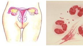
Botals Dol (Figure 1, video 1) is a vessel that normally operates by the fetus and connects the two mainstream heart vessels - the aorta and the pulmonary artery. It exists in order for the blood could bypass by the lungs that do not function in intrauterine. During the first days of the life of the newborn baby Botallov, the duct is closed. Sometimes it happens that the open arterial duct is not closed, which leads to a number of unpleasant problems. The duct that did not close during the month of the child's life is considered congenital heart disease.
Natural vice flow. Or what will the Open Botalov Doc?
The fact is that this vessel still connects two large heart vessels - the aorta and the pulmonary artery. Pressure in the aorta is much higher than the pressure in the pulmonary artery. Therefore, through the open arterial duct with the aorta, an excessive amount of blood falls into the lungs, which will first lead to frequent bronchopile diseases, and with very large Botallails - to irreversible changes in the vessels of lungs and inoperability. In addition, large Botals Doc significantly increases the load on the heart, especially on the left ventricle. Therefore, it is impossible to delay with the treatment of this vice.
Treatment of open arterial duct.
Currently, there is no such Botallian duct, which could not be closed by a non-immutable endovascular method, which will avoid cut, scars and long-term rehabilitation. Surgery This vice remained in the past, surgeons close the Botalas duct only to premature children or in countries where medicine has insufficient funding. In all developed countries of Europe and America, this vice eliminates exclusively endovascular in Rengen-operative. In addition, the probability of complications in endovascular treatment is much less.
Endovascular closing procedure.
In case of endovascular closure through a small puncture into the femoral vessels in the heart vessels and in Botals, thin tubes, the so-called catheters are breeded. Using x-ray and contrast agent, the doctor estimates the size and shape of the Botallian duct, after which it selects the most suitable occlusive (from the English. Occlusion - blockage) device. Occludes can be used as such devices (Fig. 2; video 1, 2, 3) or spirals (Fig. 3; video 4, 5, 6).
The selection of a closure device occurs during the operation, and depends on the size and shape of the Botallian duct. As a rule, occluders are used for large ducts, for small-spirals. During the six months, occlusive devices completely finish with their own heart cells, the so-called endothelization occurs. Dumping through Botallals in 90% of cases is terminated immediately after the procedure, in other cases, at the end of the endothelization period of the device.
Rehabilitation after the procedure
1. Patients are discharged, as a rule, the next day after the procedure.
2. Within 6 months, it is recommended to carry out antibiotic-pylintics of infectious endocarditis.
We have the highest experience in Ukraine endovascular treatment of open arterial ducts - more than 300 operations. We have access to the equipment to close the Botallastic duct of any size and shape. Also, specializing in the treatment of defects between interdestrial and interventricular partitions. In order to get to us on a consultation or hospitalized by call one of the phones or sign an online reception.
|
Video 1 - Botals Dol |
|
Video 2 - In this colorful animation, you can see how the Botals are closed |
|
Video 3 - video from the operating room: blood through the open Botallals duct (vessel in the center) gets from aorta (large vessel on the right) into the pulmonary artery (vessel on the left) |
|
Video 4 - video from the operating room: the duct is blocked by an occluder. Blood reset stopped |
|
Video 5 - And in this video you can see how Botallah Cottage Spiral |
|
Video 6 - Video from the operating room: blood through the open Botallov duct (vessel in the center) gets from the aorta (large vessel on the right) into the pulmonary artery (vessel on the left) |
|
Video 7 - video from the operating room: the duct is blocked by a spiral. Blood reset almost stopped |
Myths and Reality About Edovascular Surgery
Congenital heart defects
Currently, X-rayland surgery attracts more and more attention to almost all media, including printed publications, Internet and television. We are daily confronted with a massive flow of information on various aspects of this modern field of medicine. Every day they write about it and say, unfortunately, not everything is not always objective. There are many erroneous statements, rumors or even myths that need to be corrected using actual information.
Myth 1. This is a very new, almost experimental area of \u200b\u200bcardiovascular surgery.
This is not true! Endovascular surgery has a rich history and has long been widely used in medical practice. For the first time, the catheterization of the heart was performed in 1929 by R. Forsman (Germany), for which in 1956 he received the Nobel Prize. In 1964, the first balloon angioplasty was carried out and since then endovascular surgery ceased to be a purely diagnostic area of \u200b\u200bmedicine. Next, the discovery and inventions of the devices were followed by one one: 1975 - Spiral, 1976 - occluders, 1979 - Emboli, 1986 - coronary stents, 1994 - stents for large vessels, 2005 - endovascular heart valves! To date, all the above devices have evolved to more advanced counterparts. The most common occluder in the world became an amplucer - more than half a million implants since 1995. At the AMOSOV Institute, the amplucer occluders are their analogues since 2003. The trend in the world is such that the diagnosis moved from the x-ray-operating in the cabinets of echocardiography and computed tomography, and the treatment of heart defects moved from the operating room to X-ray. In developed countries of the world (USA, Canada, Australia, Europe) Botallas, the defects of partitions, and the aortic coarctations practically do not operate surgically. Our institute takes into account all modern global trends in the treatment of patients.
Myth 2. Devices, with the help of the treatment of defects (occluders, spirals, stents) are foreign bodies And they can turn back.
All these devices are made of modern high-tech biocompatible materials that do not cause reactions of rejection. Six months after the operation, these devices are completely covered by endothelium (germinate with their cells) and do not differ from the inner surface of the heart. All devices are non-magnetic, after their implantation, MRI can be performed. They do not publish sound signals on metal detectors at the airport, a shopping center, etc.
Myth 3. Occludes are displaced (fly away).
Indeed, in our and world practice such cases happens, but their frequency is about 1%. Complication is unpleasant, but not critical. There was not a single case in the world when the interchangeable occluder would lead to a fatal outcome. As a rule, such an occluder extract endovascularly and set again or replaced with more. The greatest number of displacements fall on the first hours or days after surgery, when the patient is still in the clinic. Next, the probability of this sharply decreases, remote offsets are casual.
Myth 4. Defects of the interpresent partition with the absence of edges or thinned edges are not subject to endovascular closure.
The absence of the aortal edge of the partition is not a contraindication to the layout of an occluder. The same applies to the thinned or aneurysmatic partition. Remember that the usual (transstorakal) echocardiography does not give a complete picture of the defect. Even if the lack of edge is diagnosed, it does not mean that it is not there. Only a clear anatomy of defect can be judged only after conducting transpisco echocardiography, which is a gold standard in the selection of patients for endovascular treatment.
Myth 5. Occludes with time require replacement.
Neither with an increase in the patient nor over time, the device is not needed. An occluder after 6 months will grow into a partition and creates the basis for its further growth. In the case of vascular stenting, it is possible an endovascular increase in the lumen of the stent with an increase in the vessel without replacing the implant.
Myth 6. It is expensive ...
Endovascular surgery is high technologies that really cost more than ordinary operations. In some cases, an implantation device buys a patient, however, there is a queue for free implants that the institute purchases. In addition, we cooperate with numerous assistance funds, which for relatively short time collect funds for the purchase of devices for children. In most cases, there are no urgency in the operation, and patients have enough time to assemble funds for the implant, wait for their turn or find a sponsor. Therefore, if the patient wishes to operate endovascular, barriers for this today does not exist.
Frequently asked Questions
The average time staying in the hospital is 3-4 days. As a rule, on the day of receipt in the morning you pass a survey that includes clinical and biochemical analysis blood (you need to come on an empty stomach), make x-ray, ECG, ultrasound procedure Hearts and Consultation of the cardiologist and cardiac surgeon. If all the indicators are normal, the next day is carried out to eliminate the vice. On the third day we are conducting test research and write you down.
For hospitalization to our hospital, you will need a passport or a child's birth certificate.
If patient children's age, I need a certificate of sanitary-formation (that recently the child has not in contact with infected patients), which you get in the clinic at the place of residence.
It is desirable to have previous advisory conclusions, an ECG and an X-ray snapshot of the breasts.
Direction from cardiologist at the place of residence is not required. You can come to a consultation and subsequent treatment in self-defense. If you are more than 30 years old or you felt the interruptions in the work of the heart, it is advisable to hold Halter monitoring at the place of residence. Such a study can be carried out with us, but it will increase your stay in hospital for 1-2 days.
If you suffer chronic gastritis, ulcerative disease Stomach or duodenal gut It is necessary to make fibrogastroduodenoscopy. In case of confirmation of the disease, you need to undergo a course of treatment at the place of residence. Such a study can be carried out with us, but it will increase your stay in the hospital for 1-2 days in the absence of ulcers and erosion.
All adult patients are carried out under local anesthesia. The patient can observe the course of the operation and communicate with the staff. The exceptions are patients with a defect of the inter-subsensity partition, which during the operation requires control of transpishery ultrasound and for the patient's comfort, the operation is carried out in a state of drug sleep. All endovascular operations in children and constant patients are conducted under general anesthesia.
It is necessary to limit strong physical Load for 6 months. Prevention of respiratory infections, tonsillitis, caries are needed. In the event that the disease began to develop, I need to include in the treatment regimen antibacterial drugs, After consulting a doctor. Within the first month after the operation, it is also necessary to limit sex life.
Openbotallovduct (arterial) - This is one of the most common patterns of the "white" type. In normal conditions, the battles of the Dol, immediately after birth, ceases to function and is bred in the first 2-3 months, rarely in a later date. If the duct is unforgettable, vice develops,
in which through the hole in the duct, the blood from the aorta is reset into the pulmonary artery. As a result of B. big circle The blood circulation is income the amount of blood, and in a small circle - excessive.
Symptoms of open battal
Clear clinical signs open arterial protocol Usually appear on the 2-3rd year of life. In the first phase, frequent respiratory infections, pneumonia, subfebrile temperature can be observed.
Schematic image open arterial duct defect
Children with the unguardment of the Batalov of duct of pale, fragile physique. When palpation over the base of the heart, trembling is felt. The boundaries of the heart are somewhat extended both to the right and left. In the region of the vascular beam to the left of the sternum, the purlization of the percussion sound is determined (Zone Gerhardt). Here, in the second-third intercostal, systolic noise is listened. In the future, the noise becomes a systra-diastolic ("machine") and is heard throughout the heart of the heart. Minimum blood pressure reduced. When x-ray is noted an increase in the left sections of the heart and ascending aorta, signs of lung vessels overflow. With the development of hypertension of a small circulation of blood circulation, an increase in the right heart departments is found.
On the ECG in the first and second phases of the disease, the left type of ECG is noted, in the third phase there are signs of hypertrophy of both ventricles, also indicating the lesion of myocardium. With the help of heart probing determine increased content Oxygen in the blood from the pulmonary artery, in addition of the pulmonary artery, the probe can be held in the aorta. The diagnosis can be confirmed and angiocardiography.
Complications. Frequent pneumonia pulmonary hypertension, bacterial endocarditis, insufficiency of blood circulation.
Forecast. With timely operational treatment - Favorable. Without surgery, children often die from complications.
Treatment of Batalov Protoka
Treatment of an unnecessary duct battal is operational in the event that its diameter is more than 3 mm. Indications for the operation are set depending on clinical flow Diseases at any age. The basic meaning of the battalock operation is a duct bandage, which contributes to the normalization of hemodynamics.
If the diameter of the Botallian duct is less than 3mm, that is, the possibility of its closing by an occluder without surgery. Occlode is a kind of "rivet", which is delivered to the hole that you need to overlap and, thanks to its design, it closes this hole. In the future, the occluder is covered with blood closures, calcium, etc., which reliably fixes it in the heart muscle.
- functioning pathological communication between the aorta and the pulmonary barrel, which is normally provided by embryonic blood circulation and subjected to obliteration in the first hours after birth. Open arterial duct is manifested by the lag of a child in development, increased fatigue, tachipne, palpitations, and heartfelt interruptions. Diagnose open arterial ductors help data of echocardiography, electrocardiography, radiography, aortography, heart catheterization. Treatment of vices surgical, including dressing (ligation) or intersection of open arterial duct with the embedding of aortic and pulmonary ends.
MKB-10
Q25.0.

General
Open arterial (Botalles) Doc - the incoming of an additional vessel connecting the aorta and a pulmonary artery that continues to function after the expiration of its obliteration. The arterial duct (Dustus ARTERIOSUS) is the necessary anatomical structure in the system of embryonic blood circulation. However, after birth, due to the advent of pulmonary respiration, the need for arterial duct disappears, it ceases to function and is gradually closed. Normally, the functioning of the duct is stopped in the first 15-20 hours after birth, the full anatomical closure continues from 2 to 8 weeks.
Complications of open arterial duct can serve as bacterial endocarditis, aneurysm of duct and its gap. The average lifespan with the natural flow of duct is 25 years. Spontaneous obliteration and closure of open arterial duct occurs extremely rarely.
Diagnosis of open arterial duct
When examining a patient with an open arterial duct, the deformation of the chest (heart hump), reinforced pulsation in the projection of the top of the heart is often detected. The main auction of the open arterial duct is the coarse systra-diastolic noise with the "machine" component in the II intercostal left.
The mandatory minimum studies in the open arterial duct includes radiography of the chest, a defect of the aorthopic partition, a common arterial barrel, valsalviva aneurysm, aortic insufficiency and arteriovenous fistula.
Treatment of open arterial duct
Premature children use conservative maintenance of open arterial duct. It implies the introduction of inhibitors of the synthesis of prostaglandin (indomethacin) in order to stimulate the independent duct obliteration. In the absence of an effect from the 3-fold repetition of the drug course in children over 3 weeks, the surgical closure of the duct is shown.
In children's cardiac surgeages, open and endovascular operations are used in the open arterial duct. Open interventions may include a dressing of open arterial duct, complaining it with vascular clips, crossing the duct with the east of the pulmonary and aortic ends. Alternative methods for closing open arterial duct are its consumption in the process of thoracoscopy and catheter endovascular occlusion (embolization) with special spirals.
Forecast and prevention of open arterial duct
Open arterial ducts of even small size is associated with an increased risk of premature death, as it leads to a decrease in compensatory myocardial reserves and pulmonary vessels, the addition of serious complications. Patients who have suffered a surgical closure of the duct have the best hemodynamic records and a greater life expectancy. Postoperative mortality is low.
To reduce the likelihood of the child's birth with an open arterial duct, you must eliminate all possible risk factors: smoking, alcohol, drugs, stress, contacts with infectious patients, etc. In the presence of a PRD, a consultation of genetics is needed at the pregnancy planning stage.