Cytosis of the spinal fluid. The composition of the liquor in various nosologies. Set for analysis
Lumbal lycvore is normal.
Table 17.
Purulent meningitis
Serous Meningitis
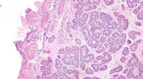
Tuberculous meningitis.
Epidemic Encephalitis.
Brain-brain injury
Tumor CNS.
1) red a) norm
3) Yellow c) stagnation
d) purulent meningitis.
1) Norma a) 0.033
4. Terms denoting inflammation:
d) arachnoiditis
e) meningitis.
2) Pandi b) Samson
d) sulfosalicylic acid
e) Azur Eosin.
2) CITOZ B) in the counting chamber
d) nonnet-opt.
Publication date: 2014-11-02; Read: 16554 | Violation of copyright page
The cerebrospinal fluid is involved in the nutrition of brain cells, in the creation of osmotic equilibrium in brain tissues and in the regulation of metabolism in brain structures. Various regulatory molecules that change the functional activity of various CNS departments are transferred according to the liquor.
Supports a certain concentration of cations, anions and pH, which ensures the normal excitability of the CNS (for example, changes in the concentration of Ca, K, magnesium change blood pressure, heart rate).
Introduction
The spinal fluid (cerebrospinal fluid, liquid) is a liquid that is constantly circulating in the ventricles of the brain, liquor-conducting paths, subarachnoid (subpautented) space of the head and spinal cord
The role of the spinal fluid in the vital activity of the central nervous system Great. The spinal fluid protects the head and spinal cord from mechanical influences, ensures maintenance of constant intracranial pressure and water-electrolyte homeostasis. Supports trophic I. exchange processes Between blood and brain.
Bibliography.
- Anatomy of man / ed. MG Lake - 9 ed., From 542.
- Kozlov V.I. Anatomy of the nervous system: Tutorial For students / V.I. Kozlov, T.A. Cechmistrenko. - M.: Mir: Act Publishing House, 2004. - 206 p.
- Man Anatomy: Tutorial in 2 volumes / Ed. M.R.Sapina.
- Anatomy of the central nervous system. Reader. (Tutorial for students). The authors are compilers: i.e.rosolimo, L.B. Rybalov, I.A. Moskvina-Tarkhanova.
- Reader on the anatomy of the central nervous system: studies. Manual / Red.-Cost. L.K. Khludova. -M.
The composition of the liquor in various nicologies
: Ros. psychologist. General, in, 1998. - 360 p. - Decree. anatomist. Terms: s. 342-359.
- http://knowledge.allbest.ru; http://www.kazedu.kz; http://medbiol.ru.
- Spinal fluid (liquor), its composition, functions, circulation paths.
- The composition of the spinal fluid (liquor).
- Ways of circulation of spinal fluid (liquor).
Karaganda State Medical University
Department of Anatomy.
Topic: Circulation of the spinal fluid.
Performed: Student 246 OMF Groups
Kosilova E.Yu.
Checked: teacher G.I.Tugambayeva
Karaganda 2012 year.
Pages: ← Previous12
Lumbal lycvore is normal. In healthy people, obtained in lumbar puncture, is a colorless and transparent, like water, a low-alkaline reaction liquid (pH 7.35-7.4) with a relative density of 1.003-1.008. Contains 0.2-0.3g / l protein; 2.7-4.4 mmol / l glucose; 118-132mmol / l chlorides. In microscopic examination, 0-5 cells are detected in 1 mk (mainly lymphocytes).
Under a number of diseases of the CNS, the liquor has similar properties, which made it possible to distinguish three laboratory pathological lycvore syndrome: serous liquor syndrome, purulent liquor syndrome and hemorrhagic liquor syndrome (Table 17).
Table 17.
Basic pathological lycvore syndromes
Purulent meningitisit may be caused by meningococci, streptococci and other global coccal. Often develops as a complication of purulent otitis, during the injuries of the skull. On the second or third day of the disease, pronounced pleaocytosis appears (up to 2000-3000 · 106 / l), which is growing very quickly. Likvor becomes muddy, purulent. When defending a rough fibrinous film is formed. The overwhelming majority of uniform elements are neutrophils. The protein content sharply increases (up to 2.5-3.0g / l or more). Globulin reactions are positive. The content of glucose and chloride is reduced from the first days of the disease.
Serous Meningitiscan cause tuberculosis mycobacteria, cokes and enemy viruses, epidemic vapotitis, herpes, etc. The most severe form of serous meningitis is tuberculous meningitis.
Tuberculous meningitis.A characteristic feature is to increase the pressure of the spinal fluid. Normally, the liquor is highlighted at a speed of 50-60 drops per minute, with elevated pressure, the liquor flows into a jet. The liquid is more often transparent, colorless, sometimes opales. In most patients, a thin fibrinous mesh is formed in it. Citosis in the midst of the disease reaches 200 · 106 / l and more, lymphocytes prevail. The level of protein is increased to 0.5-1.5g / l. Globulin reactions are positive. The concentration of glucose and chlorides is noticeably reduced. Deciding in the diagnosis of tuberculosis meningitis is the detection in the fibrinic film of mycobacterium tuberculosis.
Epidemic Encephalitis.The spinal fluid is more often transparent, colorless. Plequitosis moderate, up to 40 · 106 / l, lymphoid character. The protein level is normal or slightly elevated. Globulin reactions are weakly positive.
Brain-brain injury. One of the leading signs of the cranial injury is a blood admixture in the CSC (red color of different intensity). Blood adherence can be a symptom of other lesions of the CNS: the aneurysm of the vascular vessels of the brain, hemorrhagic stroke, subarachnoid hemorrhage, etc. In the first day after hemorrhage, the liquid after centrifuging becomes colorless, xanthromy appears on the second day, which disappears in 2-3 weeks. An increase in protein content depends on the amount of spending blood. With massive hemorrhages, the protein content reaches 20-25 g / l. A moderate or pronounced pleyocytosis is developing with the predominance of neutrophils, which are gradually replaced by lymphocytes, macrophages. Normalization of the liquor occurs after 4-5 weeks after injury.
Tumor CNS.Changes in liquor depend on the localization of the tumor, its size and contact with the likvorn space. The liquid can be colorless or xanochromic at a block of subarachnoid space. The protein content increases slightly, but with a block of liquor paths, the tumors of the spinal cord is revealed by a sharp increase in protein content, globulin samples are positive. Cytosis does not exceed 30 · 106 / l, mostly lymphoid. When localizing the tumor, the CSH can be unchanged from the liquor pathways.
5.4. Control questions to the chapter "Study of the cerebrospinal fluid"
Set the correspondence between the elements in columns. One element in the left column corresponds to only one element in the right column.
1. Number of liquor (ml), which:
1) is produced per day a) 8-10
2) circulates simultaneously b) 15-20
3) extracted when puncture in) 100-150
2. The color of the cerebrospinal fluid is normal and with pathology:
1) red a) norm
2) colorless b) subarachnoid hemorrhage (1 day)
3) Yellow c) stagnation
d) purulent meningitis.
1) Norma a) 0.033
2) spinal cord tumor b) 0.2-0.3
2.4 Laboratory Liquor Research Methods
Terms denoting inflammation:
1) brain a) Pleocytosis
2) solid cerebral shell b) stroke
3) Cellic shell c) Encephalitis
d) arachnoiditis
e) meningitis.
5. Reagents used for:
1) Cottosis a) ammonium sulfate
2) Pandi b) Samson
3) determination of the amount of protein c) carbolic acid
d) sulfosalicylic acid
e) Azur Eosin.
6. The predominant type of cell elements in the liquor in the diseases of the CNS:
1) neutrophils a) tuberculous meningitis
2) Erythrocytes b) purulent meningitis
c) hemorrhage (first day).
7. Methods for defining in Likvore:
1) ratio of protein fractions a) with sulfosalicylic acid
2) CITOZ B) in the counting chamber
3) quantities of protein B) in painted drugs
d) nonnet-opt.
Publication date: 2014-11-02; Read: 16555 | Violation of copyright page
studopedia.org - Studdiadia.org - 2014-2018. (0.002 s) ...
Product Catalog
| 38.02 | Blood Clinic No. FSR 2008/03535 of 10/29/2008 Set for holding general analysis blood with unified methods: fixation and coloring of blood smears (4000 ODA), the number of erythrocytes (4000 ODA), the number of leukocytes (4000 ODA), the number of platelets (4000 ODR), SE micrometeode Panchenkova (4000 o |
— |
| 38.03 | Clinic-Cal. Set number 1 (general) No. FSW 2010/09420 dated 08.12.2010 A set of reagents for clinical analysis of Cala: Hidden Blood (1000 ORD), Sterkobilin (50 ORD), Bilirubin (200 ORD), Microscopic Study (Neutral Fat, fatty acid, soap, starch, helminth eggs) (2000 ORD.) |
— |
| 38.03.2 | Clinic-Cal. Set number 2 Definition of hidden blood |
1000 |
| 38.03.3 | Clinic-Cal. Set number 3 Definition of Sterkobilo Reagent set for clinical analysis |
50 |
| 38.03.4 | Clinic-Cal. Set number 4 Definition of bilirubin Reagent set for clinical analysis |
200 |
| 38.03.5 | Clinic-Cal. Set number 5 Microscopic examination | 2000 |
| 38.04 | Clinic-uro. Set number 1. |
Set for clinical analysis of urine No. FSW 2010/09509 dated December 17, 2010
Acidness (pH) (1000 ORD), glucose (1000 ORD), ketones (1000 ODR.), Bilirubin (400 ORD), Urobilinoids (1000 ORD), Protein shared: - Qualitative ODA. (1000), - quantitative ODA. (330)
- Qualitative ODA. (1000) - Quantitative ODA. (330)
Reagents set for clinical analysis of sputum: acid-resistant mycobacteria (CUM) (200 ODR), alveolar macrophages with hemosiderian (reaction to Berlin Azure) (100 ORD), cells of malignant neoplasms (300 ORD)
The set for the analysis of the spinal fluid: cytosis (Samosone reagent) (200 ODR.), Protein shared: Quality reaction of Pandi (200 ORD), quantitative ODA. (sulfosalicyl. K-TA and sodium sulfate) (200 ODR.), Globulins (200 ODR.)
A set for detecting in the feces of helminths and their eggs by a thick smear. CATO reactive - 1 vial (50 ml.) Cellophane coating plates - 500 pcs. Silicone rubber plug - 1 pc.
A set of reagents to determine the content of protein in the urine and the spinal fluid with pyrrogalol red. Reagent - a solution of pyrogalla red in a succinate buffer. Calibrator 1 - Calibration Squirrel Solution
100 ml
a set of reagents for the aspirational color of the urine precipitate (modification of the Stergeemer method)
Microscopic examination (quantity and morphological structure of cell elements)
The amount and morphological structure of cell elements are essential to establish the nature of inflammatory processes in the brain and its shells.
According to the nature of the liquor changes, purulent and serous meningitis (meningoencephalites) differentiate. The serous includes meningitis (meningoencephalitis), in which the liquor is transparent, sometimes slightly mightly, opalesse; The number of cellular elements increased to 500-600 in 1 μl, lymphocytes prevail.
The purulent includes meningitis (meningoencephalites), in which the number of leukocytes exceeds 0.5 - 0.6 * 109 / l and can reach 20 * 109 / l and more. Colorless, transparent or opalesing liquor should be specially investigated in order to identify the fibrin film specific for tuberculosis meningitis ("mesh"), which can be formed in a test tube after 12 - 24 hours.
Very often in such a film microscopically detected tuberculosis sticks.
Microscopic examination of liquor
In meningitis, meningoencephalitis, septic thrombosis of the brain sinus, changes in the liquor are inflammatory.
The number of cell elements (predominantly neutrophils) increases to a much greater extent than the protein content increases - cell-protein dissociation.
In pathological processes accompanied by an edema of the brain, an increase in intracranial pressure and leading to the blockade of liquor-conductive paths, a significant increase in protein content with a non-trunk or normal number of cell elements (protein-cell dissociation) is more characteristic.
Such ratios are observed with sharply manifest brain tumors, large epidural and subdural hematomas and some other pathological processes that cause edema and dislocation of the brain.
As a result of the microscopic study of the likvore smears, determine the pathogen of meningitis (bacteria, mushrooms, the simplest, tumor cells) is far from constantly - at 35 - 55% of cases. Thus, the role of microscopy in establishing the etiology of inflammatory lesions of cerebral shells is limited.
Equally, this refers to the possibilities of bacteriological diagnosis of the etiology of meningoencephalitis, brain abscesses and septic thrombosis of cerebral sines. The sugar content in the lycvore is reduced in many pathological processes by reducing its transport through the hematorecephalic barrier.
"Emergency condition in neuropathology", B.S.Vilensky
The human body is a perfect, well-working, coordinated biological mechanism. Each cell structure, fabric, system of organs and metabolites are necessary for certain purposes and in a specific amount.
The compounds produced by our body include biological substances that perform a mass of important functions: protective and regulatory. The outcomed volume, composition, color and other characteristics can tell, healthy person or it is worth thinking about a visit to the doctor. The most significant essences consider breast milk, colostrum, blood, sperm, saliva, urine, vaginal selection, as well as a liquor, which today will be discussed.
What is liquor, definition of liquor
Spinal, or cerebrospinal fluid (SMF, or CES) - this is a liquid medium that fills the space in the brain ventricles, flows through the liquor-conducting path, circulates in the subarachnoid segment. Alternative name -likvor.
The synthesis and selection of the substance is due to the process of filtering the plasma (liquid blood) through the capillary wall and the subsequent secretion of substances into the exudate from ependium and secretory cellular structures.
If there is any pathological condition with a violation of the integrity and structure of the bone and soft tissue of the cranial box, it occurslikvorea - Isolation of spinal fluid from ears, nose or defective, damaged skull and spine places. Probable reasons:
card and brain injury;
hernia neoplasms or tumors;
non-accuracy of medical manipulations;
postoperative weakness of seams.
Any deviation from the norm in the functioning of the system of organs affects the thickness, transparency and the number of substance allocated, therefore, at its state, some pathologies can be determined.
Functions of lycvore
Like each substance in the human body, the CMF performs a lot of vital functions:
Mechanical protection. Ensuring the shock absorbing effect with sharp movements or shocks head - aligning intracranial pressure,spinal fluid Protects the brain from damage, providing its integrity and normal operation even in honeycomb situations.
The excretion of metabolites. Some substances can accumulate in the brain space, which will negatively affect its functioning - the lycvore is responsible for their allocation (excretion) and outflow.
Transportation of the necessary compounds. Hormones, biologically active substances and metabolites that are responsible for the central performance are transferred to a gray substance with the help of a cerebrospinal substance.
Breathing (performing respiratory function). Neuronal clusters that are responsible for the respiratory function of the body are located at the very bottom of the fourth ventricle GM and washed by the liquor. It is worth a slightly changed component ratio (for example, an increase in the concentration of potassium or sodium ions), the change in the amplitude and frequency of breathing / exhalations will follow.
The role of the regulator, stabilizing structure for the central nervous system ... It is SMF that supports certain acidity, salt and cationic and anionic composition, the constancy of osmotic pressure in the tissues.
Maintaining the stability of the brain environment. This barrier must be practically insensitive to changes in the chemical composition of blood so that the brain continues to work and during the way a person is sick or fights pathology.
Work natural immunoregulators. Assess the state of the nervous system and trace the course of the disease will be able to evaluate only with the help of a detailed analysis of the point, the study of which will help clarify the diagnosis or predict the patient's health state.
The composition of the liquor
The cerebrospinal substance is produced, on average, at a rate of about 0.40-0.45 ml per minute (in an adult). Volume, product speed, and the most important thing - the component composition of the CCH directly depends on the metabolic activity and age of the body. Typically, analyzes reflect that the older man is, the stronger the production is reduced.
This substance is synthesized from the plasma part of the blood, however, the substrate and the producer differ significantly in ion and cellular content. Main components:
Protein.
Glucose.
Cations: sodium, potassium, calcium and magnesium ions.
Anions: Chlorine ions.
Cytosis (the presence of cells in the liquor).
The increased content of protein and cell clusters indicates a deviation from the norm, which means it is a state that requires further analyzes and mandatory consultation with the attending physician.
Analysis and study of liquor
The study of cerebral-spinal point is a method that is used to identify and diagnose various brain structures and shell disorders, the central nervous system. These pathologies include:
meningitis, tuberculosis meningitis;
inflammatory processes in the shell;
tumor education;
encephalitis;
syphilis.
Conducting the procedure for analyzing and studying by the CM liquid requires the sample fence as a punctate from the lumbar spinal cord. The fence is made through a small point puncture in the desired spine.
IN complete analysis The CSC includes a macroscopic and microscopic examination, as well as cytology, biochemistry, bacterioscopy and bacterial sowing on the nutrient medium.
Explore the cognition puncture will be in several parameters:
Transparency.
Likvor healthy man It is absolutely transparent as clean water, therefore, with a macroscopic analysis, it is compared with the benchmark - distilled highly purified water in good lighting. If the sample taken is not enough or there is a strong, obvious cloudy, that is, the reason to look for the disease. After detecting the non-compliance with the standard, the test tube is sent to the centrifuge - the procedure will allow to determine the nature of the clouding:
If after centrifuging the sample is still muddy, then it indicates bacterial contamination.
If the precipitate dropped to the bottom of the flasks, then the cloudiness was given uniform elements of blood or other cells.
Color.
The liquor produced by a healthy organism must be absolutely colorless. The change shows the presence of any compounds in it, which normally should not be there - many pathological conditions of the body provokes xanthromhromy, that is, its staining in shades of red and orange. Xanthromy is caused by the hit of hemoglobin and its types in the sample, for example:
yellowishness - the presence of a bilirubin fraction isolated during the decay of hemoglobin;
light pink, red-pink stencil indicates oxymemoglobin (hemoglobin saturated with oxygen) in the liquor;
orange shades - there are bilirubin compounds in the sample, which appeared due to the decay of oxymemoglobin;
brown colors - reflect the presence of methemoglobin (oxidized hemoglobin form) - such a state is observed in tumor phenomena, strokes;
muddy green, olive - presence of pus with purulent meningitis or after opening the abscess.
red reflects blood availability.
If a little sucrovitz got into the sample during the fence of the point, then such a mixture is considered "traveling" and does not affect the result of macroscopic analysis. This impurity is observed not throughout the volume of the point, but only on top. The impurities can be pale pink, muddy pink or grayish-pink.
The KSTanochromic sample intensity is estimated at the plus laboratory assistant during visual assessment:
the first degree (weak).
second degree (moderate).
third degree (strong).
fourth degree (excessive).
Blood fractions or strong saturation of the point allow us to assume one of the diagnoses: the gap of the aneurysm vessels and the subsequent intracranial hemorrhage, hemorrhagic encephalitis or stroke, the middle and strong, hemorrhage into the brain tissue.
Cytology.
The state of the cerebrospinal fluid of a healthy person admits a minor cell content, but within the limits of the established values.
Leukocytes in one cubic mm:
up to 6 units. (in adults);
up to 8-10 units. (in children);
up to 20 units. (Babies and babies up to 10 months).
Plasma cells should not be. The presence indicates infectious diseases of the central nervous system: multiple sclerosis, encephalitis, meningitis or recovery after surgery with a wound, which has not heal for a long time.
Monocytes are observed in an amount of up to 2 on cubic mm. If the number is growing, it is a reason to suspect chronic pathology CNS: Ischemia, neurosofilis, tuberculosis.
The neutrophilic component is present only with inflammatory processes, modified shapes - when recovery after inflammation.
Cell-macrophages of grainy type can be in the SMF only when the brain tissue of the body breaks down, as with a tumor. Epithelial cells fall into the point only in the case of the development of the TSS tumor.
Norm, focus indicators in a healthy person
In addition to the components, transparency and color characteristics,normal Likvor Must correspond to other indicators: medium reaction, cells, chlorides, glucose, protein, maximum cytosis, no antibodies, etc.
Deviation from the above indicators can serve asidentifier diseases - for example, immunoglobulins andantibodies Oligoclonal type in the sample may indicate the presence or risk of developing sclerosis.
Protein in Likvore: Lubal - 0.21-0.33 g / liter, ventricular - 0.1-0.2 g / liter.
Pressure in the range of 100-200 mm of water art. (sometimes indicate the values \u200b\u200bof 70-250 mm - in countries outside the post-Soviet space).
Glucose: 2.70-3.90 mmol per liter (some sources indicate: two thirds of the total glucose in plasma).
Glorides of SMG: from 116 to 132 mmol per liter.
The optimal indicators of the reaction of the medium are values \u200b\u200bwithin 7.310 - 7.330 pH. The change in the acidity is extremely negatively affecting the performance of biological functions, the quality of the SUM and the rate of its flowing paths on the leacure paths.
Cytosis in Likvore: Lumbal - up to three units. on μl, ventricular - to one to μl.
What should be in the point of a healthy person should not?
Antibodies and immunoglobulins.
Tumor, epithelial, plasma cells.
Fibrinogens, fibrinogenic film.
Sample density is also defined. Norm:
The total density should not exceed 1,008 grams per liter.
Lumbal fragment - 1,006-1.009 g / l.
Ventricular fragment - 1,002-1.004 g / l.
Suboccipital fragment - 1,002-1.007 g / l.
The value can decrease with uremia, diabetes or meningitis, and increase - with hydrocephalic syndrome (increasing the size of the head due to the accumulation of the fluid and its difficult removal).
Violation of liquor. Causes and symptoms
Among the main painful states associated with the CMF, they allocate liquor, liquorodynamic imbalance, the "water" of the brain and increased intracranial pressure. Their development mechanism varies like a symptom complex.
Likvorea
It is the most pathogenetic simple disease, because its mechanism is understood: the integrity of the bones of the base of the cranial box or brain shell is disturbed, which provokes the release of the spinal substance.
Depending on the symptoms and visual manifestations, the liquor is called:
Hidden - Likvor expires on the nasal move, which is not noticeable visually due to aspiration or random swallowing.
A clear - transparent liquid or an admixture of Sukrovitsy is intensively allocated from the ears, the fractures seats, which is noticeable to flow the banding head bandage.
Also allocate:
The primary nature of the disease - the expiration is manifested immediately after injury, after operational intervention.
Secondary, or liquor fistulas - the expiration is observed in the late period of strong complications of infectious diseases.
If primary pathology is not treated for a long time, and then enjoy inflammation (meningitis or encephalitis), then this is fraught with the development of fistula.
Common causes of the expiration of SMG:
strong bruises with cranial and brain injury;
injuries and serious wound injuries;
complicated hydrocephalus;
hernia neoplasms and tumors in dangerous proximity or directly in brain tissue;
neaccurability of medical manipulations - washing or drainage of the ENT profile;
the weakness of the seams of the hard shell after the operations of the neurosurgical profile;
spontaneous Likvorea - very rarely.
Likvorodynamic violations
Likvorodynamics is broken in case of difficulty or incorrect circulation of the spinal fluid. Disease flows can be hypertensive (associated with increased pressure) or hypotensive (on the contrary - with reduced).
Hypertensivethe form occurs when:
excessive allocation - due to the strong excitability of vascular plexuses, which are responsible for the products of the CSC;
insufficient suction, elimination.
Licvor is produced in large quantities or simply not absorbed, which provokes such symptoms:
pronounced headaches, especially intense in the morning hours;
nausea, frequent vomit urge, periodically vomiting;
the head is spinning;
slow heartbeat - bradycardia;
sometimes nistagm - frequent involuntary movements of the eyes, "jitter" of pupils;
symptoms characteristic of meningitis.
Hypotensivethe form arises less frequently, with hypofunction, or weak activity of vascular plexuses, consequence - reduced products of the Likvorn substance. Symptoms:
strong headache in the occipital and dark areas;
unpleasant sensations, injection of pain in sharp movements, excessive physical activity;
hypotension.
Disturbance of Flushing and Resorption
When the body fails in the body, then the outflow of the cerebrospinal substance and its resorption may be impairedfrom the brain - Due to this, deviations develop, which are different in adults in different ways and in children.
The adult will react to a deviation with an increase in intracranial pressure due to a strong, "overgrown" cranial box. The bones of the child's skull immature and have not yet grown, so the excess accumulation of the spinal substance provokes hydrocephalus (Vyanka GM) and other unpleasant manifestations.
Calculation of the liquor in the brain - increased VCH in adults
In the cranial box there is not only brain tissue and a great many neurons - a significant part of the volume is busy. His big share is in the ventricles, and the smaller - heams the GM and moves between its web and soft shells.
Intracranial pressure directly depends on the volume of the skull and the amount of fluid circulating in it. The products of the substance increases or its resorption is reduced - the body immediately responds to it with an increase in the PCF.
This indicator reflects how much pressure inside the skull exceeds the atmospheric - the norm is the value from 3 to 15 mm mercury column. Minor fluctuations lead to deterioration of well-being, but the height of the PCD to a mark of 30 mm Hg. Art. Already threatens with a fatal outcome.
Manifestations of elevated GFD:
constantly clone in sleep, low performance;
pronounced headaches;
deterioration of visual acuity;
forgetfulness, scattering, low concentration of attention;
the "jumps" of pressure - hypertension is regularly replaced by hypotension;
bad appetite, nausea, vomiting;
emotional instability: mood swings, depressiveness, apathy, severe irritability;
vertebral pains;
chills;
raising sweating;
respiratory failures, shortness of breath;
the skin is more sensitive;
muscular paresis.
Availability of 2-3 symptoms is not a reason to suspect an increased GFD, but a practically full complex is a weighty reason to contact a specialist.
The brightest sign of the disease is a slouching headache, not expressed in any separate area. Cough, sneezing and sharp movements only provoke the strengthening of pain, which are not even stopped by analgesics.
The second important sign of an increased ICF is vision problems. The patient suffers from doubles in the eyes (diplopia), notes a worsening of vision in the dark and in bright lighting, sees, as in the fog and suffers from blindness attacks.
The pressure can also increase in a healthy body, but immediately comes to normal - for example, during physical and emotional loads, stress, cough or sneezing.
Cutting of the liquor in the brain - Children's watercolor GM
Small children cannot report their own well-being, so parents should be able to determine the violation of the liquor outflow on the external signs and the behavior of the infant. These include:
noticeable vascular grid on the skin of the forehead, the nape;
night anxiety, bad sleep;
frequent crying;
vomiting;
spring, his ripple;
convulsions;
an increase in the sizes of the head;
the uneven muscular tone is part of the tense, and some relaxed.
The most serious feature of the increased GFDthe child has It is a hydrocephalus that occurs with a frequency to one case for a couple of thousand newborns. Male kids are sick of the brain water more often, and the defect itself is diagnosed with doctors usually during the first 3 months of life.
Do not confuse "Brain Watermark" as an independent disease, with a diagnosis of "hypertensive-hydrocephalic syndrome". It reflects that the newborn is slightly elevated by the CFD, but this does not require therapy, as well as surgery, as it is eliminated by itself.
Children's form of illness can be congenital or acquired depending on the cause of development, which, according to medical professionals, may be up to 170. Congenital disease provoked:
child injury during childbirth;
hypoxy during childbirth (insufficient oxygen flow);
genetic failures;
infectious diseases transferred to the fruit during their stay in the womb (cytomegalopathy, sharp respiratory viral infections, infection with mycoplasma and toxoplasma, syphilis, rubella, vapotitis and herpes virus).
Genetic deviations that cause a congenital form:
underdeveloped leachelving ducts;
kiari syndrome - a child skull in volume is greater than his brain;
narrowed liquor;
other chromosomal pathologies.
The acquired form arises due to toxic poisoning, the development of tumors, brain hemorrhages, transferred infectious diseases outside the maternal womb - These include otitis, meningitis and encephalitis.
Speaking about the hydrocephalus in newborns, it is worth considering that in the normal kids head circumference increases rather quickly (one and a half centimeters per month), but if the growth exceeds the indicators, then this is a significant reason to examine the child ..
The skull is a soft, not an infraced, but excess of the liquor slows down the springing of the spring, "spreads" the bone and prevents the normal development of the cranial box - due to this, the head increases disproportionately. Accumulatingin subarachnoid spacewhich shares the brain shells, the lycvore squeezes some brain departments. Despite the support of children's cranial bones, this manifestation of the disease is dangerous and requires immediate treatment. An increase in the size of the head is not the only sign of difficulty lycvaric outflow in children. Characteristic is:
the specific sound of the "broken pot", hearing with a slight tapping of the skull;
difficulties with raising and holding the head in one position;
chin trembling, hands.
It is important to pay attention to the eyes of the baby, because some signs are indicative:
involuntary, chaotic eye movements;
periodic Eye Rolling;
eyes "kost";
syndrome of the "Settlement Sun" - when blinking, a thin white strip between the pupil and the upper age is noticeable.
Under 2, the hydrocephalus is manifested by this symptom complex, and later it is combined by vomiting, nausea, problems with coordination, irritability, diplopia or even blindness.
Sometimes hydrocephalic syndrome is developing in adults as a result of transferred infections, but this is a rare phenomenon.
How to improve the outflow of liquor
On the pathology of Likvorn Flowock, the baby usually recognizes from the neuropathologist, the examination of which takes place in the first month after birth. Primary examination and identification of signs requires medical correction, since this disease will prevent the normal development of the child.
If the condition of the small patient is complex, then specialists using surgical intervention create "bypass tracks" for SMF and eliminatebad outflow artificially. If the situation does not threaten the life of the baby, then the treatment can also take place at home with medication therapy. In order to appoint optimal medicines to a child, you need to understandwhat can interfere with the outflow of the liquor at hydrocephalus. The reason, origin and complications - all factors will play a role in the selection of treatment.
Pharmacological correctiondisturbations of outflow In children includes:
preparations that improve and stimulating blood flow (actovegin, pantogam, cinnarizine);
drugs that contribute to the removal of excess fluid (triampur or diakarb);
preparations - neuroprotectors (cerakson).
Treatment of disintegration disorders
Children's diseases of liquorodynamics are most often corrected by pharmacotherapy, and the adults are required to assign physiological procedures:
Currency electrophoresis with euphilline (ten visits) - a dosage "feeding" will allow you to activate the delivery of oxygen into a brain tissue suffering from hypoxia with an elevated HD. The state of the vessels comes to normal, which will ensure normal resorption.
15 collar zone massage sessions - the procedure is simple, therefore, with the time of the patient, it can also carry out such manipulation. With it, the hypertonus muscles is reduced, spasm is removed and the outflow is settled.
Magnetic effect on the collar zone is a decrease in swelling and vascular spasm, an improvement in innervation.
Medicinal swimming or supporting physical. charging.
The value of the cerebrospinal fluid in osteopathy
The developing direction in medicine is craniosacral osteopathy. As of the state and composition of the spinal fluid, it is possible to determine many illness in the body. In the lycvore, mediators regulating are falling:
respiratory activity;
sleep and wake modes;
stability of endocrine systems;
the work of the cardiovascular complex.
For the normal human functioning, the liquor should be increedable to circulate according to its "path" and maintain component constancy. The slightest violation of the integrity of the cranial seams leads to the pinching of the brain tissue, then the effect applies to the underlying structures.
Craneosacral osteopathy is desirable after serious bruises, road accidents, cranknogo and generic injury. Consultation from a specialist will allow us to reveal a parable at an early stage, and for babies it is especially important. Plastic disorders of the craniosacral system of the newborn directly affect the subsequent development of cognitive functions, CNS and the musculoskeletal system.
Adults complain about Nistagm, violations of vision and respiration, reduce the ability to memorize information, concentrate on the subject of thought, failures in menstrual cycle, sharp weight changes, psycho-emotional instability, intense tear, saliva, and sweating. Typically, such complaints are attributed to other diseases, but an experienced osteopite doctor will be able to carry out a thorough analysis of the state of the patient, his skull and spine, after which it will find out and eliminate the initial cause.
The basis of tuberculosis meningitis lies inflammatory process Shells and vessels. To a lesser extent, this process is expressed in the brain tissue itself. More than with other forms of meningitis, choroid plexuses and ependa of the ventricles, especially III and IV suffer in tuberculosis meningitis. In addition, it should be borne in mind that a serous-fibrinous exudate is always observed for tuberculosis meningitis and a tendency to the formation of adhesive processes in the circulation fluid circulation system. All this leads to the fact that under tuberculosis meningitis, there are always pronounced quantitative and qualitative changes in the spinal "fluid, having quite typical and constant character.
The amount of spinal fluid due to the early damage to the liquid-producing systems and disorganization disorders of the spinal fluid is always increased by 4-6 times and more against the norm, i.e. may be in the amount of 400-600 ml and more. In connection with this, the pressure is usually 300-400 mm of water column and above.
Usually, the opalescence of the spinal fluid is more or less expressed due to a constant increase in protein and cytosis. With a very high cytosis, the liquid may already be at the very beginning of a mercury. In some cases, we also observed at the very beginning of the disease xanthromy. In rare cases there may be hemorrhagic spinal fluid. This is mentioned in the literature.
The amount of cells is noticeably increased, reaching 200-300 in 1 mm 3, and sometimes sharply increased to 600-800 or more. According to S. M. Zilberchid, 173 cases of tuberculous meningitis in the amount of cytosis were distributed as follows: Pleocitosis from 20/3 to 50/3 was noted in 3 cases, from 50/3 to 100/3 - at 5, from 100/3 to 5 200/3 - at 35, from 200/3 to 300/3 - at 39, from 300/3 to 400/3 - at 24, from 400/3 to 500/3 - at 32, from 500/3 to 1000 / 3 - in 31 cases.
According to D. A. Shamburova, the number of cells reached 45-800 in 1 mm 3 per 5-7th day of the disease, and ordinary oscillations did not go beyond 100-300 cells in 1 mm 3.
As for the composition of the cells, at the beginning of the disease, up to 70-80% of neutrophils and 30-20% of lymphocytes are usually available. But in some cases the number of neutrophils can be even more. We observed this in particular during the exacerbation of the disease. Sometimes the amount of lymphocytes can approach 100%. When analyzing the plealosis, it should be borne in mind that it may vary and under the influence of subarachnoid administration of streptomycin or salub. In such cases, an increase in the number of cells holds a short period. Mixed lymphocytic-neutrophilic Plequitosis is typical for tuberculosis meningitis. Plasma cells and monocytes are 1-3%. A large number of cells with some oscillations remains for a long time - 3 months and longer.
The protein with tuberculosis meningitis is increased. This increase occurs due to changes in the permeability of the vessels. In later periods, this may be associated with the destruction of the nervous system. The presence of tuberculous meningitis for a long time serous-fibrinous exudate also leads to the formation of a delicate fibrinous mesh or film that keeps standing and disappears usually along with a decrease in cytosis and protein.
The amount of protein in the initial period of the disease occurs within 0.66-0.99-1.32%. Sometimes at the very beginning of the disease, protein can reach high numbers - 6.6% or more. With early diffuse tuberculous leptopamenengitis, we already at the very beginning of the disease observed very high squirrel numbers - up to 16.5-33%. If there is a dissociation between the amount of protein in a large tank, where the protein levels stand on moderate numbers and in the lumbar department, where it, on the contrary, is sharply increased, it may indicate a wounded blockade of the subarachnoid space.
The reactions of Panda and the nonn-appendix are always sharply expressed. Weichbrodt reaction is weakly positive or negative. According to S. M. Zilberish, the Takata-Ara reaction from 79 cases in 9 was a normal type, in 30 - degenerative, in 15 - meningeal, in 25 - meningeal-degenerative. The Lange reaction more often has a meningeal or meningeal-degenerative character.
The amount of protein with some oscillations remains resistant for a long time, similar to Plarocytosis. Very pathognomonic for tuberculosis meningitis is to reduce the number of gayer, an average of up to 15-30 mg. There may be oscillations in the other side. So, we observed a decrease of up to 7 mg% and even up to 2 mg%, which coincided with the deterioration of the patients. In the treatment, it is necessary to "be murdered in mind that under the influence of ACTH, as shown by Loos and Lierinz, the amount may increase. With tuberculosis meningitis, the amount of chlorides is reduced to 600-500 mg%, and sometimes below.
Tuberculosis bacteria are found in 60-70% at the beginning of the disease and less often (40-50%) in its later periods. Currently, when analyzing the composition of the spinal fluid, the electrophoretic method is also applied. It allows you to determine the ratio of individual fractions of proteins in the liquor in different periods of the disease. According to erdesa, benose and acne at the beginning of the disease, the concentration of albumin in liquor is sometimes low, but usually normal, while the amount of U-globulin in percentage is at the highest level, and the number of A-globulin is somewhat reduced. In the second stage of the disease, the amount of albumin increases, and the U-globulin decreases and there is an obvious ratio between the amount of protein in serum and protein in the liquor. In the third stage of the disease, the amount of albumin and U-globulin can be higher than the norm. The ratio of albumin and globulin may remain modified for several years after recovery from tuberculosis meningitis.
The electrophoresis method was also used to detect tuberculous bacteria in the spinal fluid. The leaf of all patients found mycobact. Tuberculosis, and 2 infection was mixed - tuberculosis bacteria and cocci were found in the liquor. In the early stage of tuberculosis meningitis, tuberculosis bacteria move to the cathode, in treated cases - simultaneously to the anode or only to the anode. At the same time, they detect morphological changes that the author explains the change in the viability of bacteria due to the treatment of tuberculosis by bacteriostatic means. This assumption was confirmed on the experiments of electrophoresis with mycobact solutions. tuberculosis (strain H-37 Rn). The results showed that electrophoresis is the most reliable method for detecting tuberculous bacteria in liquor with basilar tuberculosis meningitis. With this method, other pathogenic agents in the liquor can be found, which is extremely important in mixed infections.
All indicated changes are characteristic of the initial period of the disease and remain quite persistent for a long time. The main in the chronic period is inflammatory syndrome, but changes in the composition of the spinal fluid can occur during treatment. While in a large tank, the composition of the spinal fluid is gradually normalized, the amount of protein may be higher in a stable plea-virocytosis. This happens in 4-5% of patients with a blockade of the subarachnoid space in the region of a large tank or in-upper spinal cord sections .. less often in early and more often in the late period, you can observe the protein-cell dissociation syndrome. This may indicate the sink of the inflammatory process with the remaining elevated permeability of the vessels, which is observed in terms of cases with meningovascular tuberculous meningitis syndrome.
Occasionally the composition of the spinal fluid is similar to that in serous meningitis. In these cases, the fibrin retina and sugar can remain for a long time and sugar can remain on relatively high numbers.
Between the dynamics of the composition of the spinal fluid and the clinical picture with a favorable current tuberculosis meningitis usually there is a mismatch: while clinical symptoms They can almost completely disappear, the spinal fluid may remain inflammatoryly modified, and usually, as indicated above, for a long time (4-6 months and more). In assessing these analysis of the spinal fluid, it is necessary to take into account the fact that its composition does not always correspond to an anatomical picture. The normalization of the composition may be observed with the deliberate, but severe changes. Muller calls it a "dumb phase" of the composition of the spinal fluid.
Currently, the normalization of the composition of the spinal fluid can be observed in 2-3 months from the onset of the disease (about 20% of cases).
Likvor is a cerebrospinal fluid that is necessary for the functioning of the central nervous system. Laboratory study of fluid is one of the most important diagnostic methods. According to the results, the diagnosis is established and treatment is assigned. Licvor during meningitis allows you to establish the degree of development of the disease and the condition of the body.
Likvor is a spinal or cerebrospinal fluid (SMG). This is a biological fluid controlling the work of the nervous system. Laboratory study consists of several stages:
- Pre-analytic. Patient preparation is carried out, collecting material using puncture and delivery of samples into the laboratory.
- Analytical. Conducting research.
- Predasy. Decoding received data.
The quality of analysis depends on the correct conduct of each of the steps. Likvor begins to form in the plexus of vessels of the ventricles of the brain. At the same time, an adult organism can circulate from 110 to 160 ml of liquid in subarachnoid spaces. In the spinal channel, there may be 50-70 ml of fluid. It is formed constantly at a speed of 0.2-0.8 ml per minute. This indicator depends on intracranial pressure. For the knocks, about 1000 ml of liquor can be formed.
The sample of the cerebrospinal fluid is obtained using a lumbar puncture through the spinal channel. The first drops of the liquid are removed, and the rest is collected in two test tubes. The first is centrifted for the chemical and overall analysis of the liquor. The second test tube is sterile and is used to carry out bacteriological analysis of the liquor. On a special form, the specialist indicates not only the name and patient patient, but also the diagnosis and analysis task.
Meningochamia is characterized not only by a severe course of the disease, but also in the blood toxic substancesaffecting all organs and systems of the body. That is why together and the study of the liquor is prescribed blood test.
Decoding indicators

Spinal fluid in the absence of violations and various diseases It has no color and transparent.
In the case when a variety of bacteria and other pathogenic microorganisms are present in the lycvore, it acquires gray-green. In this case, leukocytes are also detected.
Erythrichromia, in which the cerebrospinal fluid acquires a red shade, due to the presence of hemorrhages. Also installed in the injury of the brain.
In cases where inflammatory processes are beginning to develop in the body, the liquor becomes yellow-brown, the spree products of hemoglobin can be traced. In medicine, this condition is called xanthromia. But there is a false type when the change in the shade of the fluid occurs as a result of a long-term intake of drugs.
In rare cases, the green color of the cerebrospinal fluid is installed. It is often observed with purulent meningitis or brain abscess. When breaking through cysts when its contents penetrate into the likvorn paths, it acquires brown.
Lanyaling fluid can occur when blood cells or microorganisms are kept. Cytosis of protein compounds makes the liquor opalescent.
The density of the cerebrospinal fluid is 1.006-1.007. In cases of the development of the pathological process affecting the brain shell or skull injuries, the relative density increases to 1.015. But at hydrocephalus, it begins to decline.
When an increased fibrinogen content is established, the formation of a fibrous bunch or film is observed. Typically, this process is provoked by tuberculous meningitis.
CMZH with purulent meningitis
With purulent meningitis, the liquor does not have homogeneity. A distinctive feature This form of the disease is that the amount of cells begins to increase rapidly. If the purulent form of pathology is suspected, a laboratory study of the spinal fluid should be carried out no later than an hour after taking the sample.
The biological fluid is muddy and can have a greenish, milky white or xanthomic shade. In the study, the liquor contains a large number of neutrophils, and the number of forming elements varies in wide ranges.
On a favorable flow of pathology, there is a decrease in the number of neurocils and an increase in the level of lymphocytes in the spinal fluid. But in the correlation, which is expressed quite clearly, the differences between the play-virocytosis and the severity of purulent meningitis may not be installed. The sharpness of the pathological process is established by the nature of cytosis. There may also be cases in which there are insignificant playful plates. According to scientists, this is associated with a partial blockade of the subarachnoid space.
In the purulent form of meningitis, the protein is elevated, but when the spinal fluid rehanging is beginning to decrease. A large number of protein compounds in the liquor is observed most often when severe course pathology. In cases where the increase in its quantity is established already during recovery, this indicates the presence of intracranial complications. An unfavorable forecast is established also when combined with pleyatosis and high level squirrel.
Purulent meningitis is also characterized by changing biochemical indicators. The glucose level is reduced to 3 mmol / l and below. A favorable attribute is considered to increase the level of glucose in the spinal fluid.
CD with tuberculosis meningitis
Laboratory study on the content of bacteria with tuberculous form of meningitis always gives a negative result. The percentage of the detection of tuberculosis sticks in the liquor increases with a more thorough analysis. With this disease, the precipitate is observed within 12-24 hours after the fence procedure. The precipitate has the appearance of the fibrin-shaped mesh, in some cases it can be in the form of flakes. Pathogenic microorganisms may not be detected in the spinal fluid, but the presence of them in the sediment is established.
Cerebrospinal fluid with purulent meningitis is colorless and transparent. Citosis is observed in a wide range and depends on the stage of development of pathology. The number of cells in the liquid is constantly growing if etiotropic therapy is not carried out. When re-puncture, which is performed after the day after the first procedure, and the study of the material is noted a decrease in the number of cells.
The liquid marks a large number of lymphocytes. An unfavorable feature is the presence of a significant number of monocytes and macrophages in Likvore.
A distinctive feature of the meningitis of tuberculosis form is a variety of cellular composition. In addition to lymphocytes, neutrophils, giant lymphocytes and other cells are established during the study.
In the lycvore with tuberculous meningitis, the protein is elevated, and its indicator ranges from 2 to 3 g / l. The number of protein substances is growing to Pleustosis, and begins to decline only after it is reduced.
In the study of the liquor, a decrease in glucose is observed to 1.67-0.83 mmol / l. In certain cases, there is a decrease in the concentration of chlorides in the spinal fluid.
SMM with meningococcal meningitis
When meningococcal meningitis, a bacteriological study of the cerebrospinal fluid is an accurate method for establishing the growth of pathological organisms. The simultaneous study of the liquor and liquor gives a positive result in 90% if the patient's survey was carried out in the first day after hospitalization. On the third day of the development of the disease, the percentage of microorganisms in the liquor decreases in children to 60%, in adults can be completely absent.
Meningococcal meningitis develops in several stages:
- Increase intracranial pressure.
- Detection of low-degraded neutrophilic cytosis.
- The development of certain changes characteristic of the purulent form of meningitis.
That is why in every fourth case, in the study of the spinal fluid, the first hours of the development of the disease are not characterized by deviations from the norm.
With incorrect treatment with a course of some time, there is a purulent type of spinal fluid, the content of protein substances and increased neutrophilic plea-satellosis increases. The protein content from the cerebrospinal fluid reflects the degree of development of pathology. With proper treatment, the playlocytosis decreases and is replaced by lymphocytic cytosis.
CMZ in serous meningitis
In the case of the establishment of the serous type of meningitis, the spinal fluid is transparent, a slight lymphocytic plea-virocytosis is noted. In certain cases on initial stage The development of pathology is observed neutrophilic Pleasitosis. This indicates a serious course of the disease and is characterized by an unfavorable forecast.
In the study of the liquor in the case of the serous form of meningitis, there is a slight excess of the norms of the protein, but most often the indicators are normal. In a certain group of patients, a decrease in protein substances is observed, which is due to a decrease in the hyperproduction of the spinal fluid.
Under meningitis, the study of liquor is one of the most informative diagnostic methods. The results of the analysis make it possible to estimate the patient's condition, determine the forecasts and therapy scheme
Citation:Deconenko E.P., Karetkin G.N. Viral and bacterial meningitis // RMW. 2000. №13. P. 548.
Institute of Poliomyelitis and Viral Encephalitis named after M.P. Chumakov Ramne, Moscow
MGMS named after N.A. Semashko
Meningitis is a group of diseases characterized by lesion of cerebral shells and inflammatory changes in cerebrospinal fluid.
Normal cell number in the cerebrospinal fluid (CSW) It is no more than 5 in 1 μl, the amount of protein is not more than 0.45 mg / l, sugar - at least 2.2 mg / l. Cells in normal lycvore are represented by lymphocytes.
In the composition of the forming elements in the lycvore and etiology meningitis are divided into purulent (bacterial) with the predominance of neutrophilic leukocytes and serous (usually viral) with mainly lymphocytic pleaocytosis. For some bacterial meningitis The predominance of lymphocytic (serous) composition of the lycvore (tuberculosis, syphilitic, with borroeliosis, etc.) is characterized. Meningitis can be primary or secondary (develops against the background of an already existing common or local infectious process); By the nature of the flow - sharp, chronic, sometimes lightning.
In pathogenesis Meningitis plays a role of a complex of factors: first of all the properties of the pathogen, the reaction of the host organism and the background, on which the contact of micro and macroorganism occurs. Great importance It has the virulence of the causative agent, its neurotropy and other features. In the host response, age, nutrition, socio-domestic factors, transferred injuries and diseases, the nature of the preceding treatment, immune status, etc., is played; environmental conditions include the impact of physical cooling factors, overheating, insolation; Contacts with animals, carriers and sources of infection, etc.
Certain faces have an increased risk of infectious damage to the nervous system. These include people with some concomitant diseases and chronic infections, such as skull injuries, the consequences of neurosurgical interventions and shunting of the liquor system, chronic purulent processes in the chest cavity, septic endocardits, lymphoma, blood disease, diabetes, chronic diseases putty sinuses Skulls, alcoholism, long-term therapy with immunosuppressors, etc. To the increased risk group also include patients with congenital and acquired defects of immunity, pregnant women, patients with unrecognized diabetes, etc. In such persons, due to the defect of immune protection, an increased risk of viral infections is noted, with which they have already come across early childhood. This is primarily among the diseases caused by the herpes group: cytomegalovirus, the Epstein-Barr virus, Virus Varizella - Zoster.
The pathogen can penetrate into the brain shell in various ways: hematogenic, lymphogenic, perineural or contact (in the presence of a purulent focus, directly in contact with cerebral shells, is otitis, sinusitis, brain abscess).
Meningitis hyperproduction of Meningitis is essential in the pathogenesis, disruption of intracranial hemodynamics, directly toxic action The causative agent for the substance of the brain. The permeability of the blood capillar stakes is grown, the endothelium of brain capillaries is damaged, microcirculation is disturbed, metabolic disorders are developing, aggravating brain hypoxia. As a result, there is a brain swelling, the progression of which can lead to the dislocation of the brain and death from stopping respiration and cardiac activity.
Viral meningitis The etiological classification of viral meningitis most fully meets epidemiological and practical requirements. One of the frequent types of viral meningitis, most authors consider enterovirus. Enterovirus family (Picornaviridae family) includes poliovirus 1-3rd types, Coxica A (1-24th Types) viruses and (1-6 types), Echo viruses (1-34th types), Enteroviruses 68 -71-th types. All representatives of enteroviruses cause meningitis, but the most often Coxica viruses and Esno. Often, the causes of viral meningitis are also paramixes (epidemic vapor, paragrippa, respiratory-sycitial), viruses of herpes family (simple herpes of the 2nd type, Viocella Zoster, Epstine-Barr, Gerpes 6-th type), Arbovirus (tick-borne encephalitis) , lymphocytic choriomeningitis, etc.
Clinic
Meningitis, including viral, characterized by sharp start with high temperature, headache, nausea and vomiting, common malaise and weakness . Typical for meningitis is the presence of meningeal symptoms, indicating the irritation of brain shells. The meningeal symptom complex includes, except for headaches, the rigidity of the occipital muscles, the symptoms of Kerniga and the Burutsinsky, light-friendly, hyperesthesia of the skin. In children younger age The sprout and voltage of the spring is observed, tympanitis when the skull is harvested, the symptom of "hanging" (leszaza).
In some types of pathogens, an erased clinical picture with a subfebrile temperature and moderate headache, a lack of vomiting, meningeal monosimptoms or reduced symptomatomy is noted.
General-level symptoms in the form of a violation of consciousness, convulsion and signs of focal defeat nervous system under meningitis are absent, And their presence testifies to encephalitis, but some authors allow them a short presence in the debut of the disease, as a manifestation of brain edema.
The main criterion of meningitis is to increase the number of cells in the CES. With viral meningitis observed the lymphocytic composition of the CES. Cytosis is represented by a two-three-digit number, as a rule, not more than 1000 in 1 μl. The percentage of lymphocytes is 60-70% of the total number of cells in the liquor. The level of protein and sugar within the normal range. In the presence of meningeal signs, but in the absence of inflammatory changes in Likvore, they talk about meningism. In some meningitis, there are signs of a general viral infection (Table 1).
The duration of viral meningitis is 2-3 weeks. In 70% of cases the disease ends with recovery But at 10% the current longer and may be accompanied by complications.
Features depending on the causative agent
Although in most cases, with viral meningitis, there is no clear clinical correlation with a certain causative agent, some features may be observed. So, often koksaki group viruses in cause diseases proceeding with severe malgic syndrome (the so-called epidemic pleurody or bornholm disease); Diarrhea may be observed. Both groups of coking viruses can cause pericarditis and myocardits .
Adenoviral meningitis accompanied by inflammatory reaction from the top respiratory tract, conjunctivitis and keratoconjunctivitis .
Parotitis often flows with the defeat of the parole , stomach pains and increase amylase levels and diastases (pancreatitis), orchitis and ooforitis. At the beginning of the disease, the composition of the CCH may be neutrophile with low level Sahara. Often the disease takes a protracted character with a delay in the shanction of the liquor.
The protracted flow can take and lymphocytic choriomeningitis, and meningitis caused virus of a simple herpes of the 2nd type. With these types of disease at the beginning of the disease, the sugar level in the CCC may be below the norm, which causes differentiate them with tuberculous meningitis.
Herpety meningitis It is often observed against the background of the primary genital infection - in 36% of women and 13% of men. Most patients with herpetic rashes on average over the week are preceded by signs of meningitis. Herpetic meningitis can cause complications in the form of disorders of sensitivity, root pain, etc. In 18-30% of cases, recurrences of the disease are described.
Meningitis with herpes hazing In some cases, it flows with minimal meningeal syndrome or asymptominously. As a rule, it is not a monosyndrome damage to the nervous system, but develops against the background of concomitant radicular phenomena, sensitivity disorders, etc.
Meningitis kleschiv Encephalitis It is observed nearly half of the fallen. The beginning is acute, accompanied by high fever, intoxication, pain in the muscles and joints. The hyperemia of the face and the upper part of the body is characteristic, severe headache, repeated vomiting. In 20-40% of cases there is a two-wave fever with a recreation period in 2-6 days. In the first day of illness, neutrophilic leukocytes may prevail in the first days of illness, which can be preserved for several days. Inflammatory changes in the CSW hold a relatively long time - from 3 weeks to several months, accompanied by poor well-being. At the same time, absentful neurological symptoms may be observed. Asthenic syndrome after a disease characteristic of tick-borne encephalitis is observed about 40% of the breakdown and persist from 1-3 months to 1 year. In 2-6% later, a transition can be observed in the progrement of the disease.
Diagnosis
The diagnosis of viral meningitis is difficult, especially in cases of sporadic diseases. In some virus meningitis, assistance can assist collection of anamnesis or concomitant bodies (Table 1). But the focus is on laboratory diagnostics: isolation of the virus from the CSZH and the determination of the 4th multiple growing of specific antibodies in the dynamics of the disease . Currently, large medical centers are used polymerase chain reaction (PCR), which has high sensitivity and specificity.
Treatment of the overwhelming number of viral meningitis symptomatic . In the acute period, prescribe disinfection therapy : solutions of glucose, ringer, dextrens, polyvinillirrolidone, etc. Apply moderate dehydration: acetasolamide, furosemide). Symptomatic means (Analgesics, vitamins A, C, E, Group B, Antiagregants, etc.).
Under meningitis caused by a variece 2 type virus, recommended intravenous administration acyclovir 10-15 mg per 1 kg per day for 10 days with the calculation of 3 times the introduction.
Bacterial meningitis Meningococci, pneumococci, hemophilic wand, staphylococci, salmonella, lemary, tuberculosis, spirochetes, etc., can be causative agents, and others. Developing in brain shells. The inflammatory process is usually purulent. In recent years, the etiological structure of purulent bacterial meningitis (GBM) has changed significantly. In adults in more than 30% of cases, Streptococcus Pneumoniae is the pathogen, in persons over 50 - S.pneumoniae and gram-negative bacteria of the intestinal group (E. coli, Klebsiella Pneumoniae, etc.), in children under 5 years old in more than 30% of GBM Called Haemophilus influenzae type IN . However, according to the forecast of epidemiologists, in a few years the next rise in the incidence of meningococcal infection is expected.
Clinically GBM is characterized by a more acute principle of illness, more pronounced intoxication and high fever, compared with viral meningitis, more severe flow . CSZH with GBM muddy, with high neutrophil pleaocytosis, increased protein content; Sugar level is reduced.
Meningococcal Meningitis
Meningococcal meningitis meets mainly in children and young people. Almost half of the patients are preceded by name-pharyngitis, it is often mistakenly diagnosed as an ARVI. Against this background, or among full health, meningitis begins sharply with chills, increasing body temperature up to 39-39.50 s, headache, the intensity of which increases with each hour. In the first day vomiting, photophobia, hyperactus, hyperesthesia, meningeal symptoms are joined. There is a revival or inhibition of tendon reflexes, their asymmetry. A little later, signs of the growing edema of the brain may appear: attacks of psychomotor excitation, aligning drowsiness, then coma. Focal symptoms are possible: diplopia, ptosis, anisocaria, squint, etc. With a non-separation of meningochamia, a characteristic hemorrhagic rash is detected on the skin, the appearance of which is usually preceded by symptoms of meningitis.
Possible atypical formsespecially in patients receiving antibacterial drugs. The flow of meningitis in these cases is subacute, the body temperature is subfebrile or normal, the headache is moderate, there is no vomit, meningeal symptoms appear late and weakly expressed, but encephalitis is developing, ventriculite and may have a fatal outcome.
In children of chest The beginning of meningitis, meningococcal, including, is manifested by common concern, crying, scraping, refusal from sucking, with a sharp excitement from the slightest touch, convulsions.
In the first hours of meningitis, the CSC is either not completely changed, or inflammatory changes are poorly pronounced. From the end of the 1st day, the CSC becomes typical of purulent meningitis. With microscopy of the likvore sediment smears in most cases, gram-negative diplococci detects, mainly intracellularly. Invoice, adequate therapy ensures recovery in most cases ; In the absence of such mortality reaches 50%.
Pneumococcal Meningitis
Pneumococcal meningitis happens both primary and secondary (in this case, it is preceded by the average otitis or mastoid, pneumonia, sinuit, crank and brain injury, liquor fistula, etc.). It often occurs in persons with a burdened premorbid background: alcoholism, diabetes, splenectomy, hypogammaglobulinemia, etc.
The beginning can be both rapid (25%) and gradual, within 2-7 days. Meningheal symptoms Received later than with meningococcal meningitis, and with very difficult course there are no no. Most patients already in the first days of the disease there are convulsions and violation of consciousness. The clinical course is characterized by exceptional severity due to the involvement of the brain substance in the pathological process. Developing due to this, encephalitis is manifested by focal symptoms in the form of paresis and paralysis of the limbs, PTOs, and eye disorders, etc. In cases where meningitis is one of the manifestations of pneumococcal sepsis, a petechial rash is observed on the skin, similar to that of meningochacemia.
CSZH is very turbid, greenish, the amount of cells range from 100 to 10,000 or more than 1 μl, and cases with low cytosis proceed especially hard. The protein level rises to 3-6 g / l and above, the sugar content decreases. In microscopy, the smear can be detected by gram-positive diplococci, located extracellularly.
Forecast for pneumococcal meningitis is worse than during meningococcal: even with early therapy due to the rapid consolidation of a pno, the process progresses and mortality reaches 15-25%.
Meningitis caused by hemophilic chopstick type in
Meningitis caused by the hemophilic chopstick type B, most often affects children up to 1.5 years, but it can be more older children, in adults after 65 years, sometimes in people of young and middle age. According to some data, in recent years, up to 95% of all cases of GBM are caused by a pneumococcal and hemophilic chopstick type B (HIB).
Symptoms of HIB-meningitis depends on the age of the patient and the duration of the disease. The beginning may be sudden, with a sharp increase in body temperature to 39-400 s, repeated vomiting, strong headache. A few hours later convulsions, violation of consciousness, coma and death can come. Perhaps the gradual development of the disease, at the same time the symptoms associated with the primary focus of the NIB infection (epiglotte, cellulite, purulent otitis, arthritis, etc.) appear, and then meningeal, international and focal symptoms are also attached. CQJ muddy, green. The discrepancy between the clove of the liquor is characterized (it is due to the large concentration of the causative agent in the CSW) and relatively low cytosis. Meningitis can flow sluggishly, waving, with alternating periods of improvement and deterioration. Untime and / or inadequate antibiotic therapy leads to a fatal outcome, the frequency of which reaches 33%.
The purulent meningitis of other etiologies (stafilo and streptococcal, klebseyellese, salinelose, caused by a blue rod, etc.) are usually secondary (different and rinegenic, septic, after neurosurgical operations) and are relatively rare.
Diagnosis
The acute principle of the disease, a combination of fever, intoxication, meningkeal syndrome, characteristic CCED changes (high neutrophily plea-virocytosis, an increase in protein content and a decrease in glucose levels) give reason to diagnose purulent meningitis.
The etiology of the GMB can be approximately established in the bacterioscopy of the MAI MAIA and clarified with the help of bacteriological research of CES and blood. However, in patients who have already received antibiotics, the likelihood of detecting the pathogen by these methods is small. Therefore, various immunological methods are used, detecting antigens of the pathogen and antibodies to them (WEF, LATEX-agglutination). The most accurate etiology of meningitis is established when using PCR. Differential diagnosis Both with purulent and serous meningitis, it is carried out between meningitis of various etiologies, as well as with other diseases, accompanied by meningeal syndrome and neurological strokes: hemorrhage into the brain and subarachnoid, brain injuries, brain abscess and other volume processes, cerebrovasculites, infectious diseases with meningal syndrome, etc.
Treatment
At GBM, unlike viral, is carried out antibacterial therapy which is urgent. At the first stage, before establishing etiologyGBM, one of the following antibiotics is recommended: ampicillin / oxacillin (200-300 mg / kg per day); Ceftriaxone (100 mg / kg / day) or cefotaxim (150-200 MHK); In early age children, the combination of ampicillin with ceftriaxone. In the future, antibacterial therapy is corrected depending on the etiology of meningitis and the sensitivity of the pathogen. Antimicrobial drugs must be appointed in maximum dosesproviding bactericidal concentration in the CSW. In patients with secondary GBM, a primary focus is needed.
Tuberculosis Meningitis
Tuberculosis Meningitis more often amazes children and elderly . The disease in most cases is secondary in nature, spreading from primary foci in internal organs (Light, lymph nodes, kidneys). It is also possible to defeat the shells from subependimal caseous foci, which existed for a long time without manifestations. Provoking factors are immunodeficiency states, alcoholism, depletion, drug addiction.
In the shells of the base of the brain, the formation of dense infiltrates with the squeezing of the cranial nerves and vessels of the Willisian circle. The disease develops gradually, weakness, adamine, sweating, increased fatigue, emotional lability appear. The headache is joined, increasing intensity, subfebrile temperature, vomiting. Early appear lesions of glasses.
In the CSZH - lymphocytic pleaocytosis, protein-cell dissociation, hypogly scary. The diagnosis is based on the definition of antigen and antibodies to Mycobacterium tuberculosis in the CCH method of immununimal analysis (ELISA), the use of the PCR method.
In the treatment, isoniazide (5 mg / kg / day) is used in combination with rifampicin (10 mg / kg / day) and pyrazinomide (15-30 mg / kg / day). Duration of treatment 9-12 months.
Meningitis during syphilis
Meningitis during syphilis is observed in all stages of clinical manifestations of the disease and with asymptomic flow. It may have a manifest or erased clinical picture. From 10 to 70% of persons with an early form of syphilis have lymphocytic pleaocytosis in the CSC, which is often combined with the increase in protein. In the diagnosis, given the polymorphic clinical picture, the main role is allocated to laboratory research: a complex of serological reactions with cardiolipin and vertonem antigens in serum and CSW; The reactions of the microhemagglutination of pale treponama. Treatment is carried out by penicillin (2-4 million units in an internally every 4 h) or 2.4 million units / day intramuscularly with a problinicide (500 mg inside 4 times / day). Course of treatment 10-14 days.
Meningitis with Light Borreliosis
Meningitis with Lime Burreliosis is a frequent complication of the disease. He may be observed in combination with migratory erythema - A characteristic marker of the disease. The disease is usually preceded by ticks when visiting the forest. The flow of meningitis polymorphic, meningeal signs can be expressed moderate. In the CSC lymphocytic pleaocytosis. Seric tests are played in the diagnosis of a decisive role: immunofluorescence or an antigen reaction with an antigen V.Burgdorferi. . The treatment is carried out by Penicillin intravenously 24 million units / day for 14-21 days or ceftriaxone 1 g 2 times a day.
Specific prophylaxis
Specific prevention of bacterial meningitis. Currently, there are vaccines for the prevention of meningococcal meningitis, hemophilic and pneumococcal infections. Vaccination is carried out in high-risk groups, as well as on epidemiological indications.
The list of references can be found on the site http: //www.Syt
Literature
1. Menkes J.h. Textbook of Child Neurology, 4ED. Lea & Feiberg, London. 1990; 16-8.
2. Roos K.L. Meningitis. 100 Maxims in neurology. ARNOLD, LONDON, 1996.
3. Pokrovsky V.I., Favorova L.A., Kostyukova N.N. Meningococcal infection. M., Medicine, 1976.
4. Lobsin V.S. Meningitis and arachnoidits. L., Medicine, 1983.
5. Acute neuroinfection in children. Ed. A.P. Zinchenko. L., Medicine, 1986.
6. Sagar S., Mak Gir D. Infectious diseases. Neurology. Ed. M.Samuels. M., 1997; 193-275.
7. Deconenko E.P., Lobov M.A., Idrisova Zh.R. The lesions of the nervous system caused by herpes viruses. Neurological magazine. 1999; 4: 46-52.
8. Demin A.A. Epidemiological supervision of meningococcal infection and purulent bacterial meningitis. Epidemiology and infectious diseases 1999; 2: p.25-8.
9. Sorokina M.N., Skrinchenko N.V., Ivanova K.B. et al. Meningitis caused by hemophilic chopstick type in: diagnosis, clinic and treatment. Epidemiology and infectious diseases. 1998; 6: 37-40.
10. Platonov A.E., Shipulin G.A., Queen I.S., Shipulin O.Yu. Prospects for the diagnosis of bacterial meningitis. Journal of Microbiology. 1999; 2; 71-6.
11. Methodical instructions on the clinic, diagnosis and treatment of meningococcal infection. Annex to the Order of the Ministry of Health of the Russian Federation, 1998.
12. Padea E.N. Antimicrobial preparations for the treatment of purulent bacterial meningitis. RMG, 1998; 6 (22): 1416-26.
13. Marra C.M. Neurosyphilis. Current Therapy in Neurologic Disease, Ed. R.T. Johnson, J.W.griffin. MOSBY, 1997; 136-40.