Care behind the central catheter. Standard "Care for peripheral venous and connective catheter." Video: Catheterization of the central veins - training film
High-quality catheter care is the main condition for the success of the treatment and prevention of complications. It is necessary to clearly follow the rules of operation of the catheter.
Each catheter connection is a gate to penetrate the infection. Touch the catheter as much as possible, strictly observe the rules of aseptics, work only in sterile gloves.
More often to change the sterile plugs, never use the plugs, the inner surface of which could be infected.
Immediately after the introduction of antibiotics, concentrated glucose solutions, blood preparations, it is necessary to rinse it with a small amount of physiological solution.
For the prevention of thrombosis and extension of the functioning of the catheter in Vienna, to additionally rinse with its physiological solution between infusions. After the introduction of a physiological solution, it is necessary to introduce a heparin solution (prepared in the ratio of the heparin per 100 parts of the physiological solution).
Monitor the state of the fixing bandage, if necessary to change it.
Regularly inspect the place of puncture with the aim of early detection of complications.
When changing the leucoplasty dressing, it is forbidden to use scissors, since it can be cut off the catheter, and it will fall into the circulatory system.
For the prevention of thrombophlebitis on Vienna above the place of puncture with a thin layer, thrombolytic ointments are imposed (heparin, thrombuszine)
Algorithm for the removal of the venous catheter.
Collect a standard set to remove a catheter from Vienna:
thrombolytic ointment;
skin antiseptic;
tray for garbage;
sterile test tubes, scissors and tray (used if the catheter is affected or with suspected infection).
sterile gloves;
sterile gauze balls;
leucoplasty;
Wash your hands.
Stop infusion, remove the protective bandage.
Treat hands with antiseptic, put on gloves.
Moving from the periphery to the center, remove the locking bandage without scissors.
Slowly and gently print the catheter from Vienna.
Caution for 2-3 minutes. Press the catheterization site with a sterile gauze tampon.
Process the place of catheterization of the skin antiseptic.
Take a sterile gulling bandage to the catheterization and fix it with adhesive plane.
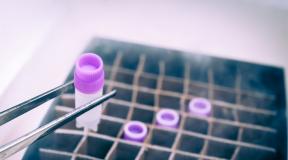
Check the integrity of the catheter cannula. If there is a thrombus or suspicion of infection of the catheter, the tip of the cannula cut the sterile scissors, put in a sterile test tube and direct to the bacteriological laboratory for research (as a doctor's appointment).
Note in the documentation time, date and cause of the removal of the catheter.
Dispose of waste in accordance with the safety regulations and the sanitary and epidemiological regime.
Complications in parenteral administration of drugs
Technique of any manipulation, including parenteral administration medicines It must be clearly observed, since the effectiveness of medical care depends on the quality of the manipulation. Most of the complications after parenteral administrations arise as a result of not fulfilling the necessary requirements for compliance with aseptic, methods for conducting manipulations, preparation of the patient to manipulation, etc. Exceptions constitute allergic reactions to the administered drug.
Infiltrate
Infiltrate is the local response of the body associated with limited irritation or fabric damage.
Infiltrate, the most common complication after subcutaneous and intramuscular injection, occurs when performing a stupid needle, using short needles with intramuscular injection, improperly determining the injection site, the injection in the same place.
Infiltrate is characterized by the formation of the seal at the injection site, which is easily determined during palpation (feeling).
For infiltrate characteristic local signs Inflammation:
hyperemia;
swelling;
palpation pain;
local temperature rise.
When the infiltrate occurs, local warming compresses in the shoulder area and the heater to the area of \u200b\u200bthe buttocks.
Abscess
In case of violation of aseptics during injection, patients develop abscess - purulent inflammation of soft tissues with the formation of a cavity filled with pus.
The cause of injecting and post-adjusting abscesses is the insufficient treatment of the hands of a medical worker, the treatment of syringes, needles, the skin of patients at the injection site.
The appearance of an abscess of the patient's aggressive state is considered one of the most serious violations.
The clinical picture of the abscess is characterized by general and local signs.
Total features include:
fever at the beginning of the disease constant, and later a touching type;
pulse care;
intoxication.
To local features include:
redness, swelling at the injection site;
temperature increase;
palpation soreness;
symptom of fluctuations over the softening center.
Drug embolism
Drug embolism can occur with the injections of oil solutions subcutaneously or intramuscularly. Oil, being in the artery, borders it, and this leads to a violation of nutrition of surrounding tissues, their necrosis.
Signs of necrosis:
amplifying pain in the field of injection;
redness or red-shine skin staining;
increase body temperature.
When oil gets into a vein, it falls into pulmonary vessels.
Symptoms of emblem of pulmonary vessels:
sudden attack of suffocation;
cough ;
cyanosis of the upper half of the body;
feeling confrontation in the chest.
Necrosis(tissue homes)
Tissue necrosis develops with unsuccessful venopunction or erroneous administration under the skin of a significant amount of strongly irritating drug. Most often it happens with inept intravenous administration 10% calcium chloride solution. When the veins and the expiration of the medicinal substance in the tissue around the vessel, hematoma, swelling, pain in the injection site are observed.
Thrombophlebit
Thrombophlebitis is acute inflammation of blood vessels, accompanied by the formation of infected thrombus.
The process begins in the lumen of the inflamed venous wall and applies to the periphery with the involvement of the surrounding tissues, causing the formation of a thrombus fixed on the vein wall.
In case of inspection in the affected place, a clearly bounded tumor is determined in the form of a serpentuous vessel. The skin shine slightly. The tumor is well moving towards the tissues to be tissues, but the skin with the skin. There is a local temperature increase, but the soreness is small and does not prevent the function of the limb.
Hematoma
Hematoma - hemorrhage under the skin with intravenous injection.
The cause of hematoma is ineptly venopunction. At the same time, the bugger spot appears, the bloody vein at the injection site from the puncture of both walls of veins and the washing blood penetrated into the tissue.
Anaphylactic shock
Anaphylactic shock develops with the introduction of antibiotics, vaccines, healing sera. The development time of anaphylactic shock is from a few seconds or minutes from the moment of administration of the drug. The faster the shock develops, the worse the forecast. The lightning current of shock ends fascinated.
Most often, anaphylactic shock is characterized by the following sequence of signs:
reduced blood pressure, heartbeat, arrhythmia.
overall redness of the skin, rash;
cough attacks;
severe concern;
breath rhythm violation;
Symptoms can manifest in various combinations. Death comes from acute respiratory failure due to bronchospasm and pulmonary edema, acute cardiovascular failure.
The development of a patient of an allergic reaction to the introduction of a drug requires emergency assistance.
Allergic reactions
Allergic reactions include:
local allergic reaction,
hives,
swelling quinque,
Local allergic reaction can develop as an answer to subcutaneous or intramuscular injection. A local allergic reaction is expressed by the tissue seal at the injection site, hyperemia, swelling, but necrotic changes of tissues in the injection area may occur. There are general signs such as headache, dizziness, weakness, chills, increase body temperature.
Hives
It is characterized by an edema of the papillary layer of the skin, which manifests itself in the form of rashes on the skin of itching blisters. The skin around the blisters is hyperemic. The rashes of blisters are accompanied by a pronounced itch. Rales can spread throughout the patient's body. There is chills, an increase in the body temperature of the patient, insomnia. The urticaria may occur as a response to entering the body of various allergens (drugs, cosmetics, food products).
Sweet Qincke
Agrio-erect edema with the propagation of skin, subcutaneous tissue and mucous membranes. Sweet is dense, pale, itching is not marked. Most often, the swelling captures the eyelids, lips, the mucous membranes of the oral cavity, can spread to the larynx, cause a suffocation. In this case, the peeing cough appears, witness voice, difficultness of both inhalation and exhalation, shortness of breath. With further progression, breathing becomes stridorous. Death can come from asphication. When localizing edema on the mucous membrane of the gastrointestinal tract, severe abdominal pains that stimulate the clinic of acute abdomen may occur. In engaging in the process of brain shells, meningial symptoms, inhibition, the rigidity of the occipital muscles, headache, convulsions appear.
Damage to nerve trunks
Damage to the nerve trunks occurs during intramuscular and intravenous injections or mechanically when the injection site is incorrect: chemically, when the drug depot is near the nerve. The severity of the complication may be different - from the neuritis (inflammation of the nerve) to paralysis (loss of the limb function). The patient is prescribed thermal procedures.
Sepsis
Sepsis is one of the complications arising from gross violations of the aseptic rules during intravenous injection, as well as using non-sterile solutions with intravenous inflows.
Whey hepatitis. HIV infection.
Remote complications arising from non-compliance with anti-epidemic and sanitary and hygienic measures in manipulation include serum hepatitis - hepatitis B and C, as well as HIV - infection, incubation period which ranges from 6-12 weeks to several months.
Treatment of these complications is carried out in specialized medical institutions.
Survey of surgical patients. Preparation of patients with radiological and instrumental research
Preparation of patients
endoscopic research
In the surgical clinic, one of the most common instrumental methods of diagnostics are endoscopic studies, which consist in visual inspection (sometimes accompanied by manipulations) of hollows internal organs and cavities with tools equipped with an optical system. Schematically, any endoscope is a hollow tube with a light bulb, which is injected into the clearance of the underlying organ or cavity. The design of the corresponding endoscope, of course, depends on the shape, magnitude, the depth of the occurrence of one or another organ. Diagnostic and therapeutic endoscopy, depending on the degree of invasiveness, is carried out in specialized cabinets, as well as in the operating or dressing.
Laryngoscopy(Large Inspection) spends most often anesthesiologist. This manipulation is one of the first stages of endotracheal anesthesia (the tube in the trachea is administered under the control of the laryngoscope). Folangologists enjoy laryngoscopy. Usually, these methods are owned by surgeons and sisters - anesthetics.
Bronchoscopy. It is made with the diagnostic (in these cases through the bronchoscope, the mucous tracheobronchial tree is examined up to subsegimentary bronchi, and biopsy is conducted) and therapeutic (evacuation of the secretion of tracheobronchial wood, toilet it, the introduction of medicinal substances, removal foreign languages) Goals.
Ezophagoscopy(inspection of the esophagus), gastroscopy. (inspection of the stomach) and duodenoscopy(Inspection of the duodenum) are produced to verify the diagnosis visually or using biopsy, as well as for the purpose of therapeutic procedures (the removal of foreign bodies, stop the bleeding, removal of polyps, installation of endoprostheses). Since B. clinical practice The inspection of the esophagus, the stomach and duodenum of the ocean flexible fibroscope is also most often carried out, usually use the term fibroezophagastrododenoscopy (FEGDS).
While doing rectorOnososcopya rigid or flexible endoscope is inspected direct and sigmid gut with diagnostic I. medical goals (To remove polyps, coagulation of ulcers, cracks, biopsy, etc.). For total inspection, the colon is carried out colonoscopyflexible fibroscope.
In urological practice, routine research is cystoscopy(inspection of the mucous membrane of the urethra and bladder) with diagnostic and therapeutic goals. In gynecological departments, an endoscopic examination of the uterine cavity is produced - hysteroscopy.In the pathology of large joints, one of therapeutic and diagnostic methods is arthroscopy.
For inspection of the abdominal and pleural cavity Produced respectively laparoscopyand thoracoscopy.It should be emphasized once again that in a large percentage of cases, all endoscopic procedures are not only diagnostic, but also therapeutic. Currently, the development of endoscopic technologies has led to the creation of laparoscopic, arthroscopic surgery.
Most endoscopic manipulations in complexity and tolerance can be compared with operations, the success of which largely depends on the proper preparation, since the hollow organs through which the endoscope passes and which are subject to inspection should be as free of content.In addition, on the entire path of the endoscope, the musculature should be relaxed, and the pain areas are anesthetized.
The attending physician, assigning a patient endoscopy under local anesthesia, in a preliminary conversation shows him a posture in which a study is committed. These poses are very different even with the same form of endoscopy and depend on a number of reasons, including the anesthesia. Naturally, under the anesthesia procedures produced in a lying position of the patient. Ladies inspection, respiratory tract, esophagus and stomach are carried out either under anesthesia, or under local anesthesia, which consists in irrigating the mucous membranes of 10% aerosol of lidocaine. The specified procedures make an empty stomach. 30 minutes before laryngo, bronchoscopy, laparo- and thoracoscopy are premedicated: atropine, narcotic analgesic. These studies are produced in a special endoscopic office, in dressing or in the operating room, where the patient will be taken on a catal (it is necessary to remove the dentures). Lapro- and thoracoscopy are, in fact, operational intervention and require the same training as an extensive operation.
Before recooked cystoscopy, you can resolve the patient to drink a glass of sweet tea. Cistoscopy often does not require other preparation, except for good intestinal cleansing. For reccroscopy, the patient is prepared for several days: limit carbohydrates in food, they put daily cleansing enemas in the morning, in the evening and, moreover, early in the morning on the day of the study, which is sent to the patient. For a full-fledged and more comfortable patient, a colonoscopy requires adequate preparation of the colon. Optimal (with the exception of patients with the stenosis tumors of the colon) is the use of forttrans (macrogol) - the laxative means most effectively exempting the thick intestine from kALOV MASS.. The action of macrogol is due to the formation of hydrogen bonds with water molecules and withholding it in the intestine. Water dilutes the contents of the intestine and increases its volume, reinforcing peristaltics and thereby providing a laxative effect. The drug is completely evacuated from the intestine along with its contents. Fortrans is not absorbed in the intestine and is not metabolized in the body, is excreted unchanged. The preparation of the colon using Fortrans is carried out as follows.In the morning, a day before the study, the patient takes a light breakfast. Subsequently, the patient does not dine and does not dinner (only sweet tea). Owl the patient prepares 3 liters of cool boiled water and dissolves in it 4 Ford Package. The solution is taken by portions of 100ml with such a calculation so that 100-200 ml of the solution remains in the evening. This portion of the solution is taking the patient in the morning on the day of study with such a calculation so that the reception of the drug is finished 3 hours before the procedure. A light breakfast is allowed.
It is not recommended to prepare patients with colonoscopy with vaseline oil as a laxative, since the oil, falling on the endoscope optics, causes it to be stitched and worsens the quality of inspection. It should be remembered that after cysto - and rectoscopy patients may experience pain, unpleasant sensations when urinating and defecation, and sometimes there is a blood admixture in the urine and feces. In these cases, painfully relieves candles with anesthesia, belladonnaya.
Some are different preparation of patients with emergency endoscopic studies. So, when conducting an emergency FEGDS for gastroduodenal bleeding, the most rapid release of the stomach from the blood, dietary masses is required. To this end, it makes the installation of a thick gastric probe and wash the stomach icewater (hemostasis means) until it removes liquid blood and its clots. Water in the probe is introduced by a syringe of Jean, the evacuation of water from the stomach occurs in a samoter or when creating a minor discharge with a syringe. To effectively prepare the stomach in this situation requires at least 5-10 liters of water.
For emergency colonoscopy, laxatives are not used because of the expectation of the effect. After their reception, there are several cleaning enema for the preparation of the colon, and with their ineffectiveness, the siphon enema before the disheaval of carts and gases in significant quantities.
Preparation of patients
to radiological studies
A frequently used study method in the surgical clinic is to perform x-ray or radiography. In some cases (X-ray chest) No special preparation is required, and the informationality of the study depends on the proper preparation of the patient.
Careful preparation is needed for the x-ray study of the gastrointestinal tract. For 2-3 days it is necessary to exclude black bread, cereal, vegetables, fruits, milk to limit the formation of slags and gases; With the same purpose, the sick-suffering delay in intestinal gases should be prescribed activated carbon or espeamizan, to do enemas from chamomile in the morning and in the evening, give drinking a warm chamomile infusion (1 tablespoon of chamomile on a glass of hot water) 1 tablespoon 4-5 times in day. In no case in order to the x-ray study of the gastrointestinal tract, it is impossible to use saline laxatives, since they enhance the accumulation of gases in the intestine and irritate the intestinal wall. In the evening, on the eve of the study, they put a cleansing enema, and in a number of institutions, another enema is necessarily in the morning, but not less than 3 hours before x-ray.
The study of the upper departments of the gastrointestinal tract is performed on an empty stomach. Having received a light dinner in the evening, he does not eat sick in the morning, does not drink, does not take any medicines, does not smoke. Even the slightest pieces of food and several sips of the liquid prevents the uniform distribution of the contrasting suspension on the walls of the stomach, they interfere with its filling, and nicotine enhances the secretion of the gastric juice, excites the peristaltics of the stomach. In patients with disturbed evacuation from the stomach, before shipping to X-ray Cabinet the stomach empty (but not washed!) Tolstish probe. A full-fledged study can only be produced if the stomach is empty.
Preparation for the study of a large intestine by irrigoscopy (the introduction of a contrast agent directly into the intestines) is slightly different from the preparation of the colonoscopy described above. For 2-3 days, the patient gives a semi-liquid, not irritant intestine and easily absorbed food. At 6 o'clock in the morning, a day of study put another cleansing enema, in addition, a light breakfast is allowed: tea, egg, white Sukharik with butter. If the patient suffers from constipation, it is advisable to prepare it with siphon enemas or intake castor Oil (OL. ricini. 30 g., per. oS.), not salting laxatives. Possible a variant of the preparation of the colon using Fortrans. In preparation for the X-ray study of the large intestine, the appointments of antispasmodics or prokinetics are canceled, since these drugs, acting on muscle elements of the intestinal wall, can change the relief of the mucous membrane.
A contrast agent that makes it possible to visualize the lumen of the digestive tube, usually introduced in an X-ray office. In the study of the upper departments of the gastrointestinal tract, the patient gives the barium suspension of various consistency, separating the bary powder with the corresponding amount of water, and in the study of the thick bowel, it is introduced into the enema. In addition, there are ways to study providing for pre-taking contrast substances inside. So, sometimes the patient in the department (it is necessary to clarify the time of the summer of the contrast substance) give a drink barium suspension (in each case it is important to know how many grams of barium and in which volume of water it is necessary to dilute), and on the other day at a certain time they send it to X-ray Cabinet: By this time, the barium suspension should fill out the studied intestinal departments. So investigate the ileocecal angle of the intestine or place the place of obstacles in intestinal obstruction. Usually after the study, the doctor - the radiologist speaks the patient if he needs to come again on the same day or tomorrow. In some cases, the patient is warned that it is still a time for the time (for example, when the evacuation delayed from the stomach or duodenum) or abstained from defecation (in the study of the colon) and again came at a certain hour to the X-ray office. Sometimes a radiologist asks for a patient to lie down in a specific pose (for example, on the right side).
Urinary Training Study (Urography)includes a survey (without contrast) urography, excretory or excretory (intravenously introduced a contrast agent, which is highlighted by the kidneys and makes visible urinary Ways: kidneys with lochanks and cups, ureters and bladder), as well as retrograde (the contrast drug is administered through the catheter directly into ureters or even in the renal pelvis in order to fill all urinary system - From the kidney to the bladder inclusive).
Urography requires careful intestinal training (cleansing enema in the evening and early in the morning) so that the accumulations of gases and carts do not interfere with the identification of the stones of the urinary tract. In the morning on the day of the study, you can allow the patient to drink a glass of tea with a piece of white bread. Before studying the urinary tract, it is not necessary to force the patient to lie down, but on the contrary, to recommend to stroll. As before other radiological studies, the patient must purle. This is limited to the preparation for the overview urography, the task of which consists only in identifying the renal shadow (which can be approximately judged on the position or size of the kidneys) and large stones. With an excretory urography, an intravenously slow water-soluble contrast drug is introduced in an X-ray office. Intravenous administration of the drug carries out a procedural sister of the ward compartment. When conducting an emergency urography, in addition to the x-rayologist, a doctor should be near the patient, ready to assist in the event of a non-separation of an allergic reaction to a contrast agent. Usually, with intravenous administration of contrast, a patient feels weak pain or burning in the course of veins, sometimes bitterness in the mouth. These sensations quickly pass. It should be remembered that the random extravasive introduction of some contrast agents can lead to thrombophlebitis phenomena, fatty tissue necrosis.
For X-ray study, the skull preparation is not required (women should take out hairpins and hairpins). With a snapshot of the bones of the limbs, it should be removed from the skin of iodine, replace massive oil bandages with light aseptic, remove the strips of the sticky plaster. If there is a gypsum bandage, it is necessary to clarify the doctor if the snapshot in the bandage or should be removed. This is usually done in the presence of a doctor who, after an inspection, a short shot is solved by the question of further immobilization. It is necessary to assimilate well that the accompanying staff without special instructions of the doctor can not be removed the gypsum bandage, to give the limbs necessary for a picture, to carry the patient, without fixing the limb. These rules are of particular importance for traumatological or orthopedic patients, but they should be aware of the personnel careing for sick surgical departments, where interventions on the bones and joints are sometimes produced. For a snapshot of the shoulder belt (blade, clavicle), sternum, ribs, cervical and breast spines of special preparation is not required. On the contrary, for high-quality x-ray studies of the lumbling spine, a preliminary exemption of the intestines is required, so the enemas and the restriction of the food regime on the eve of the study are necessary.
Care rules for the central venous catheter
Intravenous injections
The final stage of manipulation.
22. Drain the face of the child with a napkin.
23. Used probe, syringe Jean (funnel), apron to be disappeared into the appropriate tanks with a disinfectant solution.
24. Remove gloves, disinfect them. Wash and dry hands, treat cream at extremely important
1) disorders of water and electrolyte metabolism;
2) perforation of the esophagus and stomach in poisoning with alkalis and corrosion poisons;
3) infection.
Appendix 4.
to instruction manual
medical and diagnostic procedures
and manipulations on the disciplines "Nursing in Pediatrics", "Pediatrics" by specialties 2-79 01 31 "Nursing", 2-79 01 01 "Therapeutic case"
Care for the central venous catheter (FAV)
Indications for the use of central veins: 1) the very importance of conducting long-term infusion therapy; 2) the introduction of vasoactive and irritating peripheral veins of substances; 3) for a rapid volume infusion of solutions; 4) conducting hemisorption and plasmaresis; 5) in the absence of venous access on the periphery; 6) monitor observation of pressure in the cavities of the heart; 7) rational, "without pain", blood collection for analysis.
General. The catheterization of the central vein is carried out by the doctor. A procedural nurse is responsible for the preparation of the workplace. "Prepares the patient to the procedure, helps the doctor getting into sterile overalls, assisters it when performing catheterization. After the procedure lays the child on the back without a pillow with turned on the side of the head (prevention of aspiration of the vomit mass). Controls its drinking mode: it allows you to drink no earlier than after 2 hours, there is 4 hours after catheterization. Conducts continuous monitoring arterial pressure, cardiac frequency, respiratory frequency. Provides care for the central venous catheter.
For the prevention of purulent complications, the rules of asepsis and antiseptics should be followed, at least 1 time in 3 days, with extremely importantness more often, to change the fixing bandage with the processing of the puncture hole and the skin around it with an antiseptic agent; Wrapping a sterile napkin space of a catheter with a system for intravenous drip injections, and after infusion - the free end of the catheter. Multiple touch to the element of the infusion system should be avoided, minimizing access inside it. To replace infusion systems for intravenous infusion of solutions, antibiotics daily, replacement of tees and conductors - once every two days (for patients with cytopenic state - daily). The use of a sterile fixing bandage ensures protection against the penetration of infection on the outer surface of the catheter.
In order to prevent the thrombing of the catheter, the blood clots are preferable to use catheters with anticoagulant coating. If the catheter is tombed, it is unacceptable to rinse it to remove the thrombus.
To prevent bleeding from the catheter, it is necessary to tightly close the plug, tightly fix it with a gauze cap, constantly monitor the position of the plug.
In order to prevent the air embolism, it is imperative to use catheters with a lumen diameter of less than 1 mm. Manipulations that are accompanied by disconnecting and attachment of syringes (droppers), it is preferable to carry out in exhale, to pre-block the catheter with a special plastic clamp, and if there is a tee to overlap its corresponding channel. Before connecting a new highway, make sure that it is completely filled with a solution. It is preferable to use small highways (the likelihood of an air embolism decreases).
To prevent spontaneous removal and migration to use only standard catheters with pavilions for needles, the catheter is fixed using a leucoplasty (special locking dressing). Before infusion, check the position of the catheter in Vienna with a syringe. Do not use scissors to remove the leucoplasty, as the catheter may be randomly cut off and migrate into the circulatory system.
Equipment of the workplace:1) a bottle with a filled system for intravenous drip injections of a single application, a tripod; 2) 5 ml heparin vial with 1 ml activity - 5000 units, ampoule (bottle) with sodium chloride solution 0.9% - 100 ml; 3) syringes with a capacity of 5 ml, injecting needles of single use; 4) sterile stubs for the catheter; 5) Sterile material (cotton balls, gauze triangles, napkins, peel rooms) in bixes or packages; 6) tray for sterile material; 7) tray for used material; 8) Hacks in the package; 9) sterile tweezers; 10) tweezers in a disinfectant solution; 11) Pilka, scissors; 12) Dispenser capacity with an antiseptic tool for treating patient's skin and staff hand; 13) Capacity with disinfectant for processing ampoules and other medicinal injection Forms; 14) the plaster (ordinary or type "tometriers") or another fixing bandage; 15) mask, medical gloves (single use), waterproof disinfeced apron, safety glasses (plastic screen); 16) tweezers for working with used tools; 17) tanks with a disinfectant to disinfection of surfaces, washing the used needles, syringes (systems), soaking used syringes (systems), soaking the needles used, disinfection of cotton balls, gauze wipes used; 18) pure rag; 19) instrumental table.
Care for the central venous catheter (FAV)
Indications for the use of central veins: 1) the need for long-term infusion therapy; 2) the introduction of vasoactive and irritating peripheral veins of substances; 3) for a rapid volume infusion of solutions; 4) conducting hemisorption and plasmaresis; 5) in the absence of venous access to the periphery; 6) monitor observation of pressure in the cavities of the heart; 7) rational, "without pain", blood collection for analysis.
General. The catheterization of the central vein is carried out by the doctor. The procedural nurse is responsible for the preparation of the workplace, prepares a patient to the procedure, helps the doctor getting into sterile workwear, assisters it when performing catheterization. After the procedure lays the child on the back without a pillow with turned on the side of the head (prevention of aspiration of the vomit mass). Controls its drinking mode: it allows you to drink no earlier than after 2 hours, there is 4 hours after catheterization. Conducts continuous observation of arterial pressure, heart rate, respiratory frequency. Provides care for the central venous catheter.
Care rules for the central venous catheter
For the prevention of purulent complications, the rules of asepsis and antiseptics should be followed, at least 1 time in 3 days, if necessary, to change the fixing bandage with the processing of the puncture hole and the skin around it with an antiseptic agent; Wrap a sterile napkin space of a catheter compound with a system for intravenous drip injections, and after infusion is the free end of the catheter. Multiple touch to the element of the infusion system should be avoided, minimizing access inside it. To replace infusion systems for intravenous infusion of solutions, antibiotics daily, replacement of tees and conductors - once every two days (for patients with cytopenic state - daily). The use of a sterile fixing bandage ensures protection against the penetration of infection on the outer surface of the catheter.
In order to prevent the thrombing of the catheter, the blood clots are preferable to use catheters with anticoagulant coating. If the catheter is tombed, it is unacceptable to rinse it to remove the thrombus.
To prevent bleeding from the catheter, it is necessary to tightly close the plug, tightly fix it with a gauze cap, constantly monitor the position of the plug.
In order to prevent an air embolism, it is necessary to use catheters with a lumen diameter of less than 1 mm. Manipulations that are accompanied by disconnecting and attaching syringes (droppers), it is preferable to carry out in exhalation, pre-block the catheter with a special plastic clamp, and if there is a tee, overlapping its corresponding channel. Before connecting a new highway, make sure that it is completely filled with a solution. It is preferable to use small highways (the likelihood of an air embolism decreases).
To prevent spontaneous removal and migration to use only standard catheters with pavilions for needles, the catheter is fixed using a leucoplasty (special locking dressing). Before infusion, check the position of the catheter in Vienna with a syringe. Do not use scissors to remove the leucoplasty, as the catheter may be randomly cut off and migrate into the circulatory system.
Equipment of the workplace:1) a bottle with a filled system for intravenous drip injections of a single application, a tripod; 2) 5 ml heparin vial with 1 ml activity - 5000 units, ampoule (bottle) with sodium chloride solution 0.9% - 100 ml; 3) syringes with a capacity of 5 ml, injecting needles of single use; 4) sterile stubs for the catheter; 5) sterile material (cotton balls, gauze triangles, napkins, diapers) in bixes or packages; 6) tray for sterile material; 7) tray for used material; 8) Hacks in the package; 9) sterile tweezers; 10) tweezers in a disinfectant solution; 11) Pilka, scissors; 12) Dispenser capacity with an antiseptic tool for treating patient's skin and staff hand; 13) a container with a disinfectant for processing ampoules and other medicinal injection forms; 14) the plaster (ordinary or type "tometriers") or another fixing bandage; 15) mask, medical gloves (single use), waterproof disinfeced apron, safety glasses (plastic screen); 16) tweezers for working with used tools; 17) tanks with a disinfectant to disinfection of surfaces, washing the used needles, syringes (systems), soaking used syringes (systems), soaking the needles used, disinfection of cotton balls, gauze wipes used; 18) pure rag; 19) instrumental table.
Preparatory stage of manipulation.1.
3. Wash hands with flowing water, twice latch. Dry them with a single cloth (individual towel). Handle hands with an antiseptic agent.
4. Wear apron, mask, gloves.
5. Process with a disinfecting solution of the surface of the manipulation table, tray, apron, bix. Wash your hands in gloves with flowing water with soap, dry.
6. Put the necessary equipment on the instrument table.
7. To cover the sterile tray by laying on it everything you need. Another version of working with sterile material is possible when it is in packages.
Connect the infusion system to the Fair. 8. Process the bottle with isotonic solution of sodium chloride.
9. Dial 1 ml of solution into one syringe, in another -5 ml.
11. Pour the catheter with a plastic clamp. Pressing the catheter warns bleeding from the vessel and an air embolism.
12. Remove the "old" pear-shaped bandage with the cannula catheter.
13. Treat the cannoun catheter and a cork with an antiseptic, while holding the end of the catheter on a weight at some distance from the cannula.
14. Put the treated part of the catheter on a sterile diaper, placing it on the baby's chest.
15. Process hands in gloves with an antiseptic agent.
16. Remove the cork with the cannula and throw away. If there are no additional sterile corks, then put it in an individual container with alcohol(Used once).
17. Attach the syringe with sodium chloride solution 0.9%,open clamp on the catheter, extract the contents of the catheter.
18. Using another syringe, rinse catheter Sodium chloride solution 0.9%in an amount of 5-10 ml.
To avoid air embolism and bleeding, you should pinch the catheter with a plastic clamp every time the syringe, system, corks are disconnected from it.
19. Attach the system for intravenous drip infusion to the cannula of the catheter "jet in a jet".
20. Adjust the speed of droplets.
21. Wrap a sterile napkin space of a catheter with a system.
Disconnecting the infusion system from the Fair. Heparin "Castle". 22. Check stickers on bottles with heparinand sodium chloride solution 0.9%(drug title, quantity, concentration).
23. Prepare bottles to perform manipulation.
24. Dial 1 ml of heparin in the syringe. Introduce 1 ml of heparin into the vial with a solution of sodium chloride 0.9% (100 ml).
25. Dial 2 - 3 ml of the resulting solution into the syringe.
26. Overlapping the dropper, to overcert the catheter with a plastic clamp.
27. Remove the gauze napkin, covering the jack of cannula catheter from the cannula system. Ship the catheter to another sterile napkin (diaper) or on the inner surface of any sterile packaging.
28. Process hands with antiseptic solution.
29. Disconnect the dropper and connect the syringe with diluted heparin to the cannula, remove the clamp and introduce 1.5 ml of the solution into the catheter.
30. Pour the catheter with a plastic clamp, disconnect the syringe.
31. Treat the cannula catheter ethyl alcohol,to remove from its surface traces of blood, another protein preparation, glucose.
32. On a sterile napkin with a sterile tweezers, lay a sterile cork and close it cannula catheter.
33. Wrap the cannuel of the catheter with a sterile marlevary napkin and secure the rubber ring or adhesive plate.
Change of dressings fixing the Fair. 34. Remove the former locking bandage.
35. Process hands in gloves with antiseptic solution (put on sterile gloves).
36. Treat the skin around the place of introduction of the catheter first 70% alcohol,then antiseptic yodobak (Betadinetc.) in the direction from the center to the periphery.
37. To cover the sterile napkin, withstand the exposition of 3-5 minutes.
38. Sleep sterile napkin.
39. To put a sterile bandage onto the entrance of the catheter.
40. To fix the bandage with the patching "TM." ("Mefix", etc.), fully covering sterile material.
41. Specify on the upper layer of the plastering date of the imposition of the bandage.
Note. If the inflammatory process occurs around the place of introduction of the catheter (redness, compaction) after consultation with the attending physician, it is advisable to use ointments (Betadine, Povidan,ointment S. antibiotics).In this case, the change of the dressing is made daily, and on the plaster, except the date, is indicated - "ointment".
42. Conduct disinfection of used medical instruments, catheters, infusion systems, apron in appropriate tanks with disinfectant solution. Process with disinfecting solution surfaces. Remove gloves and disinfect them. Wash hands under running water with soap, dry, treat with cream.
43. Fight a security guard to a child.
44. Record in medical documentation With the date, the time of infusion, the solution used, its number.
Possible complications: 1) purulent complications (suppuration of the puncture channel, thrombophlebitis, phlegmon, sepsis); 2) blood clots of the catheter; 3) bleeding from the catheter; 4) air embolism, thromboembolism; 5) spontaneous removal and migration of the catheter; 6) sclerosation of central veins in case of frequent change of catheter; 7) infiltration; 8) Allergic reaction to medications and etc.
Puncture and catheterization of peripheral veins
General. The use of peripheral venous catheter (PVC) gives the possibility of long-term infusion therapy, makes a painless catheterization procedure, reduces the frequency of psychological injuries associated with numerous punctures of peripheral veins. The catheter can be introduced into surface veins of the heads, upper and lower extremities.
The duration of operation of one catheter is 3-4 days. Patients receiving long-term treatment, vein catheterization by peripheral catheter it is advisable to start a brush or foot. In this case, with their obliteration, it is possible to use a higher vein spaced. When operating the peripheral venous catheter, the rules of aseps and antiseptics should be strictly observed. Catenter connectivity with a system for intravenous drip infusion, connector, cork thoroughly clean from blood residues, cover with a sterile napkin. Control the state of veins and skin in the field of puncture. To prevent bleeding from the catheter, the air embolism firmly fix the cork on the cannula of the catheter, press the vein to the top of the catheter every time before removing the cork, the system shutdown, the syringe. If a connector (conductor) with a tee is attached to the catheter, overlapping the appropriate tee channel. To avoid thrombing of the catheter with a blood clock, temporarily not used for infusion catheter, it is necessary to fill in a heparin solution (see PP. 20-31 "Care for the central venous catheter"). To prevent outdoor migration of the catheter with the formation of subcutaneous hematoma or (and) paravasant administration of the medicinal substance, constantly monitor the reliability of the catheter fixation, check its position in Vienna with a syringe. When setting a catheter in the joint area to use Longetu.
Equipment of the workplace: 1) the bottle (ampoule) with a solution of sodium chloride 0.9%; 2) peripheral venous catheter, cork for catheter; 3) syringes with a capacity of 5 ml, injection needles one-time; 4) sterile material (cotton balls, gauze napkins, diapers) in bixes or packages; 5) tray for sterile material; 6) tray for used material; 7) Hacks in packages; 8) sterile tweezers; 9) tweezers in a disinfectant solution; 10) Pilochka, scissors; 11) harness; 12) Dispenser capacity with an antiseptic tool for treating patient's skin and staff hand; 13) a container with a disinfectant solution for the processing of ampoules and other drug injection forms; 14) the plaster (ordinary or type "tometriers") or another fixing bandage; 15) mask, medical gloves (single use), waterproof apron, safety glasses (plastic screen); 16) instrumental table; 17) tweezers for working with used tools; 18) a container with a disinfectant for disinfection of surfaces, washing used syringes (systems), soaking used syringes (systems), soaking the needles used, disinfection of cotton and gauze balls used; 19) pure rag.
Preparatory stage of manipulation. 1. Inform the patient (close relatives) about the need to fulfill and the essence of the procedure.
2. To obtain the consent of the patient (close relatives) to perform the procedure.
3. Wash hands with flowing water, twice latch. Dry them with a single cloth (individual towel). Handle hands with an antiseptic agent.
4. To put on the apron, mask, gloves.
5. Treat the surface of the manipulation table, tray, apron, bix to disinfecting solution. Wash hands in gloves with flowing water with soap, dry, handle with an antiseptic agent.
6. Put the necessary equipment on the instrument table. Check the shelf life, packaging integrity.
7. To cover the sterile tray by laying on it everything you need. Another version of working with sterile material is possible when it is in packages.
8. Process the bottle with sodium chloride solution 0.9%.
9. Dial in syringe 5 ml of solution.
10. Wear protective glasses (plastic screen).
The main stage of the execution of manipulation. 11. To impose harness above the alleged location of the catheter. Children of early age it is better to use the finger pressed veins (performed by a nurse-assistant). 12. Processing the antiseptic agent in the veins of the rear of the brush or the inner surface of the forearm of the child (two balls, wide and narrow).
13. Proceed with an antiseptic means of hand.
14. Take the catheter in hand with three fingers and, pulling the skin in the vein area by another hand, punctured it at an angle of 15-20.
15. When blood appears in an indicator chamber, slightly pull over the needle, at the same time pushing the catheter in Vienna.
16. Remove harness.
17. Press the vein to the top of the catheter (through the skin), extract the needle completely.
18. Connect the syringe to the catheter with an isotonic solution of sodium chloride, rinse with a catheter with a solution.
19. In the same way, pressing the vein with one hand, to disconnect the syringe and close the catheter with a sterile cork.
20. Clean the outer part of the catheter and the skin under it from blood traces.
21. Fix the catheter with the plaster.
22. Wrap the cannula of the catheter with a sterile marlevary cloth, consolidate its adhesive plaster, bandage.
23. To transfer the child to the Chamber, connect the dropper (syringe pump). If, in the near future, intravenous infusions through the peripheral venous catheter will not be carried out, fill it with it with a solution of heparin (see paragraphs. 22-33 "Care for the central venous catheter").
Final stage of manipulation. 24. Conduct disinfection of the used medical instrument of catheters, infusion systems, an alarm in the appropriate tanks with a disinfectant solution. Process with disinfecting solution surfaces. Remove gloves and disinfect them. Wash hands under running water with soap, dry, treat with cream.
25. Ensure the guardianship to the child.
26. To record in medical records with the date, time of infusion, the solution used, its quantity.
Possible complications
Puncture of the vessel of the skull
Needle- "Butterfly" with a catheter
General.Children early age Medicinal substances can be administered to the surface veins of the heads. During the procedure, the child is fixed. His head holds a medical sister-assistant, hands to the torso and feet fixed with a diaper (sheet). In the presence of hair cover In the place of the alleged puncture, the hair shave.
Equipment of the workplace:1) Needle - "Butterfly" with a catheter of one-time application; 2) the bottle with a filled system for intravenous drip injections of a single application, a tripod; 3) ampoule (bottle) with a solution of sodium chloride 0.9%; 4) a syringe of one-time use of 5 ml, injection needles; 5) sterile material (cotton balls, gauze triangles, napkins, diapers) in packages or bixes; 6) tray for sterile material; 7) tray for used material; 8) Hacks in the package; 9) sterile tweezers; 10) tweezers in a disinfectant solution; 11) Pilka, scissors; 12) Dispenser capacity with an antiseptic tool for treating patient's skin and staff hand; 13) a container with a disinfectant solution for the processing of ampoules and other drug injection forms; 14) the plaster (ordinary or type "tometriers") or another fixing bandage; 15) medical gloves (one-time); Mask, safety glasses (plastic screen), waterproof disinfeced apron; 16) tweezers for working with used tools; 17) containers with a disinfectant for surface treatment, washing the needles used, syringes (systems), soaking the used syringes (systems), needles, disinfection of cotton balls and gauze wipes used; 18) pure rag; 19) instrumental table.
Preparatory stage of manipulation.1. Inform the patient (close relatives) about the need to fulfill and the essence of the procedure.
2. To obtain the consent of the patient (close relatives) to perform the procedure.
3. Wash hands under running water, twice latch. Dry hands with a single cloth (individual towel). Handle hands with an antiseptic agent. Wear apron, gloves, mask.
4. Process with a disinfectant solution of the surface of the manipulation table, tray, apron, a system tripod. Wash hands in gloves under running water with soap, dry, handle with an antiseptic agent.
5. Put the necessary equipment on the instrument table.
6. Loose sterile tray.
7. Print packages with a butterfly catheter, syringes, lay out on the tray. Another option of working with sterile material is possible when it is in packages.
8. Process the ampoule (bottle) with sodium solution of chloride 0.9%.
9. Dial 2 ml in syringe connect to the catheter, fill it out and lay out on the tray.
10. Fix the child (performs a nurse-assistant). Put a sterile diaper next to the baby's head.
11. Wear protective glasses (plastic screen).
The main stage of the execution of manipulation.12. Select a vessel for puncture and process the injection place with two balls with an antiseptic (one - wide, different - narrowly) in the direction from the ground-made area. For better blood supply of veins, it is convenient to use a special elastic tape that overlapped around the head below the punctured area (above the eyebrows). Local finger relief of Vienna is ineffective due to the abundance of venous anastomoses of the skull of the skull. Child crying also contributes to the swelling of the head of the head.
13. Process hands in gloves with an antiseptic agent.
14. Stay the skin in the area of \u200b\u200bthe intended puncture to fix the veins.
15. Punify Vienna with a "butterfly" with a catheter in three stages . To do this, send the needle to the current of blood at a sharp angle to the skin surface and make it puncture. Then the needle to promote approximately 0.5 cm, pierce the vein and send it to its go. If the needle is not in Vienna, return it back, without pulling out of the skin, and re-puncture Vienna.
The introduction of the needle to the vessel immediately after the skin puncture can lead to the puncture of both walls of the vessel.
16. Pull the syringe piston connected to the catheter. The appearance of blood indicates the correct position of the needle. If an elastic ribbon is used to enhance the blenification of the vein, remove it.
17. Enter 1 - 1.5 ml sodium chloride solution 0.9%,to avoid blood clots of needle blood clots and eliminate the likelihood of extravasive administration of the medicinal preparation.
18. Fix the needle with three stripes of the leukoplower: 1st - across the needle to the skin. The 2nd "Wings" needles - "Butterflies" with a crossover over them and fixation to the skin, 3rd - across the wings of the needle-"butterfly" to the skin.
19. Collapse the rings catheter and fix the leukoplasty on the head of the head to avoid his offset.
20. If necessary, if the angle of the needle in relation to the bending of the skull is great, to put a gauze (cotton needle) for the cannula ball.
21. Pull the syringe piston connected to the catheter, to re-check the needle position in Vienna.
22. Disconnect the syringe, connect the dropper on the jet of the solution.
23. Adjust using a clamp the rate of administration of the drug substance.
24. Cover the sterile marlevary napkin the junction of the catheter and dropper.
The final stage of manipulation.25. After the infusion is completed, it takes a dropper tube. Carefully rejuvenate the leucoplasty from the skin. Put a ball with an antiseptic needle entrance place in Vienna. Extract the needle (catheter) together with the leunkoplasty.
26. To impose a sterile napkin on the place of puncture, from above - a gouring bandage.
27. Conduct disinfection of used medical instruments, catheters, infusion systems, apron in appropriate tanks with disinfectant solution. Process with disinfecting solution surfaces. Remove gloves and disinfect them. Wash hands under running water with soap, dry, treat with cream.
28. Favorite the guard of the child.
29. To record in medical records indicating the date, time of infusion, the applied solution, its number.
Possible complications: 1) purulent complications (suppuration of the puncture channel, thrombophlebitis, phlegmon, sepsis); 2) blood clots of the catheter; 3) bleeding from the catheter; 4) air embolism; 5) spontaneous removal and migration of the catheter; 6) sclerosis of veins in case of frequent change of catheter; 7) infiltration; 8) Allergic reaction to drugs, etc.
Appendix 5.
to instruction manual
medical and diagnostic procedures and manipulations on the disciplines "Nursing in Pediatrics", "Pediatrics" in the specialties 2-79 01 31 "Nursing", 2-79 01 01 "Therapeutic case"
For the prevention of purulent complications, the rules of asepsis and antiseptics should be followed, at least 1 time in 3 days, if necessary, to change the fixing bandage with the processing of the puncture hole and the skin around it with an antiseptic agent; Wrap a sterile napkin space of a catheter compound with a system for intravenous drip injections, and after infusion is the free end of the catheter. Multiple touch to the element of the infusion system should be avoided, minimizing access inside it. To replace infusion systems for intravenous infusion of solutions, antibiotics daily, replacement of tees and conductors - once every two days (for patients with cytopenic state - daily). The use of a sterile fixing bandage ensures protection against the penetration of infection on the outer surface of the catheter.
In order to prevent the thrombing of the catheter, the blood clots are preferable to use catheters with anticoagulant coating. If the catheter is tombed, it is unacceptable to rinse it to remove the thrombus.
To prevent bleeding from the catheter, it is necessary to tightly close the plug, tightly fix it with a gauze cap, constantly monitor the position of the plug.
In order to prevent an air embolism, it is necessary to use catheters with a lumen diameter of less than 1 mm. Manipulations that are accompanied by disconnecting and attaching syringes (droppers), it is preferable to carry out in exhalation, pre-block the catheter with a special plastic clamp, and if there is a tee, overlapping its corresponding channel. Before connecting a new highway, make sure that it is completely filled with a solution. It is preferable to use small highways (the likelihood of an air embolism decreases).
To prevent spontaneous removal and migration to use only standard catheters with pavilions for needles, the catheter is fixed using a leucoplasty (special locking dressing). Before infusion, check the position of the catheter in Vienna with a syringe. Do not use scissors to remove the leucoplasty, as the catheter may be randomly cut off and migrate into the circulatory system.
Equipment of the workplace:1) a bottle with a filled system for intravenous drip injections of a single application, a tripod; 2) 5 ml heparin vial with 1 ml activity - 5000 units, ampoule (bottle) with sodium chloride solution 0.9% - 100 ml; 3) syringes with a capacity of 5 ml, injecting needles of single use; 4) sterile stubs for the catheter; 5) sterile material (cotton balls, gauze triangles, napkins, diapers) in bixes or packages; 6) tray for sterile material; 7) tray for used material; 8) Hacks in the package; 9) sterile tweezers; 10) tweezers in a disinfectant solution; 11) Pilka, scissors; 12) Dispenser capacity with an antiseptic tool for treating patient's skin and staff hand; 13) a container with a disinfectant for processing ampoules and other medicinal injection forms; 14) the plaster (ordinary or type "tometriers") or another fixing bandage; 15) mask, medical gloves (single use), waterproof disinfeced apron, safety glasses (plastic screen); 16) tweezers for working with used tools; 17) tanks with a disinfectant to disinfection of surfaces, washing the used needles, syringes (systems), soaking used syringes (systems), soaking the needles used, disinfection of cotton balls, gauze wipes used; 18) pure rag; 19) instrumental table.
4. Wear apron, mask, gloves.
5. Process with a disinfecting solution of the surface of the manipulation table, tray, apron, bix. Wash your hands in gloves with flowing water with soap, dry.
6. Put the necessary equipment on the instrument table.
7. To cover the sterile tray by laying on it everything you need. Another version of working with sterile material is possible when it is in packages.
The main stage of the execution of manipulation. Connect the infusion system to the Fair. 8. Process the bottle with isotonic solution of sodium chloride.
9. Dial 1 ml of solution into one syringe, in another -5 ml.
11. Pour the catheter with a plastic clamp. Pressing the catheter warns bleeding from the vessel and an air embolism.
12. Remove the "old" pear-shaped bandage with the cannula catheter.
13. Treat the cannoun catheter and a cork with an antiseptic, while holding the end of the catheter on a weight at some distance from the cannula.
14. Put the treated part of the catheter on a sterile diaper, placing it on the baby's chest.
15. Process hands in gloves with an antiseptic agent.
16. Remove the cork with the cannula and throw away. If there are no additional sterile corks, then put it in an individual container with alcohol(Used once).
17. Attach the syringe with sodium chloride solution 0.9%,open clamp on the catheter, extract the contents of the catheter.
18. Using another syringe, rinse catheter in an amount of 5-10 ml.
To avoid air embolism and bleeding, you should pinch the catheter with a plastic clamp every time the syringe, system, corks are disconnected from it.
19. Attach the system for intravenous drip infusion to the cannula of the catheter "jet in a jet".
20. Adjust the speed of droplets.
21. Wrap a sterile napkin space of a catheter with a system.
Disconnecting the infusion system from the Fair. Heparin "Castle".22. Check stickers on bottles with heparinand sodium chloride solution 0.9%(drug title, quantity, concentration).
23. Prepare bottles to perform manipulation.
24. Dial 1 ml of heparin in the syringe. Introduce 1 ml of heparin into the vial with a solution of sodium chloride 0.9% (100 ml).
25. Dial 2 - 3 ml of the resulting solution into the syringe.
26. Overlapping the dropper, to overcert the catheter with a plastic clamp.
27. Remove the gauze napkin, covering the jack of cannula catheter from the cannula system. Ship the catheter to another sterile napkin (diaper) or on the inner surface of any sterile packaging.
28. Process hands with antiseptic solution.
29. Disconnect the dropper and connect the syringe with diluted heparin to the cannula, remove the clamp and introduce 1.5 ml of the solution into the catheter.
30. Pour the catheter with a plastic clamp, disconnect the syringe.
31. Treat the cannula catheter ethyl alcohol,to remove from its surface traces of blood, another protein preparation, glucose.
32. On a sterile napkin with a sterile tweezers, lay a sterile cork and close it cannula catheter.
33. Wrap the cannuel of the catheter with a sterile marlevary napkin and secure the rubber ring or adhesive plate.
Change of dressings fixing the Fair.34. Remove the former locking bandage.
35. Process hands in gloves with antiseptic solution (put on sterile gloves).
36. Treat the skin around the place of introduction of the catheter first 70% alcohol,then antiseptic yodobak (Betadinetc.) in the direction from the center to the periphery.
37. To cover the sterile napkin, withstand the exposition of 3-5 minutes.
38. Sleep sterile napkin.
39. To put a sterile bandage onto the entrance of the catheter.
40. To fix the bandage with the patching "TM." ("Mefix", etc.), fully covering sterile material.
41. Specify on the upper layer of the plastering date of the imposition of the bandage.
Note. If the inflammatory process occurs around the place of introduction of the catheter (redness, compaction) after consultation with the attending physician, it is advisable to use ointments (Betadine, Povidan,ointment S. antibiotics).In this case, the change of the dressing is made daily, and on the plaster, except the date, is indicated - "ointment".
42. Conduct disinfection of used medical instruments, catheters, infusion systems, apron in appropriate tanks with disinfectant solution. Process with disinfecting solution surfaces. Remove gloves and disinfect them. Wash hands under running water with soap, dry, treat with cream.
43. Fight a security guard to a child.
44. To record in medical records indicating the date, time of infusion, the solution used, its number.
Possible complications: 1) purulent complications (suppuration of the puncture channel, thrombophlebitis, phlegmon, sepsis); 2) blood clots of the catheter; 3) bleeding from the catheter; 4) air embolism, thromboembolism; 5) spontaneous removal and migration of the catheter; 6) sclerosation of central veins in case of frequent change of catheter; 7) infiltration; 8) Allergic reaction to drugs, etc.
Puncture and catheterization of peripheral veins
General. The use of peripheral venous catheter (PVC) gives the possibility of long-term infusion therapy, makes a painless catheterization procedure, reduces the frequency of psychological injuries associated with numerous punctures of peripheral veins. The catheter can be introduced into surface veins of the heads, upper and lower extremities.
The duration of operation of one catheter is 3-4 days. Patients receiving long-term treatment, vein catheterization by peripheral catheter it is advisable to start a brush or foot. In this case, with their obliteration, it is possible to use a higher vein spaced. When operating the peripheral venous catheter, the rules of aseps and antiseptics should be strictly observed. Catenter connectivity with a system for intravenous drip infusion, connector, cork thoroughly clean from blood residues, cover with a sterile napkin. Control the state of veins and skin in the field of puncture. To prevent bleeding from the catheter, the air embolism firmly fix the cork on the cannula of the catheter, press the vein to the top of the catheter every time before removing the cork, the system shutdown, the syringe. If a connector (conductor) with a tee is attached to the catheter, overlapping the appropriate tee channel. To avoid thrombing of the catheter with a blood clock, temporarily not used for infusion catheter, it is necessary to fill in a heparin solution (see PP. 20-31 "Care for the central venous catheter"). To prevent outdoor migration of the catheter with the formation of subcutaneous hematoma or (and) paravasant administration of the medicinal substance, constantly monitor the reliability of the catheter fixation, check its position in Vienna with a syringe. When setting a catheter in the joint area to use Longetu.
Equipment of the workplace: 1) the bottle (ampoule) with a solution of sodium chloride 0.9%; 2) peripheral venous catheter, cork for catheter; 3) syringes with a capacity of 5 ml, injecting needles of single use; 4) sterile material (cotton balls, gauze napkins, diapers) in bixes or packages; 5) tray for sterile material; 6) tray for used material; 7) Hacks in packages; 8) sterile tweezers; 9) tweezers in a disinfectant solution; 10) Pilochka, scissors; 11) harness; 12) Dispenser capacity with an antiseptic tool for treating patient's skin and staff hand; 13) a container with a disinfectant solution for the processing of ampoules and other drug injection forms; 14) the plaster (ordinary or type "tometriers") or another fixing bandage; 15) mask, medical gloves (single use), waterproof apron, safety glasses (plastic screen); 16) instrumental table; 17) tweezers for working with used tools; 18) a container with a disinfectant for disinfection of surfaces, washing used syringes (systems), soaking used syringes (systems), soaking the needles used, disinfection of cotton and gauze balls used; 19) pure rag.
Preparatory stage of manipulation. 1. Inform the patient (close relatives) about the need to fulfill and the essence of the procedure.
2. To obtain the consent of the patient (close relatives) to perform the procedure.
3. Wash hands with flowing water, twice latch. Dry them with a single cloth (individual towel). Handle hands with an antiseptic agent.
4. To put on the apron, mask, gloves.
5. Treat the surface of the manipulation table, tray, apron, bix to disinfecting solution. Wash hands in gloves with flowing water with soap, dry, handle with an antiseptic agent.
6. Put the necessary equipment on the instrument table. Check the shelf life, packaging integrity.
7. To cover the sterile tray by laying on it everything you need. Another version of working with sterile material is possible when it is in packages.
8. Process the bottle with sodium chloride solution 0.9%.
9. Dial in syringe 5 ml of solution.
10. Wear protective glasses (plastic screen).
The main stage of the execution of manipulation. 11. To impose harness above the alleged location of the catheter. Early children are better to use finger pressed Vienna (performed by a nurse-assistant). 12. Processing the antiseptic agent in the veins of the rear of the brush or the inner surface of the forearm of the child (two balls, wide and narrow).
13. Proceed with an antiseptic means of hand.
14. Take the catheter in hand with three fingers and, pulling the skin in the vein area by another hand, punctured it at an angle of 15-20.
15. When blood appears in an indicator chamber, slightly pull over the needle, at the same time pushing the catheter in Vienna.
16. Remove harness.
17. Press the vein to the top of the catheter (through the skin), extract the needle completely.
18. Connect the syringe to the catheter with an isotonic solution of sodium chloride, rinse with a catheter with a solution.
19. In the same way, pressing the vein with one hand, to disconnect the syringe and close the catheter with a sterile cork.
20. Clean the outer part of the catheter and the skin under it from blood traces.
21. Fix the catheter with the plaster.
22. Wrap the cannula of the catheter with a sterile marlevary cloth, consolidate its adhesive plaster, bandage.
23. To transfer the child to the Chamber, connect the dropper (syringe pump). If, in the near future, intravenous infusions through the peripheral venous catheter will not be carried out, fill it with it with a solution of heparin (see paragraphs. 22-33 "Care for the central venous catheter").
Final stage of manipulation. 24. Conduct disinfection of the used medical instrument of catheters, infusion systems, an alarm in the appropriate tanks with a disinfectant solution. Process with disinfecting solution surfaces. Remove gloves and disinfect them. Wash hands under running water with soap, dry, treat with cream.
25. Ensure the guardianship to the child.
26. To record in medical records with the date, time of infusion, the solution used, its quantity.
Possible complications
Puncture of the vessel of the skull
Needle- "Butterfly" with a catheter
General.Early children medicinal substances can be administered into surface veins of the heads. During the procedure, the child is fixed. His head holds a medical sister-assistant, hands to the torso and feet fixed with a diaper (sheet). If there is a hair cover in the place of the alleged puncture, the hair shakes.
Equipment of the workplace:1) Needle - "Butterfly" with a catheter of one-time application; 2) the bottle with a filled system for intravenous drip injections of a single application, a tripod; 3) ampoule (bottle) with a solution of sodium chloride 0.9%; 4) a syringe of one-time use of 5 ml, injection needles; 5) sterile material (cotton balls, gauze triangles, napkins, diapers) in packages or bixes; 6) tray for sterile material; 7) tray for used material; 8) Hacks in the package; 9) sterile tweezers; 10) tweezers in a disinfectant solution; 11) Pilka, scissors; 12) Dispenser capacity with an antiseptic tool for treating patient's skin and staff hand; 13) a container with a disinfectant solution for the processing of ampoules and other drug injection forms; 14) the plaster (ordinary or type "tometriers") or another fixing bandage; 15) medical gloves (one-time); Mask, safety glasses (plastic screen), waterproof disinfeced apron; 16) tweezers for working with used tools; 17) containers with a disinfectant for surface treatment, washing the needles used, syringes (systems), soaking the used syringes (systems), needles, disinfection of cotton balls and gauze wipes used; 18) pure rag; 19) instrumental table.
Preparatory stage of manipulation.1. Inform the patient (close relatives) about the need to fulfill and the essence of the procedure.
2. To obtain the consent of the patient (close relatives) to perform the procedure.
3. Wash hands under running water, twice latch. Dry hands with a single cloth (individual towel). Handle hands with an antiseptic agent. Wear apron, gloves, mask.
4. Process with a disinfectant solution of the surface of the manipulation table, tray, apron, a system tripod. Wash hands in gloves under running water with soap, dry, handle with an antiseptic agent.
5. Play the necessary equipment on the instrument table.
6. Loose sterile tray.
7. Print packages with a butterfly catheter, syringes, lay out on the tray. Another option of working with sterile material is possible when it is in packages.
8. Process the ampoule (bottle) with sodium solution of chloride 0.9%.
9. Dial 2 ml in syringe connect to the catheter, fill it out and lay out on the tray.
10. Fix the child (performs a nurse-assistant). Put a sterile diaper next to the baby's head.
11. Wear protective glasses (plastic screen).
12. Select a vessel for puncture and process the injection place with two balls with an antiseptic (one - wide, different - narrowly) in the direction from the ground-made area. For better blood supply of veins, it is convenient to use a special elastic tape that overlapped around the head below the punctured area (above the eyebrows). Local finger relief of Vienna is ineffective due to the abundance of venous anastomoses of the skull of the skull. Child crying also contributes to the swelling of the head of the head.
13. Process hands in gloves with an antiseptic agent.
14. Stay the skin in the area of \u200b\u200bthe intended puncture to fix the veins.
15. Punify Vienna with a "butterfly" with a catheter in three stages . To do this, send the needle to the current of blood at a sharp angle to the skin surface and make it puncture. Then the needle to promote approximately 0.5 cm, pierce the vein and send it to its go. If the needle is not in Vienna, return it back, without pulling out of the skin, and re-puncture Vienna.
The introduction of the needle to the vessel immediately after the skin puncture can lead to the puncture of both walls of the vessel.
16. Pull the syringe piston connected to the catheter. The appearance of blood indicates the correct position of the needle. If an elastic ribbon is used to enhance the blenification of the vein, remove it.
17. Enter 1 - 1.5 ml sodium chloride solution 0.9%,to avoid blood clots of needle blood clots and eliminate the likelihood of extravasive administration of the medicinal preparation.
18. Fix the needle with three stripes of the leukoplower: 1st - across the needle to the skin. The 2nd "Wings" needles - "Butterflies" with a crossover over them and fixation to the skin, 3rd - across the wings of the needle-"butterfly" to the skin.
19. Collapse the rings catheter and fix the leukoplasty on the head of the head to avoid his offset.
20. If necessary, if the angle of the needle in relation to the bending of the skull is great, to put a gauze (cotton needle) for the cannula ball.
21. Pull the syringe piston connected to the catheter, to re-check the needle position in Vienna.
22. Disconnect the syringe, connect the dropper on the jet of the solution.
23. Adjust using a clamp the rate of administration of the drug substance.
24. Cover the sterile marlevary napkin the junction of the catheter and dropper.
The final stage of manipulation.25. After the infusion is completed, it takes a dropper tube. Carefully rejuvenate the leucoplasty from the skin. Put a ball with an antiseptic needle entrance place in Vienna. Extract the needle (catheter) together with the leunkoplasty.
26. To impose a sterile napkin on the place of puncture, from above - a gouring bandage.
27. Conduct disinfection of used medical instruments, catheters, infusion systems, apron in appropriate tanks with disinfectant solution. Process with disinfecting solution surfaces. Remove gloves and disinfect them. Wash hands under running water with soap, dry, treat with cream.
28. Favorite the guard of the child.
29. To record in medical records indicating the date, time of infusion, the applied solution, its number.
Possible complications: 1) purulent complications (suppuration of the puncture channel, thrombophlebitis, phlegmon, sepsis); 2) blood clots of the catheter; 3) bleeding from the catheter; 4) air embolism; 5) spontaneous removal and migration of the catheter; 6) sclerosis of veins in case of frequent change of catheter; 7) infiltration; 8) Allergic reaction to drugs, etc.
Appendix 5.
to instruction manual
medical and diagnostic procedures and manipulations on the disciplines "Nursing in Pediatrics", "Pediatrics" in the specialties 2-79 01 31 "Nursing", 2-79 01 01 "Therapeutic case"
5. Immunoprophylaxis
General. Preventive vaccinations are effective tool Fighting children's infectious diseases. The vaccine preparations used contribute to the development of immunity, irrevocability to one or another infection.
Vaccinations are carried out in specially equipped vaccination offices of therapeutic and preventive institutions, medical offices of schools and other educational institutions. The vacuum office must have equipment for rendering emergency care. In order to avoid the inactivation of vaccination drugs throughout the manufacturer's institute, the cold chain must be observed before the advent of the vaccination.
Immediately before vaccination, the child should be examined by a doctor (paramedic). Without a written permission to vaccinate a nurse is not entitled to fulfill it. In the first 30-60 minutes after vaccination, the child must be under medical supervision in the clinic (school, preschool institution).
Performing vaccinations
Equipment of the workplace:1) vaccine preparations: vaccine against viral hepatitis In ("Endzheriks-B", Euvax-B, Ebribovak NV, Shenvak-B, etc.), BCG, BCG-M, ACDS, ADS-M, ADS, ADS-M, AD-M, OPV, IPV, RPM , LCV, "Rudivaks", "Trimovaks"; 2) Solvents of Vaccine BCG, PCV, IPM, Trimovaks, Rudivaks; 3) single use of syringes with a capacity of 1-2 ml, injection needles for subcutaneous and intramuscular injections; 4) syringes tuberculin (insulin), injection needles for intradermal injections; 5) dropper for polio vaccine; 6) Pilka; 7) tweezers in a disinfectant solution; 8) sterile material (cotton balls and gauze napkins) in the package; 9) Cold element with cells; 10) light-protective cone for Vaccines BCG, PCV, Trimovaks; 11) Ethyl alcohol 70% or other antiseptic means for disinfection of the patient's skin and staff hand (capacity-dispenser); 12) Capacity with a disinfectant for processing ampoules (vials); 12) Tray for placing the vaccination material on the instrumental table; 13) tray for used material (without residual vaccine or blood traces); 14) mask; 15) Medical gloves (disposable or disinfected); 16) tweezers for working with used tools; 17) Tanks with disinfectants: a) for surface treatment, b) for washing and soaking used syringes and needles, c) to disinfect used ampoules (bottles) and cotton balls (napkins) with alive vaccine residues, d) for disinfection of the used veto ; 18) pure rag; 19) instrumental table.
Note. When working with the BCG vaccine (BCG-M) use disinfecting solutions of high activity.
Preparatory stage of manipulation.1. Inform the patient (close relatives) about the need to fulfill and the essence of the procedure.
2. To obtain the consent of the patient (close relatives) to perform the procedure.
3. Wash and dry your hands. Handle hands with an antiseptic agent.
4. To put on gloves.
5. Process the disinfecting solution tray, tool table, apron. Wash and dry your arms.
6. On the upper regiment of the instrumental table, put tweezers in the container with a disinfectant solution, ethyl alcohol 70%, lay out sterile material in packages, syringes and needles of one-time application, when performing the vaccinations of OPV - packaging of droppers; When working S. bCG vaccines, PC, Trimovaks- light-protective cone, tray for placement of vaccine material, pink.
7. On the lower shelf, place a container with a disinfectant solution, tweezers for removing the needle, tray for the material used.
8. Extract from the refrigerator, to decoke with a disinfecting solution and put a cold element on the tray. Cover the cold element of a two-three-layer marlevary napkin.
9. Check for a written permission to vaccine and comply with its permissible time.
10. Get out of the refrigerator (refrigerator bags) the appropriate vaccination preparation (if necessary and solvent), check the presence of labels, shelf life, ampoule integrity (vial), appearance of the drug (and solvent).
11. Install the vaccination drug into the cell of the cold element.
12. Ampoules (bottles) with live vaccine (LCD, BCG, Trimovaks)cover the light cone.
13. Wash and dry your arms, handle the antiseptic agent. When working with alive vaccines, put on a mask.
Vaccination
Against viral hepatitis in
Endzherix-B vaccine
Village dose . The dose is for newborns and children up to 10 years - 10 μg (0.5 ml), senior children and adults - 20 μg (1 ml).
Method and place of administration.Vaccine is introduced intramuscularly. Newborn and young children in the front of the thigh, senior children and adults in the deltoid muscle.
Equipment of the workplace and the preparatory stage.P. 1 - 13 - see. Performing vaccinations.
The main stage of the execution of manipulation.14. Shake the bottle with a vaccine to obtain a homogeneous suspension.
15. Treat ball with alcohol Metal bottle cap, remove its central part, process the rubber plug by the second ball with alcohol, leave it on the bottle. Return the bottle into the cold element cell.
16. Open the packaging of the syringe, fix the needle on the cannula.
17. To gain a vaccine in the syringe: for newborns and children up to 10 years old - 0.5 ml (10 μg), for children over 10 years old - 1 ml (20 μg).
18. Change the needle. Before changing the needle movement of the piston, draw a vaccine from the needle to the syringe.
19. Power out the air from the syringe. Used ball reset into a container with a disinfectant solution. Handle hands with an antiseptic agent.
20. Process the skin with newborns and young children is an advanced surface of the hip, eldest children - the area of \u200b\u200bthe deltoid muscle with two balls with alcohol (wide and narrow).
21. Remove the cap from the needle and introduce the vaccine dose of vaccine intramuscularly.
22. Process the skin after an alcohol injection.
The final stage of manipulation.23. Rinse the used syringe and needle in the first container with a disinfectant solution and, removing the needle with a tweezers, immersed unchanged into the corresponding containers with the same solution.
24. Reset the bottle used in the tray for spent material.
25. Processing with antiseptic hand in gloves, remove and disinfect gloves. Wash and dry hands, treat with cream if necessary.
26. Register vaccination, and later information about the reaction to it in the relevant documents: in the hospital - in the history of the development of a newborn (accounting form No. 97 / y), exchange card (accounting form No. 113 / y), a journal of preventive vaccinations (accounting form No. 64 / y); in the polyclinic - in the map of prophylactic vaccinations (accounting form No. 63 / y), in the history of the child's development (accounting form No. 112 / y), in the journal of accounting for prophylactic vaccinations (accounting form No. 64 / y, Fig. 59); At school - in the individual card of the child (accounting form No. 26 / y) and the journal (accounting form No. 64 / y). At the same time, specify the date of vaccination, dose, control number, the number of the drug series, the institute manufacturer.
Possible vaccination reaction: 1) Painful sensations, erythema and sealing soft tissues at the injection site in the first 5 days after the introduction of the vaccine.
Possible unusual reactions and complications:1) fever; 2) pain in the joints, myalgia, headache; 3) nausea, vomiting, diarrhea; 4) lymphadenopathy; 5) single cases of anaphylactic shock; 6) phlegmon, abscess; 7) Infiltrate and necrosis of fabrics, hematoma, damage to the periosteum and joint.
Vaccination
Against tuberculosis BCG vaccine (BCG-M)
Grafic dose.Is 0.05 mg bCG vaccines or 0.025 mg of BCG-M vaccine. The dry vaccine is bred in a physiological solution: 0.1 ml per vaccination dose.
Method and place of administration.The vaccine is introduced strictly intraderily at the boundary of the upper and middle third of the outer surface of the left shoulder.
Equipment of the workplace and the preparatory stage,P. 1 - 13 - see. Performing vaccinations.
The main stage of the execution of manipulation.14. Extract tweezers from kraft package Two sterile balls, moistening them alcohol.Treat the neck of the ampoules with the vaccine with alcohol, to re-process with another ball, carefully pressed from alcohol (alcohol inactivates the vaccine).
15. Sleep the ampoule end with a sterile gauze cap and opened it. The top of the ampoule with the gauze cap is reset to the container with a disinfectant solution. Open the ampoule to put in the cell of the cold element. To cover with another gauze cap and light-protective cone.
16. Process with alcohol ampoule with solvent, exercise, re-process and open.
17. Open the packaging with a syringe with a 2 ml capacity, fix the needle on the cannula. Dial solvent in syringe. The amount of solvent must correspond to the number of doses of dry vaccine in the ampoule (by 20 doses of -2 ml of the solvent, 10 doses - 1 ml).
18. Remove the light-protective cone with a dry vaccine and a gauze cap, slowly introduce the solvent, thoroughly washing the particles of the sprayed vaccine from the walls of the ampoule. Stir the dissolved vaccine with a reciprocating piston movement in the syringe. If the needle performs over a cut of the ampoule and can be hermetically connected to a tuberculin syringe, leave it in an ampoule. When using a tuberculin syringe with the cannulya needle in a vaccine soldered to the approximate cone.
19. Cover the ampoule with a sterile gauze cap and light-protective cone.
20. Syringe and needle flush in a container with a disinfectant solution and immersed in a disassembled form into the corresponding containers with the same solution. Treat hands with alcohol.
21. Treat two cotton balls with alcoholskin outdoor surface The left shoulder of the child (on the border of the upper and middle third).
The skin in the field of the upcoming injection can be processed immediately before the administration of the drug, but in this case it is necessary to thoroughly flush the residues of the alcohol on the skin sterile dry ball (napkin).
22. Fix on tuberculin (insulin) syringe needle for vaccine fence. Type 0.2 ml of the vaccine in the syringe, after having stirred the vaccine with reciprocating movements of the piston in the syringe (mycobacteria is absorbed on the walls of the ampoule). The movement of the piston draw a vaccine from the needle to the syringe. The used needle is reset to the container with a disinfectant solution.
23. A ampoule with a vaccine to close with a marlevary napkin and light-protective cone.
24. Fix the syringe on the cannula a thin short needle with a cap. Follow air from the syringe and excess vaccine onto a cotton ball, tightly pressed against the cannula needles.
25. Used ball reset into a container with a disinfectant solution.
27. Process hands with an antiseptic agent.
28. Remove the cap from the needle and reset to the container with a disinfectant solution.
29. To cover the left shoulder of the child, stretching the skin of the pre-treated area (the skin should be dry).
30. Direct the needle of the tuberculin syringe a cut up into the surface layer of the skin and, making sure in the intracutaneous position, press the needle cannool big finger hands. Introduce 0.1 ml of vaccine .
With proper introduction on the skin, a whitish-colored papula is formed with a diameter of about 8 mm, disappearing usually after 15-20 minutes. The place of injection with alcohol or other antiseptic not to process (alcohol inactivates the vaccine).
The final stage of manipulation.31. Rinse a tuberculin syringe and a needle in the first container with a disinfectant solution, to remove the needle tweezers (if it is not soldered), immerse the syringe in disassembled form and the needle into the corresponding containers with the same solution.
32. Reset the solvent ampoule to the tray for the material used. The ampoule with vaccine residues, insufficient to vaccinate the next child or with an expired storage period, reset into the container with a disinfectant solution.
33. Process with an antiseptic hand in gloves, remove and disinfect gloves. Wash and dry hands, treat with cream if necessary.
34. Register vaccination, and later information about the reaction to it in the relevant documents (see p. 26).
Summer reaction: 1) After 4-6 weeks (after a revaccination of 1-2 weeks) - a stain, infiltrate, later Vesicul (Pustula), Yazveka or without it, a rutter from 2 to 10 mm in diameter.
Possible complications:1) amplification of the local reaction (ulcer more than 10 mm); 2) regional lymphadenitis; 3) Cold abscess; 4) Kellow shape; 5) generalized BCG infection; 6) eye lesions, bones, the occurrence of lupus at the site of the Avccination.
Vaccination
Copy, diphtheria, tetanus
(ACDS, ACDS-M, ADS, ADS-M, Hell-M)
Village dose . It is 0.5 ml of vaccine or anatoxine.
Method and place of introduction . Vaccine DCit is introduced intramuscularly into the overalling area of \u200b\u200bthe thigh, the anatoksins - up to 6 years of age intramuscularly, then subcutaneously into the subband area.
Equipment of the workplace and the preparatory stage of performing manipulation.P. 1 - 13 - see. Performing vaccinations.
The main stage of the execution of manipulation.14. Shake an ampoule with a vaccine to obtain a homogeneous suspension.
15. Treat alcohol,expand, re-process and open the ampoule with the vaccine. If the vaccination drug in the bottle, process the metal cap, remove its central part, process the rubber cork with the ball with alcohol, leave it on the bottle.
16. Return the ampoule (bottle) into the cell of the cold element.
17. Open the shine packaging, fix the needle on the cannula.
18. Dial in the syringe vaccine.
19. If one or more doses of the vaccine remained in the ampoule (vial), cover the ampule or the bottle with the needle with a sterile gauze cap and return to the cold element cell.
20. Change on the syringe with a vaccine needle. Before changing the needle movement of the piston, draw a vaccine from the needle to the syringe.
21. Click to the cannula needles dry cotton ball and, without removing the cap, squeeze the air out of the syringe, leaving 0.5 ml of vaccine in it.
22. Reset your cotton ball into the tray for the material used. Handle hands with alcohol or other antiseptic.
23. Treat two balls with alcohol in the field of the front-outer surface of the hip or the skin of the subband area - with subcutaneous introduction to schoolchildren ADS, ads-m, hell-m-anoxes.
24. Remove the cap with the needle and enter 0.5 ml of vaccine ACDS, ACDS-Mintramuscular Ads, ads-m, hell-mschoolchildren - subcutaneously.
25. Process the skin in the injection area with alcohol.
The final stage of manipulation.26. Rinse the used syringe and needle in the first container with a disinfectant solution and, removing the needle with a tweezers, immersed in a disassembled form to the corresponding containers with the same solution.
27. Reset the ampoule (bottle) with the residues of the vaccination drug, insufficient to vaccinate the next child, in the tray for spent material.
28. Process with an antiseptic hand in gloves, remove and disinfect gloves. Wash and dry hands, treat with cream if necessary.
29. Register vaccination, and later information about the reaction to it in the relevant documents (see Performing vaccination against viral hepatitis B,p. 26).
Summer reaction: 1) Skin hyperemia, swelling of soft tissues up to 5 cm in diameter, not more than 2 cm infiltrate at the injection site; 2) short-term temperature rise, weakness, headache in the first 2-3 days after the introduction of the vaccine
Possible complications:1) swelling and infiltrate of soft tissues more than 8 cm in diameter, phlegmon, abscess; 2) excessively severe over 3 days fever and intoxication; 3) encephalopathy, encephalitis; 4) anaphylactic shock; 5) asthmatic syndrome, croup; 6) neuritis shoulder nerve; 7) damage to the periosteum and joint.
For the timely identification of the first signs of complications, it is necessary to visit the place of the catheter daily. Wet or contaminated dressings need to be changed immediately.
Redness and swelling of tissues at the installation site of the catheter indicate a local inflammatory reaction and indicate the need for urgent removal of PVCs. During the manipulation with PVC and the infusion system, it is very important to avoid pollution and strictly adhere to the aseptic rules. The installation time of the catheter must be fixed in writing; In adult PVCs, it is necessary to change every 48-72 hours, and when using blood preparations - after 24 hours (in children, the placement is changed only in the event of complications), the infusion system is changed every 24-48 hours. For washing catheters, a ge-poinked isotonic solution of sodium chloride is used.
The purpose of the maintenance of the installed peripheral venous catheter is to ensure its functioning and prevention of probable complications. To achieve success, it is necessary to respect all items of high-quality operation of the cannula.
Each compound of the catheter is an additional gate to penetrate the infection, so you can only touch the equipment only in cases of the reasonable need. Avoid multiple touch with your hand to the equipment. Strictly observe aseptic, work only in sterile gloves.
Change sterile plugs more often, never use the plugs, the inner surface of which could be infected.
Immediately after the introduction of antibiotics, concentrated glucose solutions, blood preparations rinse the catheter with a small amount of physiological solution.
For the prevention of thrombosis and extension of the functioning of the catheter in Vienna, we additionally rinse the catheter with a physiological solution in the afternoon, between infusions. After the introduction of the physiological solution, do not forget to introduce a heparinized solution!
Follow the status of the locking dressing and change it if necessary.
Do not use scissors when careing catheter!
Regularly examine the puncture with the aim of early detection of complications. When edema, redness, local increase in temperature, obstruction of the catheter, leakage, and painful feelings With the introduction of drugs, refer the doctor and remove the catheter.
When changing the leucoplasty dressing, it is forbidden to use scissors. There is a danger to the catheter to be cut off, which will lead to the cathelene in the circulatory system.
For the prevention of thrombophlebitis to Vienna above the place of puncture with a thin layer, overlapp thrombolytic ointments (for example, "Lioton gel").
Carefully follow the small child, which unconsciously can remove the bandage and damage the catheter.
When appearance adverse Reactions On the drug (pallor, nausea, rash, breathing, temperature rise) - Call your doctor.
Interruption of infusion. In case of non-permanent use (for example, for injections, short injections, etc.) the catheter should be kept open (passable). To achieve this goal, several methods are used.
1. Slow infusion - When the actual infusion is interrupted and replaced by infusion that do not have any active action and serving exclusively to save the catheter in the open state. Need to consider additional costs when used this method - on the introduction.
2. Heparin Block: Lumens of the catheter pipe is filled with a solution of heparin in dilution 1: 100, after the solution of the solution, the catheter must be "drowning" (wreking the cap on the catheter). This prevents the inverse blood movement along the cannula and the formation of bunches in the catheter pipe. Disadvantages of this method: costs for not necessary use of heparin.
3. Schedules are specially made for appropriate plastic oatrutors, equipped with a screw-cap (Fig. 1), specially made for appropriate intravenous catheters.
Fig. 1. Short peripheral intravenous catheter G 18 with stilette on a hydrophobic stub for interruption of infusion
They are inserted into the lumens of the catheter pipe and are fixed with a screw notch. They fully occupy the space of lumen. The lug of the stylet is rounded so as not to damage the walls of the vessels. They are safe because they provide additional stabilization of catheters.
Remove catheter. Wash your hands thoroughly. Remove all fixing catheters bandages. Do not use scissors, because it can lead to a catheter sharing and embolism cutting the catheter. Cover the place of establishing a catheter with a dry sterile cotton cloth. Remove the catheter by pressing the place where it was located for 3-4 minutes. Make sure the bleeding is not. If bleeding continues - raise the patient's hand up. If necessary, impose a sterile bandage to the site where the catheter was. Always check the integrity of the retrieved catheter.
