As a pzo eyes affect vision. Front-rear axle (PZO) eyes. Indications for the application of this method
Thanks to the studies conducted, scientists found that the starting mechanism of development is to increase intraocular pressure to a level exceeding the target. An intraocular pressure is an important physiological constant of the eye. It is regulated by several mechanisms. This indicator affects some anatomy-physiological factors. The main of them is the volume eyeball and the size of the front-rear axis of the eye. Studies conducted in recent years have made it possible to conclude that glaucoma may develop due to the change in the biomechanical stability of the connective tissue structures of the fibrous capsule of the eye, and not only the area of \u200b\u200bthe optic nerve disk.
In ophthalmological studies, such diagnostic methods are used:
- tonometry;
- tonography on nesterov and elastotonometry;
In young children, the highest border of the intraocular pressure rate can be a manifestation of a disturbance intraocular fluid. The length of the head of the axis of the eyeball increases not only due to the accumulation of intraocular fluid and the violation of the hemohydrodynamic processes of the organ of vision, as well as due to the dynamics of the pathological growth of the eye with age and degree. To diagnose congenital glaucoma, it is necessary to use data such surveys such as echobiometry, gonoscopy, measurement of intraocular pressure. It should take into account the rigidity of the fibrous shell of the eye and beginner glaucomatous optical neuropathy.
Ultrasound examination (ultrasound) completes the ophthalmic examination of the patient because it is contact. And any microchannel of the cornea can distort the indications of the autorecoretometry or aberrometry.
A-scanning (ultrasonic biometric) Determines the size of the front chamber of the eye, the thickness of the lens and the front-facing segment (PZO - the front seat of the eye) with an accuracy of the hundredths of the millimeter. At my homeland, the eye increases, which is fixed by the device. PZO applies even when identifying the degree of progression of myopia. PZO normally 24 mm (Fig. 15).
Fig. 15. The dimensions of the eyeball. The length of the front-length segment of a normal eyeball almost coincides with the diameter of the coin with a nominal five rubles
B-scanning is the usual two-dimensional ultrasound eye. You can diagnose retinal detachment (urgent operation is necessary, laser correction at best is postponed for a long time), destruction fiscame body, intraocular tumors, etc.
PAHIMETRIY. Measuring the thickness of the cornea. The very indicator that most often supplies contraindications to laser correction. If the cornea is too thin, the correction is often impossible. Normal corneal thickness in the center of 500-550 micrometers (~ 0.5 mm). Now there are not only ultrasound, but also optical patchimetras, measuring the thickness of the cornea without touching it.
Conclusion
All of the above are only the main stages of ophthalmic surveys. There may be much more studies and devices, especially if you find any eye disease. There are optional, but desirable surveys, which I decided not to mention here (such as determining the lead eye, deviation, etc.).
After the end of the ophthalmological examination, the doctor puts the diagnosis and answers your questions, the main of which: "I can do laser correction? " Extremely rarely arise situations in which the laser correction is necessary on medical testimony (for example, with a large difference in the "pluses" or "minuses" between the eyes).
Features of filling out consultation
After the survey, the patient in the hands issue a consulting conclusion, which reflects the main results, diagnosis and recommendations. Sometimes quite briefly, sometimes impressive work on several sheets, including various printouts and photos. To how lucky. The volume here does not say anything. However, learn a little useful information From it you can. I will give an example.
Consultation Conclusion No. ....
Ivanov Ivan Ivanovich. Date of birth 01.01.1980.
Surveyed in the clinic "Z" 01.01.2008.
Makes complaints by poor eyesight in the distance from 12 years. The last five years of progression of myopia does not notice that is confirmed by data from an outpatient card. Preventive laser adventure of the retina was carried out on both eyes in 2007. Wears soft contact lenses Daily over the past 3 years. Removed their last time 7 days ago. Hepatitis, tuberculosis, other infectious and general somatic diseasesAllergic to medicines denies.
On a narrow pupil:
OD SPH -8.17 CYL -0.53 AX 178 °
OS SPH -8.47 CYL -0.58 AX 172 °
In the conditions of cyclopellegia (on a wide pupil):
OD SPH -7.63 CYL -0.45 AX 177 °
OS SPH -8.13 CYL -0.44 AX 174 °
Visual acuity.
5
1 UNIIF - branch of the FGBU NMITS FPI Ministry of Health of Russia, Yekaterinburg
2 LLC "Clinic" Sphere ", Moscow, Russia
3 LLC "Clinic" Sphere ", Moscow, Russia
4 LLC "Clinic of Laser Medicine" Sphere "Professor Eskiova", Moscow; FGBU "National Medical and Surgical Center them. N.I. Pirogov "Ministry of Health of the Russian Federation, Moscow
5 GBOU VPO "RNIMU them. N.I. Pirogov "Ministry of Health of Russia, Moscow; GBUZ "GKB № 15 them. OM Filatova "DRM.
Purpose: Rate morphofunctional parameters visual analyzer In patients with myopia as the length of the head of the axis (PZO) of the eye increases.
Materials and methods: 36 patients (71 eyes) took part in the study. All patients during the study were divided into 4 groups in terms of the annex of the axis of the eye apple. The first group was the patients with myopia weak degree and the value of the PZO from 23.81 to 25.0 mm; second - patients with myopia middle degree and the value of PZO from 25.01 to 26.5 mm; Third - patients with myopia high degree, PZO value above 26.51 mm; Fourth - patients with refraction close to the emmetropic and PZO value from 22.2 to 23.8 mm. In addition to the standard ophthalmologic examination, patients conducted the following diagnostic set of activities: Echobiometry, determination of the optical density of macular pigment (OPP), digital photographing of the eye dna, optical chereten tomography The front and rear segments of the eyeball.
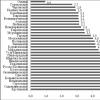
Results: The average age of patients amounted to 47.3 ± 13.9 years. With statistical processing of the obtained results of the test indicators, there is a decrease in some of them as the PZO increases: the maximum corrected acuteness of vision (p \u003d 0.01), sensitivity in Fovaa (P \u003d 0.008), the middle thickness of the retina in Fovaa (P \u003d 0.01 ), the average thickness of the choroid in the nasal and temporal sectors (p \u003d 0.005; p \u003d 0.03). In addition, all groups of subjects identified a significant statistically reliable inverse correlation relationship, between PZO and (ICC) -0.4; as well as the retinal thickness in Fovaa -0.6; thickness of the choroid in Fovaa -0.5 and sensitivity in Fovaa -0.6; (P.<0,05).
Conclusion: With a detailed analysis of the obtained mean values \u200b\u200bof the parameters under study, a tendency was detected to a general decrease in morphofunctional indicators of the eyeball as a PZO increases in groups. While the obtained correlation data of the clinical trial testifies to the close relationship between the morphometric and functional parameters of the visual analyzer.
Keywords: myopia, emmetropy, optical macular pigment density, overnight axis eye, morphometric parameters, carotenoids, heterochromatic flicker photometry, optical coherent tomography retina.
For citation: Egorov E.A., Eskina E.N., Gwetadze A.A., Belogurova A.V., Stepanova M.A., Rabadanova M.G. Morphometric features of the eyeball in patients with myopia and their effect on visual functions. // RMW. Clinical ophthalmology. 2015. No. 4. P. 186-190.
Citation:Egor E.A., Eskina E.N., Gwetadze A.A., Belogurova A.V., Stepanova M.A., Rabadanova M.G. Morphometric features of the eyeball in patients with myopia and their effect on visual functions // RMH. Clinical ophthalmology. 2015. №4. P. 186-190
Myopic Eyes: Morphometric Features and Their Influence On Visual Function.
Egorov E.A.1, Eskina E.N.3,4,5,
Gvetadze A.A.1.2, Belogurova A.V.3.5,
Stepanova M.A.3.5, Rabadanova M.G.1.2
1 Pirogov Russian State National Medical University, 117997, Ostrovityanova St., 1, Moscow, Russian Federation;
2 Municipal Clinical Hospital No. 15 Named After O.M. Filatov, 111539, Veshnyakovskaya St., 23, Moscow, Russian Federation;
3 National Medical Surgical Center Named After N.I. Pirogov, 105203, Nizhnyaya Pervomayskaya St., 70, Moscow, Russian Federation;
4 Federal Biomedical Agency of Russia, 125371, Volokolamskoe Shosse, 91, Moscow, Russian Federation;
5 LASER SURGERY CLINIC "SPHERE", 117628, STAROKACHALOVSKAYA ST., 10, MOSCOW, RUSSIAN FEDERATION;
Purpose: to Evaluate Morphofunctional Parameters of Myopic Eyes with Increase Of The Length of Eye Anteroposterior Axis (APA).
Methods: The Study Involved 36 Patients (71 Eyes). All Patients Were Divided Into 4 Groups Depending on The Apa Length. 1st GROUP INVOLVED PATIENTS WITH MILD MYOPIA AND APA LENGTH FROM 23.81 TO 25.0 MM; The 2nd -With Moderate Myopia and Apa Length from 25.01 to 26.5 mm; 3D - WITH HIGH MYOPIA AND APA LENGTH ABOVE 26,51 MM; 4th - with Emmetropic Refraction and APA Length from 22.2 to 23.8 mm. Patients Underwent Standard Ophthalmic Examination and Additional Diagnostic Examination: Echobiometry, Determination Of Optical Density Of Macular Pigment, Fundus Photography, Optical Coherence Tomography of the Anterior and Posterior Segments of the Eye.
Results: The Mean Age WAS 47.3 ± 13.9 years. Statistic Analysis SHOWED THE REDUCTION OF SOME PARAMETERS WITH APA LENGTH "S INCREASING: BEST CORRECTED VISUAL ACUITY (BCVA) (P \u003d 0.01), FOVEAL sensitivity (P \u003d 0.008), Average FOVEAL RETINAL Thickness (P \u003d 0.01), Average Thickness In The Temporal and Nasal Choroids Sectors (p \u003d 0.005; p \u003d 0.03). Inverse Correlation Between Axial Length and BCVA (R \u003d -0.4); The FOVEAL RETINAL Thickness (R \u003d -0,6); The Fovail Choroidal Thickness (R \u003d -0.5) and FOVEAL SENSITITIBY (R \u003d -0.6) WERE REVEALED IN ALL GROUPS (P<0,05).
Conclusion: The Analysis SHOWED THE TENDENCY OF A GENERAL DECREASE OF MORPHOLOGICAL AND FUNCTIONAL PARAMETERS OF THE EYE WITH THE INCREASE OF AXIAL LENGTH IN ALL GROUPS. Revealed Correlation SHOWED A Close Relationship Between Morphometric and Functional Parameters of the Eye.
Key Words: Myopia, Emmetropia, Macular Pigment Optical Density, Eye Anteroposterior Axis, Morphofunctional Parameters, Carotenoids, Heterochromatic Flicker Photometry, Optical Coherence Tomography of the Retina.
For citation: Egorov E.A., Eskina E.N., Gvetadze A.A., Belogurova A.V.,
Stepanova M.A., Rabadanova M.G. Myopic Eyes: Morphometric Features and
Their Influence ON Visual Function // RMJ. Clinical ophthalomology.
2015. No. 4. P. 186-190.
The article presents data on the morphometric features of the eyeball in patients with myopia and their effect on visual functions.
In the structure of the morbidity of the organ of vision, the frequency of myopia in various regions of the Russian Federation ranges from 20 to 60.7%. It is known that among the disabilities in vision of 22% are the faces of young age, the main cause of disabilities in which is complicated by the myopic of high degree.
As in our country and abroad, adolescents and "young adults", myopia is often combined with retinal pathology and optic nerve, thereby make it difficult to predict and the course of the pathological process. The medical and social significance of the problem is exacerbated by the fact that complicated myopia amazes people at the very working age. The progression of myopia can lead to serious irreversible changes in the eye and significant loss of vision. According to the results of the All-Russian clinical examination, the incidence of children and adolescents by myopia over the past 10 years has grown 1.5 times. Among adults with disabilities in vision due to myopia, 56% have congenital myopia, the rest - acquired, including in school years.
The results of complex epidemiological and clinical and genetic studies have shown that myopia is a multifactorial disease. Understanding the pathogenetic mechanisms of violation of visual functions under myopia remains one of the topical issues of ophthalmology. Pathogenesis units during myopic disease are difficult to interact with each other. The morphological properties of the sclera play an important role in the course of myopia. It is their particular importance in the pathogenesis of the elongation of the eyeball. Dystrophic and structural changes occur in the sclera of minor people. It has been established that extensibility and deformation of the sclera of the eyes of adults with high myopia is noticeably more than with emmetropi, especially in the area of \u200b\u200bthe rear pole. An increase in the length of the eye in myopia is currently being considered as a consequence of metabolic disorders in the sclera, as well as changes in regional hemodynamics. The elastic elastic properties of the sclera and changes in the length of the axis of the axis (PZO) have long been interested in scientists. The evolution of the study of the anatomical parameters of the eyeball is reflected in the works of many authors.
According to E.Zh. The throne, the length of the Emmetropic eye axis varies from 22.42 to 27.30 mm. In relation to the variability of the length of the PZO for myopia from 0.5 to 22,0d E.Zh. The throne gives such data: the length of the axis during myopia is 0.5-6,0d - from 22.19 to 28.11 mm; At myopia 6.0-22,0d - from 28.11 to 38.18 mm. According to T.I. Yeroshevsky and A.A. Bochkareva, biometric indicators of the sagittal axis of a normal eyeball on average are equal to 24.00 mm. According to E.S. Avetisov, with emmetropy, the length of the PZO of the eyes is 23.68 ± 0.910 mm, with myopia 0.5-3.0d - 24.77 ± 0.851 mm; in myopia 3.5-6,0d - 26.27 ± 0.725 mm; At myopia 6.5-10.0d - 28.55 ± 0.854 mm. The pretty clear parameters of the emmetropic eye are given in the National Ophthalmology Guide: the length of the emmetropic eye PZO is 23.92 ± 1.62 mm. In 2007 I.A. The craftsmen created a new anatomy-optical and corresponding reduced optical scheme of an emmetropic eye with a clinical refraction 0.0d and PZO 23.1 mm.
As mentioned above, dystrophic changes in the retina take place during myopia, which is most likely caused by impaired blood flow in choroidal and peripapillary arteries, as well as its mechanical stretching. It has been proven that people with the axial myopia of high degree the average thickness of the retina and choroid in the subfiva is less than that of emmetrov. Therefore, it can be assumed that the larger the length of the PZO, the higher the "pollen" of the shells of the eyeball and below the tissue density: sclera, choroids, retina. As a result of these changes, the number of tissue cells and cell substances is reduced: for example, a layer of retinal pigment epithelium is thinned, the concentration of active compounds is reduced, possibly carotenoids in the macular region.

It is known that the total concentration of carotenoids: lutein, zeaxanthine and mesozseaxanthin in the central field of the retina is the optical density of the macular pigment (OPP). Macular pigments (MP) absorb the blue part of the spectrum and provide powerful antioxidant protection against free radicals, lipid peroxidation. According to a number of authors, a decrease in the OPP indicator is associated with the risk of developing maculopathy and a decrease in central vision.
In addition, many authors converge in the opinion that with age there is a decrease in OPP. Studies of the level of OPPs in a healthy population in various age patients and patients of all kinds of ethnic groups in many countries of the world are a very controversial picture. For example, the average value of OPPs in the Chinese population in healthy volunteers aged 3 to 81 amounted to 0.303 ± 0.097. In addition, reverse correlation with age was revealed. The average value of OPPs in healthy volunteers in Australia aged 21 to 84 years was 0.41 ± 0.20. For the Great Britain's population aged 11 to 87 years, the total average value of OPP in the group was 0.40 ± 0.165. The connection with age and the color of the iris is noted.
Unfortunately, in the Russian Federation of large-scale studies to study the indicator of OPPs in a healthy population, in patients with the anomalies of refractive, pathological changes in the macular zone and other ophthalmological diseases were not carried out. This question is still open and quite interesting. The only study of OPPs in a healthy Russian population was held in 2013. E.N. Eskigo et al. 75 healthy volunteers aged 20 to 66 took part in this study. The average OPP indicator in the multi-age groups varied from 0.30 to 0.33, and the Pearson correlation coefficient testified to the absence of the connection between the magnitude of OPPP and age with the normally occurring age-related processes in the body of vision.
At the same time, the result conducted by foreign authors of clinical research confirms that healthy volunteers, OPP values \u200b\u200bare positively correlated with indicators of the central thickness of the retina (R \u003d 0.30), measured by heterochromatic flicker photometry and optical coherent tomography (Oct), respectively.
Therefore, in particular interest, in our opinion, represents the study of OPPs not only in a healthy population in various age patients and patients of all sorts of ethnic groups, but also in dystrophic ophthalmopathy and the anomalies of refraction, in particular during myopia. In addition, the fact of the effect of an increase in the length of the PZO on the topographic-anatomical and functional indicators of the visual analyzer (in particular, on OPP, the thickness of the retina, choroids, etc.) remains curious. The relevance of the above fundamental issues determined the purpose and objective of this study.
Purpose of the study: Assess the morphofunctional parameters of the visual analyzer in patients with myopia as the length of the PZO of the eyes increases.
Materials and methods
Total examined 36 patients (72 eyes). All patients during the study were divided into groups exclusively by the PZO of the Eye Apple (by the classification of E.S. Avetisov). The 1st group consisted of patients with the myopia of a weak degree and a value of PZO from 23.81 to 25.0 mm; 2-y - with the myopia of the moderate and the value of the PZO from 25.01 to 26.5 mm; 3 - with high degree myopia and PZO value above 26.51 mm; 4th - Patients with refraction approximated to the emmetropic, and the value of the PZO from 22.2 to 23.8 mm (Table 1).
Patients did not take preparations containing carotenoids, did not adhere to a special diet enriched with Lutein and Zeaxantin. All subjects conducted a standard ophthalmological examination, which allowed the macular pathology to eliminate them, presumably affecting the results of the examination.
The examination included the following diagnostic set of measures: an autorecatter, a visometrium with the determination of the maximum corrected visual acuity (ICM), a non-contact computer pneumotonometry, anterior segment biomicroscopy with a slit lamp, a static automatic perimetry with an aetropy correction (evaluated MD, PSD indicators, as well as sensitivity in Fovaa), indirect ophthalmoscopy of the macular area and disk of the optic nerve using a lens 78 diopters. In addition, all patients conducted echobiometry on the device of the company Quantel Medical (France), defining OPP on the MPOD MPS 1000 device, Tinsley Precision Instruments Ltd., Croydon, Essex (United Kingdom), digital photographing of the eye bottom using the Fundus Camera CARL ZEISS MEDICAL TECHNOLOGY (Germany); OCT of the front segment of the eyeball on the OCT-Visante Carl Zeiss Medical Technology (Germany) (according to the Ost-Visant study, the central thickness of the cornea) was evaluated); OKT retina on the Cirrus HD 1000 Carl Zeiss Medical Technology (Germany) apparatus. According to Oct., the average retinal thickness in the Fowea region was estimated, calculated by the device in automatic mode, using the Macular Cube 512x128 protocol, as well as the average thickness of the choroid, which was calculated manually from the hypereflective boundary corresponding to the RPE, to the border of the choroid-scleral interface, clearly visible On the horizontal 9-millimeter scan formed through the Fovie Center when using the High Definition Image: HD Line Raster protocol. Measuring the thickness of the choroids was carried out in the Fowea Center, as well as 3 mm in the nasal and temporal directions from the Fovye center, at the same time from 9:00 to 12:00.
Statistical processing of clinical research data was performed according to standard statistical algorithms using Statistica software, version 7.0. The difference of values \u200b\u200bfor p was considered a reliability<0,05 (уровень значимости 95%). Определяли средние значения, стандартное отклонение, а также проводили корреляционный анализ, рассчитывая коэффициент ранговой корреляции Spearman. Проверка гипотез при определении уровня статистической значимости при сравнении 4 несвязанных групп осуществлялась с использованием Kruskal-Wallis ANOVA теста.
results
The average age of patients amounted to 47.3 ± 13.9 years. The partition distribution was as follows: 10 men (28%), 26 women (72%).
The average values \u200b\u200bof the test parameters are presented in Tables 2, 3 and 4.
When conducting correlation analysis, a statistically reliable feedback between the PZO and some parameters (Table 5) was revealed.
Of particular interest, in our opinion, represent the data of the correlation research in the group of patients with a diagnosis of "high degree" myopia. The results of the analysis are presented in Table 6.
Conclusion
With a detailed consideration of the obtained mean values \u200b\u200bof the parameters under study, the tendency towards a general reduction in the functional indicators of the eye is revealed as the PZO increases in groups, while the obtained correlation analysis data indicate a close relationship between the morphometric and functional parameters of the visual analyzer. Supposed these changes are also associated with "mechanical interpretation" of shells in patients with myopia due to an increase in the PZO.
Alternatively, I would like to note, though inaccurate, but decrease in OPPs in groups, and a small tendency to negative feedback between OPPP and PSO. It is possible, as the number of groups of subjects increases, a stronger and reliable correlation between these indicators will be marked.
Literature
1. Avetisov E.S. Myopia. M.: Medicine, 1999. P. 59..
2. Akopyan A.I. et al. Features of the optic nerve disk with glaucoma and myopia // Glaucoma. 2005. No. 4. P. 57-62. .
3. Dal N.Yu. Macular carotenoids. Can they protect us from age macular degeneration? // Ophthalmological statements. 2008. No. 3. P. 51-53. .
4. Eroshevsky T.I., Bochkareva A.A. Eye diseases. M.: Medicine, 1989. P. 414..
5. Zykova A.V., Rzaev V.M., Eskina E.N. The study of the optical density of the macular pigment in the midst of the age patients is normal: Mat-lies Vi Ross. common Ophthalmol. Forum. Collection of scientific papers. M., 2013. T. 2. P. 685-688. .
6. Kuznetsova M.V. Causes of the development of myopia and its treatment. M.: Medpress-Inform, 2005. P. 176..
7. Libman E.C., Shakhova E.B. Blindness and disability due to the pathology of the organ of vision in Russia // Bulletin of ophthalmology. 2006. No. 1. P. 35-37. .
8. Ophthalmology. National Guide / Ed. S.E. Avetaisova, E.A. Egorova, L.K. Moshetova, V.V. Nereva, H.P. Tahchidi. M.: Gootar Media, 2008. P. 944..
9. Crafts I.A. The patterns of the ratio of the sagittal sizes of the anatomical structures of the eye is normal and at a primary closed-curved glaucoma with a relative pupil unit: author. dis. ... Cand. honey. science Volgograd, 2007. P. 2..
10. Slutsko E.L. Myopia. Disturbance of refraction is a disease // Astrakhan Bulletin of Environmental Education. 2014. № 2 (28). Pp. 160-165. .
11. Eskina E.N., Zykova A.V. Early risk criteria for glaucoma development in patients with myopia // Ophthalmology. 2014. T. 11. No. 2. P. 59-63. .
12. Abell R.G., Hewitt A.W., Andric M., Allen P.L., Verma N. The Use of Heterochromatic Flicker Photometry to Determine Macular Pigment Optical DenSity in a Healthy Australian Population // Graefes Arch Clin Exp Ophthalmol. 2014. Vol. 252 (3). P. 417-421.
13. Beatty S., Koh H.h., Phil M., Henson D., Boulton M. The Role of Oxidative Stress In The Pathogenesis of Age-Related Macular Degeneration // SURV. Ophthalmol. 2000. Vol. 45. P. 115-134.
14. Bone R.A., Landrum J.T. Macular Pigment in Henle Fiber Membranes A Model for Haidinger "S Brushes // Vision Res. 1984. Vol. 24. P. 103-108.
15. Bressler N.M., Bressler S.B., Childs A.L. Surgery for Hemorrhagic Choroidal Neovascular Lesions of Age-Related Macular Degeneration // Ophthalmology. 2004. Vol. 111. P. 1993-2006.
16. GUPTA P., SAW S., Cheung Cy, Girard MJ, Mari Jm, Bhargava M., Tan C., Tan M., Yang A., Tey F., Nah G., Zhao P., Wong Ty, Cheng C. Choroidal Thickness and High Myopia: A Case-Control Study of Young Chinese Men in Singapore // Acta Ophthalmologica. 2014. DOI: 10.1111 / AOS.12631.
17. Liew S.h., Gilbert C.E., Spector T.D., Mellerio J., Van Kuijk F.J., Beatty S., Fitzke F., Marshall J., Hammond C.J. Central Retinal Thickness Is Positively Correlated With Macular Pigment Optical Density // Exp Eye Res. 2006. Vol. 82 (5). P. 915.
18. MAUL E.A., FRIEDMAN D.S., CHANG D.S., BJLAND M.V., RAMULU P.Y., JAMPEL H.D., QUIGLEY H.A. Choroidal Thickness Measured by Spectral Domain Optical Coherence Tomography: Factors Affecting Thickness In Glaucoma Patients // Ophthalmol. 2011. Vol. 118. (8). P. 1571-1579.
19. Murray I.J., Hassanali B., Carden D. Macular Pigment in Ophthalmic Practice // Graefes Arch. CLIN. EXP. Ophthalmol. 2013. Vol. 251 (10). P. 2355-2362.
20. Rada J.A et al. The SCLERA AND MYOPIA // EXP. Eye Res. 2006. Vol. 82. No. 2. P. 185-200.
21. Zhang X., WU K., SU Y., ZUO C., Chen H., Li M., Wen F. Macular Pigment Optical Density in a Healthy Chinese Population // Acta Ophthalmol. 2015. DOI: 10.1111 / AOS.12645.
Objective: To study the dynamics of the CDC, taking into account the refraction of healthy eyes in healthy children aged 1 months. up to 7 years and compare with a pzo eye with congenital glaucoma in children of the same age.
Material and methods: Studies were conducted on 132 eyes with congenital glaucoma and 322 healthy eyes. By age, children with congenital glaucoma and with healthy eyes were distributed according to the classification E.S. Avetisova (2003). So, with glaucoma was 30 newborns (55 eyes), children up to 1 year - 25 (46 eyes), up to 3 years - 55 (31 eyes). Among the studied with healthy eyes: Newborn - 30 eyes, up to 1 year - 25 eyes, up to 3 years - 55 eyes, 4-6 years old - 111 eyes, 7-14 years old - 101 eyes. The following research methods were used: Tonometry, Tonography on Nesterov and Elastotonometry, Biomicroscopy, Honoscopy, Ophthalmoscopy, A / B scanning on the ODM-2100 UltraSonik A / in Scanner for Orhthalmology.
Results and conclusions: After studying the normal PZO eyes in different age periods, we revealed a significant range of oscillations of PZO indicators, the extreme indicators of which can correspond to pathological. The increase in the size of the front-rear axis of the eye with an innate glaucoma depends not only on the violation of the hemogrodynamic processes of the eye with the accumulation of intraocular fluid, but also from the age dynamics of the pathological growth of the eye and the degree of refraction.
Keywords: the front-rear axis of the eye, congenital glaucoma.
Abstract.
Comparative Analysis of The Anterior-Posterior Axes of Eyes Of Patients With Congenital Glaucoma and Healthy
PATIENTS TAKING INTO CONSIDERATION OF THE AGE ASPECT
Yu.A. Khamroeva, B.T. Buzrukov.
Pediatric Medical Institute, Tashkent, Uzbekistan
Purpose: To Study The Dynamics of the A Apa in Healthy Children Taking Inteo Consideration The Refraction of Healthy Eyes Aged from One Monh to Seven Years, Compared to APA of Patients with Congenital Glaucoma of the Same Age.
Methods: The Study Was Performed On 132 Eyes with Congenital Glaucoma and 322 of Healthy Eyes. Patients with Congenital Glauccoma and Healthy Subjects Were Distributed by Age According to the Classification of E.S. Avetisov (2003), 30 Newborns (55 Eyes), 25 Patients under 1 Year Old (46 Eyes) of, 55 Healthy Patients Under 3 Years Old, (31 Eyes) and Newborns (30 Eyes), UNDER 1 Year (25 Eyes) , UNDER 3 YEARS (55 EYES), 4-6 YEARS OLD (111 EYES), FROM 7 TO 14 YEARS OLD (101 EYES). Tonometry, Tonography, Elastotonometry, Biomicroscopy, Gonioscopy, Ophthalmoscopy, A / B Scanning Were Performed.
Results and Conclusion: There Were Significant APLITUDE OF THE APAINDICES REVEALED IN PATIENTS OF VARIOUS AGES. The Extreme Values \u200b\u200bMay Indicate The Pathology. Increase of APA Size in Congenital Glaucoma Depends Not Only On a Disparity of Hydrodynamic Processes But Also On Age Dynamics of Eye Growth and Refraction.
Key Words: Anterior-Posterior Axis (APA) of the Eye, Congenital Glaucoma.
Introduction
Currently, it has been established that the main starting mechanism for the development of the glaucomatous process is to increase intraocular pressure (WGD) to the level above the target. BGD is an important physiological constant of the eye. There are several types of regulation of the WGD. At the same time, several anatomy-physiological factors influence the exact indicators of the WGD, especially in children, the main points of which are the size of the eye and the size of its front-rear axle (PZO). Research recent years show that one of the key factors for the development of glaucomatous damage can be a change in the biomechanical stability of the connective tissue structures of the eye not only in the area of \u200b\u200bthe optic nerve disk (DZN), but also the fibrous capsule as a whole. In favor of this statement, the gradual thinning of the sclera and cornea is evidenced.
Objective: To study the dynamics of the CDC, taking into account the refraction of healthy eyes in healthy children aged 1 months. up to 7 years and compare with a pzo eye with congenital glaucoma in children of the same age.
Material and methods
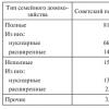
Studies were held at 132 eyes with congenital glaucoma and 322 healthy eyes. Children were distributed by age according to the classification E.S. Avetisova (2003): With congenital glaucoma: newborn - 30 patients (55 eyes), up to 1 year - 25 (46 eyes), up to 3 years - 55 (31 eyes); Children with healthy eyes: newborns - 30 eyes, up to 1 year - 25 eyes, up to 3 years - 55 eyes, 4-6 years old - 111 eyes, 7-14 years old - 101 eyes.
The following research methods were used: Tonometry, Tonography on Nesterov and Elastotonometry, Biomicroscopy, Honoscopy, Ophthalmoscopy. A / B scanning on the ODM-2100 UltraSonik A / C Scanner for Opfhthalmology. In the stages of disease and age, patients with congenital glaucoma were distributed as follows (Table 1).
Results and discussion
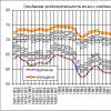
Despite the fact that there are data on the average values \u200b\u200bof the anatomy-optical elements of healthy eye, including the front-rear axis of the eye (PZO) aged from the newborn to 25 years (Avetisov E.S., et al., 1987) and from Newborn up to 14 years old (Avetisov E.S., 2003, Table 2), in the Republic of Uzbekistan, such studies have not been previously conducted. Therefore, it was decided to perform echobiometric studies of PZO indicators on 322 healthy eyes in children aged 1 months. Up to 7 years old, taking into account the degree of eye refraction and compare the data obtained with the results of similar studies in their eyes with congenital glaucoma (132 eyes) in children of the same age. Research results are presented in Table 3.
PZO rates are normal in almost all age groups, except newborns, practically coincided with the data given in the E.S. Table Avetisova (2003).
Table 4 presents the data of the eye PZO in the norm, depending on refraction and age.
The relative dependence of the degree of refraction against the shortening of the PZO eye was noted only from 2 years (by 1.8-1.9 mm).
It is known that in the study of the WGD in front of the congenital glaucoma, difficulties arise in determining how much the NWP is characterized by normal hydrodynamic processes or their pathology. This is due to the fact that the little children are soft, easily spells. As the intraocular fluid accumulate, they are stretched, the eye increases in the amount, and the WGD remains within normal values. At the same time, this process leads to metabolic disorders, damaging the fibers of the optic nerve and worsening metabolic processes in ganglion cells. In addition, it is necessary to clearly differentiate pathological and natural, associated with age, the growth of the child's eye.
Having studied the normal points of the eye of the eyes in different age periods, we found that the extreme values \u200b\u200bof these indicators may correspond to the values \u200b\u200bfor pathology. In order to clearly determine whether the ebony apple is a pathological, we simultaneously analyzed the connection of the PZO indicators with IGD, refraction, the presence of glaucomatous excavation, its size and depth, horizontal corneal size and its limb.
Thus, with the developed stage of the disease in 10 eyes of newborns at PZO \u003d 21 mm, the tonometric pressure (Pt) was 23.7 ± 1.6 mm RT. Art. (p≤0.05), disk excavation - 0.3 ± 0.02 (p≤0.05); In children up to 1 year (36 eyes) at pzo \u003d 22 mm Pt, it was equal to 26.2 ± 0.68 mm Hg. Art. (p≤0.05), disk excavation - 0.35 ± 0.3 (p≤0.05). In children under 3 years old (10 eyes) at pzo \u003d 23.5 mm PT reached 24.8 ± 1.5 mm Hg. Art. (p≥0.05), disk excavation - 0.36 ± 0.1 (p≤0.05). The size of the PZO eye exceeded the average rate by 2.9, 2.3 and 2.3 mm, respectively, in each age group.
With a far-sighted stage of glaucoma in children under 1 year (45 eyes), the Size of the PZO was 24.5 mm, PT - 28.0 ± 0.6 mm RT. Art. (p≤0.05), disk excavation - 0.5 ± 0.04 (p≤0.05), in children up to 2 years (10 eyes) at a PZO 26 mm PT reached 30.0 ± 1.3 mm RT . Art. (p≤0.05), disk excavation - 0.4 ± 0.1 (p≤0.05). In children under 3 years old (11 eyes), at a PZO, 27.5 mm Pt was equal to 29 ± 1.1 mm Hg. Art. (p≤0.05), disk excavation - 0.6 ± 0.005 (p≤0.05). Under the terminal stage (10 of the eye), at a PZO, 28.7 mm Pt was 32.0 ± 1.2 mm Hg. Art. (P≥0.05), disk excavation - 0.9 ± 0.04 (p≤0.05). In these children, the size of the PZO eyes exceeded the average rate of 4.7, 4.8, 6.3 mm, and at a terminal stage - by 7.5 mm.
conclusions
1. An increase in the size of the PZO of the eyes with a congenital glaucoma depends not only on the violation of the hemohydrodynamic processes of the eye with the accumulation of intraocular fluid, but also on the age dynamics of the pathological growth of the eye and the degree of refraction.
2. Diagnosis of congenital glaucoma should be based on examination data, such as the results of echobiometry, gonoscopy, WGD, taking into account the rigidity of the fibrous shell of the eye and beginner glaucomatous optical neuropathy.


Literature
1. Akopyan A.I., Yerichev V.P., Iomdina E.N. The value of the biomechanical parameters of the eye in the interpretation of the development of glaucoma, myopia and combined pathology // Glaucoma. 2008. №1. P. 9-14.
2. Harutyunyan L.L. The role of the visco-elastic properties of the eye in determining the pressure pressure and evaluation of the development of glaucomatous process: author. dis. ... Cand. honey. science M., 2009. 24 s.
3. Buzikin MA Ultrasonic anatomy-physiological picture of the acodational apparatus of the eye of young people in vivo: author. dis. ... Cand. honey. science SPb., 2005.
4. Volkov V.V. Three-component classification of open-hearted glaucoma // Glaucoma, 2004. №1. S.57-68.
5. Gulidova E.G., Strakhov V.V. Accommodation and hydrodynamics of myopic eye // Russian Nationwide Ophthalmological Forum: Sat. scientific papers. M., 2008. P. 529-532.
6. Kozlov V.I. A new method for studying the extensibility and elasticity of the eye when the possibility of ophthalmotonus // West. Ophthalmol. 1967. No. 2. P. 5-7.
7. EUROPEAN GLAUCOMA PREVENTION STUDY GROUP (EGPS). Central Corneal Thickness in the European Glaucoma Prevention Study Group // Ophthalmology. 2006. Vol. 22. P. 468-470.
8. Kobayashi H., ONO H., Kiryu J. et al. Ultrasound Biomicroscopic Measurement of Development AF ANTERIOR CHAMBER ANGL // Br J. Ophthalmol. 1999.vol. 83. N 5. P. 559-562.
9. Pavlin C.J., Harasiewecz K., Foster F.S. Eye Cup for Ultrasound Biomicroscopy // Ophthalmic Surg. 1994. Vol. 25, N. 2. P. 131-132.
10. Rogers D.L., Cantor R.N., Catoira Y. et al. Central Corneal Thickness and Visual Field Loss in Fellow Eyes of Patients with Open-Anle Glaucoma // AM. J. Ophthalmol. 2007. Vol. 143. N 1. P.159-161.
Eye ultrasound is an additional technique in ophthalmology, which has high accuracy when identifying hemorrhages and evaluating the front-axis of the axis of the eye. The last indicator is necessary to identify the progression of myopia in children and adults. There are other applications of the technique. This method of diagnosis is characterized by the simplicity of the procedure, the lack of additional training and speed of the survey. Ultrasound is carried out with the help of universal and specialized ultrasound devices. The results assessment are made in accordance with regulatory tabular data.
Indications and contraindications
Ultrasound examination of organs of vision is a non-invasive diagnostic method used to identify many ophthalmic diseases.
Indications for ultrasound eyes are:
- diagnosis of retinal detachment, vascular shell associated with a tumor process and other pathologies,
- confirmation of the presence of neoplasms, control of their growth and effectiveness of treatment,
- differential diagnosis of intraocular tumors,
- determination of the position of the lens when clouding the cornea,
- scanning the character of vitreous body
- identification of invisible foreign bodies in the eye (after injury), refinement of their size and localization,
- diagnosis of vascular ophthalmopathologies,
- cyst detection,
- diagnosis of congenital diseases,
- identification of pathological changes with a deep lesion of the eyeball in the eyeboard (determination of the nature of damage - the fracture of the orbit wall, disruption of nervous bonds, reducing the apple itself),
- clarification of the reasons for the displacement of the eyeball forward - autoimmune pathologies, tumors, inflammation, skull development anomalies, high one-sided myopia,
- determination of changes in retrobulbar space with elevated intracranial pressure, retrobulbar neuritis and other diseases.
Contraindications for ultrasound diagnostics are injuries of the eye, in which the integrity of structures and bleeding in organs is disturbed.
Methods
There are several techniques of ultrasonic eye research:
- 1. Eye ultrasound in A-mode, at which one-dimensional signal display is obtained. There are 2 kinds of varieties:
- biometric, the main purpose of which is the definition of the length of the PZO (these data is used before the operation on the cataract and for the accurate calculation of the artificial lens),
- standardized diagnostic is a more sensitive method that allows you to identify and differentiate changes in intraocular tissues.
2. Ultrasound in B-mode. The resulting display of the echo signal is a two-dimensional, horizontal and vertical axes. As a result, the form, location and size of pathological changes are better visualized. The ultrasonic sensor contacts directly with the surface of the eye (through a water bath or gel). It is the most acceptable way to study the structures of the eye, but is in little informative for the diagnosis of corneal diseases. The advantage of scanning in this mode is the creation of a real two-dimensional picture of the eyeball.
3. Ultrasonic biomicroscopy, used to visualize the front segment of the eye. The frequency of ultrasonic oscillations is higher than that of previous methods.
In rare cases, the following types of ultrasonic surveys are applied:
- 1. Immersion ultrasound in B-mode. It is done in addition to other research methods to study the pathologies of the front edge of the retina, which are located too close with standard scanning in B-mode. A small bath is installed on the eye filled with saline used as an intermediate medium.
- 2. Color Doppler. Allows you to simultaneously get a two-dimensional image and evaluate the bloodstream in the blood vessels. Since the vessels have small sizes, then the exact localization cannot be visualized. Bloodstock is encoded in red (artery) and blue (veins) color. The method also allows you to determine the growth of blood vessels in tumors, evaluate the pathological deviations of the sleepy and central artery, the veins of the retina, the defeat of the optic nerve due to insufficient blood circulation.
- 3. Three-dimensional ultrasound examination. The three-dimensional image is obtained as a result of combined with a variety of two-dimensional scans, and the sensor is set in one position, but quickly rotates. The resulting scan can be considered on various sections. Three-dimensional ultrasound is indispensable in ophthalmcology (to determine the volume of melanoma and evaluating the effectiveness of therapy).
At the initial degree of cataract, the cloud of lens does not allow an ultrasound lens. When a certain maturity of the disease is reached, the study shows various options for its echo-transparency.
In ophthalmology, both specialized and universal ultrasound devices are used. In the latter case, the resolution of the sensors should be at least 5 MHz. Sensors of universal ultrasound devices have large dimensions, which makes it impossible to impose them directly on the eye from behind its round shape. Therefore, liquid gaskets installed on the eye can be used as an intermediate environment. The small working surface of specialized ophthalmological sensors allows you to visualize the intraocular space.
Advantages and disadvantages
The advantages of the method of ultrasound examinations include:
- Lack of thermal effects.
- The possibility of obtaining information about the state of the anatomical areas located near the eyeboard.
- High sensitivity in the study of intraocular hemorrhages and discharge processes, especially when clouding Optical eyes, when traditional ophthalmologic means of diagnosis are not applicable.
- Accurate definition of retinal detachment area.
- The possibility of evaluating the volume of hemorrhage, according to which the further tactics of treatment (2/8 of the vitreous body is a conservative treatment, 3/8 - surgery).
The disadvantages of ultrasound bodies of the organs are the following:
- contact the sensor with the surface of the eyeball,
- measurement error arising from the compression of the cornea,
- inaccuracies associated with the human factor (not strictly perpendicular to the location of the sensor),
- risk of infection in eye.
Survey features in children
Eye ultrasound is carried out at any age, but young children are difficult to achieve immobility and closing of the eyelids. This method of examination helps to identify congenital deviations in the organs of vision (retinopathy premature, colobroms of the vascular shell and disk of the optic nerve, other pathologies). In children of junior and school age, the main indication for the destination of ultrasound is myopia.
In newborns, the refractive power of the optical system of the eye is weaker than in adults, and the size of the eyeball is less (16 mm versus 24 mm). In the norm after birth, there is a "reserve" of hyperopia in 2-5 diopters, which is gradually "consumed" as children and the eye apple grow. By 10 years, its value reaches the appropriate size in an adult, and the image focus falls precisely on the retina ("100%" vision).

After 7 years, the load on the visual apparatus of children increases much, which is most often associated with studying at school, burdened by heredity and weakness of accommodation - the ability of the lens to change its form in order to see equally well near and away. Ultrasound diagnosis is the main method for determining the PZO (axial size of the eye) in children when diagnosing myopia with accommodation spasm. Due to the peculiarities of growth, it is recommended to conduct an ultrasound for a child of 10 years to detect the lengthening of the front axle of the eye axis.
If the refraction violations were revealed at an earlier age, then the survey is carried out earlier. The lack of full-fledged vision correction up to 10 years leads to pronounced functional impairment and squint. Additionally, determine the transverse size of the eyeball and acoustic density of the sclera.
PZO measurement is the only reliable method for determining the progression of myopia. The main criterion is the increase in the front of the axis of the eyeball of more than 0.3 mm per year. Under the progression of myopia, all the structures of the eye are stretched, including the retina, which can lead to severe complications - its detachment and loss of vision.
Procedure
Before conducting the procedure, special training is required. When scanning orbits, women's eyes need to remove makeup cosmetics and eyelashes. The patient is placed on the back so that the headboard was near the doctor. Under the back, the roller is put in order for the head to take a horizontal position. In some cases, if it is necessary to determine the displacement of any structures of the eye or in the presence of a gas bubble in a ball, the patient is examined in a sitting position.
Scanning is made through the bottom or top closed eyelid, pre-apply gel. During the procedure, the doctor presses a little on the sensor, but it is painless. If a specialized sensor is applied, the patient's eyes can be open (at the same time local anesthesia is pre-performed).

The diagnosis of the structures of the eyeball is made in the following order:
- study of the front of the enemy (eyelids, lacrimal glands and bag) - Overview scanning,
- to obtain a cut through the front-raging axis (PZO), the ultrasonic sensor is installed on the closed top eyelid above the cornea, at this point the doctor becomes available to the central area of \u200b\u200bthe eyebound, iris, crystal, the vitreous body (partially), optic nerve, fatty fiber,
- to study all segments of the eye, the sensor is installed at an angle in several positions, while the patient is asked to transfer the look down towards the internal and outer corner of the eye,
- apply an ultrasonic head to the inner and outdoor part of the underground (the patient's eyes are open) in order to visualize the upper part of the orphanage structures,
- if it is necessary to assess the mobility of the identified formations, then the person is examined asking for quick movements of the eyeballs.

Scanning segments of the eye
The duration of the procedure is 10-15 minutes.
Results of research
During the survey, ultrasound diagnostic specialist fills the protocol with the conclusion. Deciphering the results of the ultrasound makes the attending ophthalmologist, comparing them with tabular regulatory indicators:

Normal indicators of an ultrasound examination of the eye in adults
Normal PZO values \u200b\u200bin children are shown in the table below. With different eye diseases, this indicator varies.

Normal figures in children
Normally, the image of the eyeball is characterized as a rounded form of dark color (hypoethogenic). In the forefront, two light strips are visualized that reflect the lens capsule. The visual nerve looks like a dark, hypo echogenic strip in the back of the eye chamber.

Normal indicators of blood flow with color dopplerography
Below is an example of an eye ultrasound protocol.

Read also ...
- The work of "Alice in Wonderland" in a brief retelling
- That transformation. "Transformation. Attitude towards the hero from the sister
- Tragedy Shakespeare "King Lear": the plot and the history of the creation
- Gargantua and Pantagruel (Gargantua et Pantagruel) Francois Rabl Gargantua and Pantagruel Brief