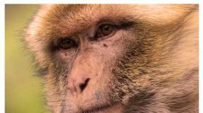
Eye diseases in dogs: classification and treatment. If the eyes of a dog fester: how to treat at home? The puppy has an enlarged eye
Sharp eyesight for a dog is not as important if it were an eagle, but still dogs rely on their eyes to a large extent. Basically, like all animals. Therefore, eye diseases in dogs should be treated immediately after they are discovered, without starting the process.
This is the name of the rapid and unconscious contraction of the muscles of the eyelid, as a result of which the animal blinks non-stop. In addition, there is photophobia, when the dog cannot look at the light at all, exudate is released from the eye. How dangerous is this condition for your pet? By itself, this pathology is not fatal, but ...
In almost all cases, blepharospasm is not an independent disease. Rather, it is a sign that extremely unfavorable pathological processes are taking place in the animal's body. So, sometimes this condition can be a “hint” to inflammation. trigeminal nerve. In general, blepharospasm can very often be observed with any injuries or inflammatory diseases of the eye. At the same time, the organ itself often swells, with palpation the dog shows signs of a pain reaction.
There are no specific methods of treatment, since this eliminates the underlying disease that caused blepharospasm. To deprive the animal of discomfort, drops with lidocaine or other anesthetic used in ophthalmology can be used. At home, these drugs do not need to be used! Many of them are used only in ophthalmology due to increased toxicity. General surgery requires doses that can easily poison a dog. So leave the use of such specific drugs to experienced veterinarians.
Prolapse of the third century, aka "Cherry eye"
A pathology in which the third eyelid slips out of its place and ends up in the corner of the eye (which is clearly visible in the photo). Prolapse most often manifests itself only on one side, but there are also cases of bilateral pathology. The disease got its second name because of the specific type eyeball, which really becomes like an overripe cherry. The causes of the "cherry eye" are far from completely identified, but usually the weakening of those tissues that usually hold the third eyelid in its "rightful" place leads to the appearance of this disease.

In some breeds, this attachment is initially weak, so cases of prolapse in these animals occur regularly. Among those, almost all, hounds, were noticed. There is information about hereditary predisposition. If at least one parent individual has a tendency to prolapse of the third eyelid, it will inevitably manifest itself in the offspring. If a description of something like this appears on the dog's veterinary card, you should not buy such a puppy.
Read also: Acral Dermatitis in Dogs: Diagnosis and Treatment
This is not a deadly disease. But! Firstly, she definitely does not add a “presentation” to the dog. Secondly, with prolapse, the functionality of the lacrimal gland is disrupted, which can already lead to keratitis and conjunctivitis. So this disease should be treated without delay.
Treatment, alas, often consists of a surgical operation, since an eyelid that has fallen out once will fall out again. The problem here is (the intervention itself is not too complicated) that the lacrimal gland suffers during surgery, and therefore the dog will have to be instilled into the eyes either with special preparations or with a simple saline solution (of course, sterile) until the end of its days.
Dermatitis of the century
Of course, it is difficult to consider dermatitis as an eye disease, but in this case, these pathologies are absolutely interconnected. The skin of the eyelids becomes inflamed, gets wet, and cases of suppuration are not uncommon. Naturally, under such conditions, the migration of pathogenic microflora into the conjunctival cavity becomes only a matter of time ... Moreover, this pathology is typical for long-haired dogs and breeds with long ears.

Clinical signs are quite characteristic: the skin on the eyelids turns red and inflamed, suppuration is possible, extremely bad smell. Souring of the eyes occurs, exudative discharge appears. Treat the disease with antibiotics a wide range. Hair in the affected area must be cut off, antiseptic ointments should be applied to the skin. Instilled into the eyes antimicrobials washed with sterile saline. To prevent the animal from rubbing and scratching its eyes, a surgical collar is used.
Conjunctivitis
It's easy to guess what they're called eye diseases in dogs, the main symptom of which is inflammation of the conjunctiva and adjacent tissues. Most often it has an infectious etiology. The clinical picture includes the following signs:
- Pinkish or even reddish tinge to all visible mucous membranes.
- These same tissues (like the eyelids) can swell noticeably.
- There are tears from the eyes and discharge, and the characteristics of the latter may vary from the usual watery discharge to pus.
- From inner corner the eye may protrude a piece of the third eyelid (which we just talked about above). At the same time, novice breeders may even think that the dog's eye popped out.
- , constant flashing. Moreover, in the latter case, the process can clearly cause pain to the dog, she constantly rubs her eyes with her paws, whines.
- Clouding of the cornea (although this may indicate).
Read also: Encephalomyelitis is an inflammation of the brain and spinal cord in cats and dogs

As for the causes of this disease, they (as already mentioned) most often have an infectious background. But this is not always the case:
- Viruses.
- Chlamydia is a fairly common cause.
- Allergic reactions.
- Inflammation or blockage tear ducts, because of which the conjunctival cavity does not receive sufficient moisture.
- Foreign body in the eye.
- Irritant substances that have entered the conjunctival cavity.
- (when the eyelashes literally scrape the delicate tissues of the eye).
- Diverse.
So how to treat this unpleasant pathology? First, it all depends on the root cause. Various antibacterial agents are usually prescribed, including drops and ointments (tetracycline, for example). It all depends on the results of the tests taken and on the recommendations of your veterinarian.
Eversion and inversion of the eyelid
Ectropion and entropion are the scientific names for eversion and respectively. Both pathologies are truly "canine", since they are incomparably less common in cats and other domestic animals. Great Danes, Newfoundlands and some spaniels are especially predisposed. Thus, these are hereditary eye diseases in dogs.

Both pathologies should be considered together, since these varieties often develop in parallel to each other. Of course, eyelid eversion, unlike eyelid inversion, rarely leads to really serious problems for the animal. But here everything rests on the fact that the eye, left without a reliable cover, becomes predisposed to the introduction of pathogenic microflora. In addition to direct surgical manifestations, sick dogs have multiple eye discharges, they constantly blink, and when the eyeball is pressed, a strong pain reaction occurs. Dogs with ectropion suffer from drying out of the conjunctiva, which is fraught with other severe disorders.
Some kind of cases should be considered a separate pathology, in which eyelashes begin to grow incorrectly, literally growing into the eye. This disease develops for a long time, sometimes for several years. The symptomatology is similar to volvulus, that is, the eyes of the animals are constantly watery, pus flows, pain occurs when pressed, but in this case the clinical picture worsens more slowly.
This eye disease in dogs is treated exclusively surgically. It is only important to note that it is advisable to perform the operation on an already adult animal, in which the process of changing the size of the eyes has already stopped. To relieve the manifestations of the disease, apply antiseptic ointments and drops, hormonal preparations and other medicines. Only in very rare cases, when eversion or inversion (which is generally rare) of the eyelids are insignificant, is it possible conservative treatment. In any case, the decision here remains with the veterinarian.
Inflammation of the eyes in dogs is quite common problem. It can be caused by many diseases, which only a specialist can correctly diagnose. Untimely access to a doctor can lead to blindness of the animal. Let's highlight the main eye diseases in dogs that can cause inflammation. Consider their symptoms and causes.
General information
Inflammation of the dog's eye is the first wake-up call that should seriously disturb the owner. Behind this symptom can be quite serious diseases, which can eventually lead to the pet's blindness or loss of an eye.
There are three types of eye problems in dogs:
- Infectious - are the result of infection of the body by a virus or bacteria. The eye itself may be infected, or its inflammation is a symptom of another infectious disease dogs.
- Non-infectious - mechanical damage to the eyes, swelling, eversion of the eyelids and ingrown eyelashes can lead to them.
Congenital - are the result of improper intrauterine development, or diseases inherent in certain breeds, as a result of selection.
Conjunctivitis
Conjunctivitis is an inflammation of the eye in a dog, particularly the inside of the eyelids and the lining of the eyeball. This is a fairly common disease, it is especially common among breeds with bulging eyes, but it is also common among other dogs. This disease is infectious and difficult to treat. In the absence of timely therapy, it can go into chronic form. Conjunctivitis may be one of possible causes Why does a dog's eyes fester.
Conjunctivitis can be caused by eye injury, blockage lacrimal ducts, ingrown eyelashes, viruses, allergic reaction.
The main symptoms of conjunctivitis:
- tears and pus stand out from the eye;
- conjunctiva swell and redden;
- the third eyelid swells;
- the dog often rubs the eye with its paw;
- the dog becomes restless and whines.
There are the following types of conjunctivitis:
- Purulent.
- catarrhal.
- Phlegmonous.
- Follicular;
- fibrinous.
Keratitis
When there is damage and inflammation of the surface layer of the cornea. Keratitis can occur due to conjunctivitis, become a symptom of an infectious disease or beriberi. If a dog has cloudy eyes, this is a reason to sound the alarm. Corneal problems cause a sharp drop in the pet's vision, and in order to avoid blindness, keratitis should be treated at the first symptoms.
Types of keratitis:
- Surface.
- Deep.
- Purulent. The cornea swells, acquires a yellowish color. There is purulent discharge from the eye. Without timely treatment may lead to corneal ulcer.
- Spot.
- Ulcerative.
- Vascular. The cornea becomes gray-red.
- Uveal.
- Fliktenulosny. Grayish nodules form on the cornea, which grow together if left untreated. The cornea becomes gray-red. This type of keratitis is typical for collies, German and East European shepherds.
- catarrhal. The cornea becomes cloudy very quickly and becomes rough. It becomes gray or blue.

Dermatitis of the century
With dermatitis in a dog, the eyelid becomes inflamed and reddened, it becomes wet. You can notice purulent unpleasantly smelling discharge. The skin of the eyelids begins to peel off. Over time, the eyes turn sour, the eyelids swell. Conjunctivitis may develop on the eyes. Eyelid dermatitis often appears with floppy ears and hanging folds of skin on the muzzle.
Eyelid dermatitis is basically an independent disease, however, if not treated in time, it can develop into other more serious diseases.
To prevent the dog from combing the eyelids with its paws, a special collar is put on it. Hair from the eyelids is cut off, and antiseptic ointments are applied to the skin.
Blepharospasm

Blepharospasm is a neurological syndrome characterized by voluntary contraction of the muscles of the eyelids, which causes rapid blinking almost without stopping. In addition, the dog's eye swells, when touched, the animal feels pain and may whine. The animal constantly squints, hiding from the light. Liquid accumulates in the corners of the eyes.
This disease may be a symptom inflammatory process occurring in the body. It can also be caused by mechanical damage to the eye, inflammation of the nerve, congenital pathologies and diseases. Blepharospasm can become defensive reaction body for severe pain in the eye.
This disease in itself does not pose a particular threat, however, it may well turn into a chronic form, due to which the animal’s eyesight may drop sharply, and in the worst case, complete blindness is possible.
The doctor prescribes treatment in connection with the diagnosed root cause.
Prolapse of the third eyelid
Third eyelid prolapse is often referred to as "cherry eye". The eyeball is very swollen and reddens, the third eyelid loses its tone and protrudes from the edge of the eye. Prolapse rarely occurs in both eyes, more often it affects only one eyelid. The main cause of this disease is an infection, although a hereditary factor is not uncommon. Most often, prolapse of the third century occurs in bulldogs, spaniels and beagles.

Due to prolapse, the mucous membranes dry out, which can lead to problems with the cornea and conjunctiva. The prolapse can only be corrected surgically. Before surgery, the dog is prescribed moisturizing eye drops.
Blepharitis
Blepharitis often accompanies other eye diseases. To avoid serious consequences, its treatment should be carried out immediately when the first symptoms occur. Most often, the doctor prescribes antibiotics, antiallergic and antimicrobial drugs to the animal. It also recommends how to drip a dog's eyes with inflammation.
Cataract

As a result of a cataract, the crystal of the eye brightens and swells, intraocular pressure rises. Cataracts can be congenital or result from exposure to toxins. This disease can lead to partial or complete loss of vision due to rupture of the tissues of the eyeball.
Cataract tendencies are often genetically transmitted. The disease develops slowly, gradually progressing and worsening the condition of the animal's vision. So if a dog white eye need to see a doctor urgently. Cocker spaniels, Yorkshire and Boston terriers, poodles and golden retrievers are most susceptible to it.
Cataracts can be treated with medication, but this is ineffective. Only surgery can really help. The effectiveness of the operation depends on the stage of cataract development:
- with cataracts in initial stage the animal's eyesight drops slightly, the crystal only becomes slightly cloudy;
- when the dog's eyesight drops quite a lot, she sees only the outlines of objects;
- cataract in the stage of maturity - the dog is able to see only light, it can hardly navigate in space;
- overripe cataract - the dog becomes completely blind and does not even see the light.
Eye inflammation in dogs after cataract surgery is common. In this case, the doctor prescribes anti-inflammatory drugs, and the animal needs special care. For the first time, it is worth providing the dog with peace, carefully monitoring her well-being and following all the recommendations of the doctor.
Dislocation of the eyeball
Sometimes a dog's eyeball can come out of its orbit behind the eyelid. The main reason is mechanical damage to the head as a result of a sharp blow or push. The eyeball is strongly pushed forward, looks swollen and inflamed. The conjunctiva swells and dries up, becoming like a hanging roller. The result of a dislocation can be blindness and necrosis of the tissues of the eyeball. Very often, dislocation occurs in Japanese Chins, Pekingese and similar breeds.
With a dislocation of the eyeball, the owner can provide first aid to the pet by irrigating the eyeball with a solution of novocaine or furacilin. This is necessary to avoid drying of the mucous membranes and to reduce pain. Only a doctor can correct the eye with the help of an operation. After that, a temporary suture is applied to the eye, which fixes it.
Uveitis

Uveitis is an inflammation of the iris and choroid of the eye. It's pretty dangerous disease which can be found in all breeds. With uveitis, the dog's eye first becomes inflamed, followed by photophobia and a sharp decrease in vision. The animal can hardly open a sore eye, tries to hide in the dark.
Uveitis may result from infection, bacterial or viral infection, keratitis, trauma, or may be a complication of internal inflammatory diseases.
Only a doctor can diagnose a pathology. When the form is advanced, uveitis can lead not only to blindness, but also to loss of an eye, which is why it is important to seek help from a specialist in time.
Diagnosis and treatment
Diagnosis of eye diseases is not an easy question, and only a specialist can find the answer to it. As soon as you notice any problems with your dog's eyes, you should immediately take your dog to the hospital.
The doctor should examine the dog, take tests, and try to find out the cause of the dog's eye inflammation.
Once the diagnosis is made, the veterinarian should prescribe treatment.

It is worth remembering that the eyes of dogs are very sensitive to drugs, and therefore, using drops, you should strictly adhere to the dosages and recommendations of the doctor. The veterinarian should appoint, from pus. This must be done before using the drops. To wash your dog's eyes, you must use clean, lint-free cloths.
For infections, your doctor may prescribe antibiotics and anti-inflammatory drugs. Vitamins may be prescribed to support the body during treatment.
Often, during treatment, a dog is put on a special collar or socks so that it does not have the opportunity to disturb the diseased areas with its paws. The animal should be put on a special diet full of all the necessary vitamins and minerals. It is worth protecting the animal from stress, providing him with calm and comfortable living conditions.
Thus, inflammation of the eyes in a dog is a symptom that may indicate that the animal has serious health problems. To avoid the negative consequences that eye diseases can lead to, you need to be attentive to the dog, occasionally examining its eyes. In case of inflammation, you need to urgently consult a specialist.
The first "bells" that inflammation of the eye of a dog is beginning to be recognized are quite difficult, especially if you are a novice dog breeder. Slight tearfulness, slight swelling of the eyelids, itching in the eye area are clear, but subtle signals. According to the rules of keeping pets and for the good of your pet, the dog must be examined daily, you are interested in the condition of the coat, mouth, teeth, ears and, of course, the eyes. Consider the main causes of inflammation and symptoms indicating an illness.
The first and most logical suspicion with reddened eyelids is that it has begun. The disease is harmless enough if treated. The dog's eyes will itch, you will notice that the pet rubs its muzzle with its paws. The eyelids stick together after sleep, and a clear, yellow or yellow-green discharge is noticeable around the eyes, the mucous membrane is swollen and reddened. At the initial stage, treatment is carried out at home:
- We wash the eyes with warm clean water or herbal infusions, if we are sure that the dog has no allergies. A clean piece of gauze or sponge is used for each eye.
- Levomycetin drops are instilled into each eye (3-6 times a day) or Tetracycline ointment is laid (2-3 times a day).
- Treatment continues for 7-10 days. After the complete disappearance of symptoms, therapy is continued for 2-3 days.
Purulent inflammation of the eyes in dogs, which has spread to the nasal passages and ears, is the next, advanced form of the disease, which requires veterinarian consultation and “aggressive” antibiotic treatment. There is also a little-known form of the disease - follicular conjunctivitis. Inflammation of the mucous membrane of the eye looks like an overripe raspberry, and the rest of the symptoms are similar. The difference is that the follicular form, most often, is chronic, aggravates from time to time and is stopped only in the clinic by cauterization.
Many eye diseases in dogs are dangerous. Some are characteristic of puppies and young animals, others develop with age. If the dog has discharge from the eyes or increased lacrimation, pay attention to this.
Symptoms
Most of all, these manifestations occur with young individuals or even puppies, sometimes throughout their lives they can occur in representatives of certain breeds.
Another set of symptoms that a dog is sick is clouding of the pupil or the appearance of white spots on the iris, inflammation or swelling of the tissues around the eyes, and depressions on the eyeball.
Occasionally, an uncharacteristic neoplasm occurs, there is a noticeable nystagmus of the eyes, manifested in the trembling of the iris.
The development of photophobia is also one of the symptoms when the animal hides from bright light, prefers dark places in the house. There is a risk of loss of vision.
Among puppies there are cases of inversion of the eyelid, more often the lower one. Cilia touch the cornea and cause irritation and tearing, there is a risk of developing corneal cancer. In representatives of breeds with skin hanging from the muzzle, the lower eyelid, on the contrary, turns out. The cause may be a birth defect, trauma, or loss of muscle tone.
find the answer
Having a problem or question? Enter in the form "Breed" or "Name of the problem" press Enter and you will find out everything about the question you are interested in.If edema is detected and initially transparent, gradually flowing into purulent discharge, the symptoms are conjunctivitis. The disease is an inflammation of the mucous membrane of the eyeball and eyelid. In the case of thick, cloudy discharge, the cause is highly likely to be an infection.
In some breeds, clouding of the surface layers of the cornea occurs, called pannus. Outwardly, it most resembles a thin film of a pinkish tint.
In addition to the list of symptoms, there are a number of other diseases:
- Benign tumor of the third eyelid;
- Violation of intraocular pressure, inflow-outflow of fluid (glaucoma);
- Corneal ulcer;
- dislocation of the lens;
- dislocation of the eyeball;
- Progressive retinal atrophy.
Video
Treatment options
With increased tearing, it is necessary to determine the source of irritation and eliminate it. Washing of the eyes and nasolacrimal canal is indicated, in some cases it is necessary to prescribe antibiotics.
Sometimes there are extra eyelashes that can irritate the eye. It is worth removing them surgically, otherwise the situation will worsen with age. 
Eyelid surgery is necessary for both inversion and eversion. Surgery is necessary to prevent infection.
Treatment of conjunctivitis begins from the moment the cause is excluded. The next stage of treatment is prescribed antibiotics in the form of drops or ointments, for example, the well-known tetracycline.
In the treatment of adenoma (tumor) of the third eyelid, antibiotics and anti-inflammatory drugs are used. The main form of treatment for the disease will be surgery to dislocate the lens, glaucoma and cataracts.
Sometimes there is a serious nuisance for the animal in the form of a prolapse of the eyeball, as a result of injury. To save vision, you need to urgently put the eye in place, provide a cold compress on the damaged area and show the dog to the veterinarian.
Keratitis is serious illness for a dog. Inflammation of the cornea can deprive her of vision, so you need to go to the clinic. 
Pannus (clouding of the cornea) is not completely cured, but application drug treatment necessary. Sometimes surgery may be indicated.
Retinal atrophy is a genetic problem. Over time, there is a gradual death of retinal cells, so loss of vision is inevitable.
trichiasis in dogs
Trichiasis is considered a condition when hair from the eyelids or muzzle enters the eye, coming into contact with the conjunctiva and cornea. Trichiasis can be primary or secondary.
Primary occurs in dogs with a medial inversion of the eyelids and a large nasolabial fold. Trichiasis is found in the following breeds - Pekingese, Pugs, English Bulldogs, English Cocker Spaniels, Chow Chows, Sharpeis.
Clinical picture. In a dog during a clinical examination, a veterinarian notes lacrimation, hair in contact with the cornea causes blinking, constant leakage from the eyes, symptoms of keratoconjunctivitis, inflammation of the skin in the area of the nasolabial fold.
Diagnosis is based on detection of hair in contact with the cornea, provided there is no other pathology.
Trichiasis is differentiated from dry keratoconjunctivitis, inversion and eversion of the eyelids, districhiasis, ectopic eyelashes.
Treatment of the disease is surgical. Temporarily, improvement can be achieved by trimming the hair that gets into the eye.
Inversion of the eyelids in an animal
Inversion of the eyelids is a pathology in which part of the organ is wrapped inward towards the eyeball. The inversion can be both upper and lower, both unilateral and bilateral.
Unilateral inversion of the edge of the eyelid often appears due to heredity and manifests itself in the first year of the animal's life. Congenital volvulus occurs in puppies after opening the eyes in some breeds with excessively wrinkled skin on the head (Chow Chow, Shar Pei).
In eyelash disease, the hair and skin of the eyelid rubs against the surface of the cornea, causing it to become inflamed and irritated. clinical picture.
During a clinical examination, the veterinarian notes the outflow of a liquid secret from the eye, photophobia (for an electric light bulb, the sun), the dog rubs his eyes with his paw, blinking, there may be an eye tick. Treatment. Treatment of inversion of the eyelids is surgical.
Eyelid eversion
When the eversion of the eyelids, the edge of the eyelid is everted outward, the mucous membrane (conjunctiva) is exposed. This pathology occurs in dogs with too large palpebral fissure and excess easily shifted skin in the head area.
Cause. Mechanical eversion of the eyelids occurs as a result of pathological changes in the eyelid itself, with scarring of tissues after injuries or surgery. Paralytic eversion occurs as a result of paralysis of the facial nerve.
clinical picture. During a clinical examination, the veterinarian notes incomplete closure of the eyelids, discharge, inflammation of the conjunctiva.
Treatment. Treatment for pathology should be aimed at eliminating the cause that caused and maintains eversion of the eyelids (removal of neoplasm, conjunctivitis, facial paralysis, surgical).
Purulent conjunctivitis
Purulent conjunctivitis develops as a result of various pathogenic microorganisms entering the conjunctiva. Purulent conjunctivitis is one of the symptoms of canine distemper.
During a clinical examination, a veterinarian in a sick animal notes redness of the conjunctiva, its swelling, the dog's eyes fester.
This form of conjunctivitis is treated eye drops and ointments containing antibiotics.
Tetracycline eye ointment, Ciprovet drops are widely used. Previously, before using eye drops and eye ointment, it is necessary to clear the diseased eyes of exudate.
Diseases of the cornea in animals
Keratitis is a disease of the cornea. Common types of keratitis in dogs include:
- Purulent superficial keratitis.
- Vascular keratitis.
- Purulent deep keratitis.
Causes of keratitis:
- mechanical injury.
- Burn injuries ocular surface.
- hypovitaminosis state.
- Infectious diseases (canine distemper, parvovirus enteritis in dogs, infectious hepatitis in dogs). Invasive eye diseases (dirofilariasis).
- Diseases endocrine system(diabetes).
- Weakened immunity.
- genetic predisposition.
- Allergic reactions.
Clinical picture
During a clinical examination, the veterinarian notes in a sick animal:
- Profuse lachrymation from the affected eye.
- Opacification of the cornea.
- Photophobia.
- Puffiness.
- The sclera and conjunctiva are hyperemic.
- There is purulent discharge from the eye.
- Gray, yellow and white spots appear in the cornea.
- Redness of the eye protein and mucous membranes.
- The eye shell is rough.
- The dog blinks frequently.
- In inner corner sore eyes appear dark smudges.
The dog becomes nervous, restless or lethargic and depressed, seeks to hide from the light, constantly rubs his eyes with his paws. If keratitis in a dog is not treated in time, then the disease begins to progress, inflamed blood vessels grow into the eye cornea, as a result, it becomes bumpy and thickened.
Sequelae of keratitis. Keratitis for a dog is fraught with the development of complications such as the development of glaucoma, cataracts, and corneal perforation. Partial or complete loss of vision.
Treatment of keratitis depends on the cause of keratitis, on the factors that provoked its development. Based on this, the veterinary specialist of the clinic prescribes treatment for the dog.
In all forms of keratitis of a sick dog, the lacrimal sacs are washed daily with solutions of furacilin, rivanol, boric acid that have an antiseptic effect.
Treatment of each type of keratitis is strictly individual. With superficial keratitis, the dog is prescribed chloramphenicol drops or sodium sulfacid, injections of novocaine and hydrocortisone.
With purulent forms of keratitis, a sick dog is treated with antibiotics. Oletetrin or erythromycin ointment is placed in the sore eye.
With allergic keratitis, treatment begins with the elimination of the effect on the body of the allergen, a special hypoallergic diet is prescribed. Apply antihistamines.
4.8 / 5 ( 13 votes)
Diseases of the organs of vision in dogs are one of the most common. The condition of the eyes and auxiliary organs is a vivid diagnostic sign. So, inflammation of the conjunctiva accompanies many infectious diseases.
For diagnosis, examination, special devices and tests are used. But owners can also identify eye diseases in a dog - the presence of discharge, swelling, the dog rubs its muzzle with its paw, the eye does not open.
Scheme for the study of eye diseases in a dog
The ophthalmic examination should be methodical and complete. If you do not perform it completely and do not specifically look for the cause of the disease, you will not be able to identify it.
- Vision and symmetry of the eyes are determined at a distance (including strabismus, which assesses the state of III, IV, VI pairs of cranial nerves).
- The blinking and protective reflex evaluates the state of II, V, VII pairs of cranial nerves.
- Auxiliary organs of the eye: examine the eyelids, third eyelid, conjunctiva, eyelashes.
- Turn off the light and use the ophthalmoscope. The pupillary reflex (PR) is checked, and then the cornea and anterior chamber are examined from one side, and then from the other side.
- Pupillary reflex.
- Cornea: vascularity, pigmentation, edema, lipid/calcium deposition, scarring, ulcers.
- Anterior chamber: check the anterior chamber for the presence and size of pathological changes. Then the iris is examined. Be sure to check the aqueous humor for pathological inclusions.
- The pupils are dilated, and then the lens and fundus are examined. First, according to the indications, a Schirmer tear test is performed. The Schirmer tear test should be done before any drops are injected into the eye. The pupil is not dilated if the animal has glaucoma or signs of dislocation (luxation) of the lens (aphakic disc or vitreous body in the anterior chamber). If glaucoma is suspected, intraocular pressure is measured. In general, if the eye is not painful and the pupil is not broken, then the pupils can be dilated.
- Other Tests: While waiting for pupillary dilation, other tests such as fluorescein test, cytology, tear duct catheterization, obstruction avoidance test can be performed.
- Fundus examination: check for opalescence. Then a retinal examination is performed.
The dog lost his sight
Blind eyes may appear perfectly healthy, in which case additional tests are needed. Most ophthalmic disorders are not pathognomonic for any etiologic agent. Even if the structure that causes blindness is identified, additional tests are needed to elucidate the ethnology.
The first step in diagnosing possible vision loss is to confirm blindness. This requires monitoring the movement of the animal in the examination room and overcoming obstacles. In a dog from a distance, one can assess the symmetry of the orbits, vision, and the condition of II, III, IV, VI pairs of cranial nerves. Assessment of visual function includes:
- whether the animal can maneuver around the room or is it moving cautiously and bumping into objects;
- it looks at the objects it hears;
- vision can be assessed with further examination.
Video - loss of vision in a Chihuahua dog
Functional trials and tests
A cotton ball is thrown in front of the dog and see if it follows the fall of the ball. You can test different fields of view by throwing a cotton ball from behind the animal in different directions. If necessary, night and day vision can be checked by walking the animal over obstacles at the end of the examination.
Obstacle test - the animal is led through obstacles in both a lighted and a dark room. If the animal has signs of nyctalopia (night blindness), it may develop progressive retinal atrophy - inherited in an autosomal recessive manner, characterized by an increase in the reflectivity of the tapetum, attenuation of the retinal vessels, a decrease in non-tapetal pigmentation, night blindness with progressive day blindness and the formation cataracts.
To avoid air currents, a plexiglass plate is placed in front of the animal or vertical hand movements are made. Vision is also assessed by visual reactions and postures. You can put the animal in front of the table and encourage him to move towards the table. If it sees a table, it will approach it and place its forelegs on the table.
Inflammation of the conjunctiva
Discharge from the eyes, whether serous or purulent, indicates irritation (eg, viral, bacterial, trauma, etc.). They can also be caused by blockage of the tear ducts. Please note: purulent discharge is not pathognomonic for bacterial infection. Any chemotactic stimulus to neutrophils can cause purulent discharge.
Most common cause conjunctivitis - mechanical injuries, irritations of a different nature caused by foreign bodies, acids, alkalis and gases, pyogenic microorganisms that have entered the conjunctival sac, as well as complications of blepharitis and keratitis.
Mucous discharge may indicate keratoconjunctivitis sicca (KCM).
Swelling of the conjunctiva
Chemosis and swelling of the conjunctiva indicate acute irritation due to chemicals foreign body, injury, etc.
In the case of severe edema in conjunctival chemosis, a mixture of 0.5 ml of a 0.5% solution of novocaine and 0.1-0.2 ml of hydrocortisone is injected under the conjunctiva of the sclera.
Diseases of the eyelids in dogs
eyelash pathology
Eyelash Growth Pathologies: Magnifiers are used to detect eyelash growth pathologies.
- Districhiasis: Abnormally located eyelashes growing from the duct of the meibomian gland (rim of the eyelid). It looks like a double row of eyelashes, when one or more eyelashes grow from the openings of the meibomian glands and are directed towards the eyeball.
- Trichiasis: A condition where the hair grows normally but perpendicular to and in contact with the eye (such as wrinkles on the face). If pigmentation in the medial corner of the eye is detected in brachycephalic breeds of dogs with pronounced folds on the muzzle, it should not be thought that this is due to trichiasis, it may be due to lagophthalmos.
- Eyelash ectopia: eyelashes growing from the meibomian gland and perforating the conjunctiva of the eyelid. The eyelid is slightly everted or the conjunctiva of the eyelid is palpated to detect ectopic eyelashes.
Districhiasis Trichiasis Ectopia
Corneal diseases
The surface of the cornea should be smooth and shiny. Using a transilluminator, examine the cornea for the presence of blood vessels, pigmentation and other opacities.
Possible pathological changes in the cornea in a dog:
- Altered vessels are a nonspecific sign of the disease. The superficial vessels are long, thin and branched, similar to tree branches. They indicate a superficial pathological process. The deep vessels are short and non-branching. They talk about the presence of intraocular pathology (for example, glaucoma, scleritis, anterior uveitis).
- Pigmentation indicates corneal irritation. Its localization indicates the source of irritation. Pigmentation is a secondary disease. Its etiological factor is revealed.
- Deposits of lipids, calcium in the stroma of the cornea indicates corneal dystrophy. The use of steroids may worsen the course of the disease. It is necessary to differentiate from cicatricial changes.
- Ulcers: for better diagnosis corneal ulcers are tested with fluorescein (for more details, see the article about).
- Corneal precipitates are accumulations of cells attached to the corneal endothelium. They may be very small and diffuse or may be large, isolated masses called sebaceous precipitates.
- Edema.
- Scarring.
Pathological accumulations
Hypopion - an accumulation of pus or white blood cells in the anterior chamber. Neutrophils accumulate with any chemotactic stimulus and are usually not caused by an infectious process. In cases where there is a deep corneal ulcer, neutrophils migrate from the iris and not from the cornea.
Hyphema - blood in the anterior chamber
Pet owners may detect hyphema. Since it is a form of anterior uveitis, episcleral injection and hyperemia of the anterior chamber of the eye are often observed in such animals. Hyphema has many causes, the most common being trauma. A single hemorrhage resolves quickly. Recurrent bleeding, which is a consequence of retinal detachment, can lead to secondary glaucoma or synechia. If examination of the retina is not possible because of a hyphema, an ultrasound of the eye is used to identify retinal detachment.
needs to be identified and treated primary disease, at the same time local wires, as for anterior uveitis. If bleeding recurs, intraocular pressure should be constantly measured and secondary glaucoma treated.
Turbidity of aqueous humor
The anterior chamber normally contains very little protein. When the aqueous humor-blood barrier is destroyed, as in anterior uveitis or iritis, protein escapes from the uveal vessels into the aqueous humor.
When there is an abnormal amount of protein in the aqueous humor, the light is partially scattered so that if you shine a narrow beam of light into the dog's eye, you can see the beam passing through the anterior chamber. This phenomenon is called aqueous humor turbidity and is indicative of anterior uveitis. In order to see the turbidity of aqueous humor, you must:
- a slit illuminating head is put on the ophthalmoscope and held less than 1 cm from the eye;
- turn off the light in the room;
- then inspect the anterior chamber.
Normally, a beam is visible passing through the cornea and lens, but not aqueous humor. When the aqueous humor becomes cloudy, a beam is seen passing through the aqueous humor.
Iris and pupil
Iris pathologies: look for iris discoloration, edema, vascular injection, residual pupillary membrane, dyscoria (change in the shape of the pupil), or cysts/tumors. Cysts, unlike tumors, pass a beam of light directed at them.
Pupil dilation - necessary for further diagnosis
For further examination of the pupil, it is necessary to expand it. If an animal needs to have a Schirmer tear test (TSS), the STS is performed first. Do not dilate the pupil if the animal has glaucoma or subluxation of the lens. In general, if the eyes are not causing discomfort and the OR is normal, the animal does not have glaucoma. If there is any doubt, the intraocular pressure is measured first!
To dilate the pupil, one to two drops are injected into the conjunctival sac/cornea of each eye (make sure to inject the same number of drops into both eyes) of 1% tropicamide. Usually the maximum expansion of the pupil occurs in 15-20 minutes. While waiting for the pupil to dilate, additional tests such as fluorescein test, Schirmer tear test, cytology and culture can be performed.
Iridocyclitis and iritis
These are inflammatory diseases of the anterior vascular tract. Isolated inflammation of the iris is relatively rare. Depending on the nature of the exudate, it can be serous, serous-fibrinous, fibrinous, purulent and hemorrhagic.
- penetrating wounds;
- inflammatory processes in the cornea;
- plague of dogs;
- infectious hepatitis.
Manifested by pain in the eye and soreness of the eyeball on palpation. The iris is always edematous, may be greenish, rusty in color, its pattern is fuzzy, the pupil is narrowed, posterior synechia often occurs. With serous iritis and iridocyclitis, clouding of the moisture of the anterior chamber is noted, with fibrinous and purulent exudate of white and white-yellow color settles at the bottom of the anterior chamber (hypopyoma).
Often fibrin is deposited on the inner surface of the cornea in the form of precipitates. Intraocular pressure often reduced. Visual acuity is reduced. With hemorrhagic iritis, blood appears in the anterior chamber.
Treatment should be aimed primarily at eliminating the cause of the disease. For this purpose, a 1% solution of atropine is used 4-6 times a day or HLP with atropine 1 time a day. Subconjunctival injection is administered 1 time in 3-4 days:
- 1 ml of a 0.5% solution of novocaine;
- 0.2 ml of a 1% solution of atropine;
- 0.1-0.2 ml of hydrocortisone.
With purulent iridocyclitis, 10-20 thousand units of a broad-spectrum antibiotic are added to the mixture. Appoint eye ointments with antibiotics, as well as inside - butadiene 0.15 g or reopyrin 2.5 g 3 times a day for 10 days, a 10% solution of calcium chloride one tablespoon 3 times a day, diphenhydramine 0.03 -0.05 g 2-3 times a day, multivitamins, sulfa drugs.
With hemorrhagic iridocyclitis, subconjunctival injections of fibrinolysin at 80-1000 units every other day are used. In the treatment of the consequences of iridocyclitis, tissue preparations are used (vitreous body, FiBS, placental suspension).
Eyeball and retina
Reflection of the fundus. With the transilluminator located approximately an arm's length from the animal and the light off, direct a beam of light into the animal's eye and look at the reflection of the tapetum. This is the most effective method identify an aphakic crescent and a violation of the transparency of the intraocular media (for example, incipient cataracts). It is also the best way to distinguish lens nucleus sclerosis from cataracts.
Retinal examination: when performing indirect ophthalmoscopy - a wider field of view and this best method examination of patients with pathologies of the fundus.
- Optic disc
- Vessels: are they pathologically tortuous, is hypertension suggested?
- Retina: retina in the center? If not, the animal may have a retinal detachment.
Retinal disinsertion
It can be partial or complete. It occurs in older animals, as well as in a number of diseases.
- Etiology. Occurs due to volume reduction vitreous body in connection with its atrophy, with severe traumatic injuries, a large accumulation of exudate, hemorrhage in the space between the retina and choroid.
- Symptoms. The animal suddenly loses sight or becomes blind. The pupils are dilated, the reaction to light is slow. Ophthalmoscopy establishes various disorders of the picture of the bottom of the eye and protrusion of the retina in the form of a white bubble.
- Treatment. With complete retinal detachment, treatment is useless. With partial detachment, 0.1-0.2 ml of hydrocortisone with novocaine is injected subconjunctivally in 3-4 days; 0.3-0.5 ml of dexazone daily. A 1% solution of atropine, a 2% solution of dionine are instilled into the conjunctival sac.