Cyst in the upper jaw what to do. Treatment of radicular, residual and follicular tooth cysts in the upper or lower jaw. Wisdom tooth cyst after removal
A jaw cyst is a dense (fibrous) capsule with an inflammatory fluid inside. Accepted in medicine, the Latin name "cysta" means "bubble", on an MRI or X-ray image, you can really see clear rounded contours, reminiscent of a deflating balloon. The diameter of the jaw cyst varies from 5 mm to several centimeters, if the size is less than 5 mm, then it is called a granuloma.
Cysts belong to benign tumors: the cells are differentiated, their growth is limited by the capsule, does not spread to neighboring organs and throughout the body as a whole (do not give metastases). However, if the cyst is not dealt with in time, it can fester or grow to a large size. The first case threatens with general intoxication, the development of osteomyelitis and sepsis, the second - with compression of neighboring organs, that is, a formation benign in its cellular composition becomes quite dangerous.
Cysts sometimes open themselves, forming a fistulous tract outward according to the principle of least resistance, for example, through soft tissue gums or into the maxillary sinus cavity. Frequent consequences of any cyst are loss of teeth, not only sick, but also absolutely healthy neighbors. Cysts are especially undesirable during pregnancy, when there are contraindications for the use of certain drugs and surgical techniques, therefore, pregnant women need to especially monitor the condition of their teeth.
What can be a cyst?
- Primary, or primordial (from Latin primordialis - original, traditional), or keratocyst, is formed in the area of the third molars - "wisdom teeth" - and the corners of the lower jaw. It is often multi-chambered, combined with malformations and teething, dense crumbling contents inside the cavities. After removal, new suppurations are frequent, cyst re-growth is possible.
- Basal, or radicular (from Latin radix - root), are found in 80% of all diagnosed cases. Radicular cyst of the lower jaw is observed two times less often than the upper one. The process begins with chronic inflammation of the tissues near the root of the tooth, then the body "turns on" the protection and a capsule forms around the inflamed area. The focus is isolated from healthy tissues, cells die inside the capsule and a cavity filled with liquid forms. In the case of a radicular cyst, repeated inflammations are frequent, the cells grow vigorously (hyperplasia) and reticular processes appear. The cyst begins to grow into the surrounding tissue, that is, into the jaw bone, acquiring one of the features of malignant tumors. Important: this problem can only occur with a basal cyst.
- The follicular, or "cyst of an unerupted tooth," inside contains, together with the liquid, one or more rudimentary teeth, sometimes even normal, fully formed teeth.
- Retromolar, for chronic inflammation caused by difficult teething, especially wisdom teeth.
- Cyst of the nasopalatine canal, located above the incisors upper jaw, it is often mistaken for a basal cyst.
- An aneurysmal (forming expansion) cyst, appears on the lower jaw next to healthy teeth, contains reddish fluid or blood. The disease is associated with puberty, other causes are unclear. Early stages pass imperceptibly, but with the growth of the cyst, "swelling" and a cosmetic defect appear - deformation of the lower jaw.
- Traumatic - the consequences of a blow, the cyst of the lower jaw is more common. Usually the diagnosis is random, the problem is revealed during a routine examination by the dentist.
Why does a cyst appear and what are its main signs
- Among the reasons for the formation of a cyst in the first place are bad teeth: either it is caries and microbes penetrate into the periodontium (the layer between the root and the jaw bone) through the open root canal, or there were flaws in the treatment of a sick tooth, more often - a canal that was not fully sealed.
- The second position is divided by inflammatory diseases: maxillary sinuses (sinusitis), gums and oral cavity. It is possible for the infection to spread through the bloodstream from a distant focus, especially if immunity is reduced.
- The honorable third place is shared by injuries received not only from a blow or during an accident, but also from an attempt to gnaw nuts, and unsuccessfully fitted crowns - food remains under them and becomes an ideal breeding ground for microbes.
- Malformations of the teeth are the least common.
Jaw cyst symptoms on initial stages do not appear, there may only be a darkening of the tooth or its slight displacement, an unpleasant feeling when biting something hard, sometimes - general weakness due to the periodic activation of the "dormant" infection, headaches from mild to painful. Fever, pain and swelling of the gums, enlargement of regional lymph nodes, flux and fistulas are already late stages when the cyst has formed and festered. An exacerbation often occurs after hypothermia and colds, prolonged hard work, is associated with stress and a decrease in immune defense.
Diagnostics and treatment
 The main method for detecting a cyst is X-ray; these can be general images in the frontal plane or local ones aimed at a diseased tooth. Orthopanthogram is becoming more and more popular, a panoramic image of the dentition is more informative and helps the doctor more accurately determine the place and degree of cyst growth, its effect on healthy teeth. Cons - a fairly serious dose of radiation, it is not recommended to do it more often than once every six months.
MRI (magnetic resonance imaging) - a scan of the skull, many layers of clear images in any planes. The patient does not receive any radiation exposure; during the scan, ionizing radiation or radiopharmaceuticals are not used. Important: before the MRI, remove all metal objects, if there are crowns or any implants - tell your doctor about it. The method is contraindicated for people with pacemakers and pumps; they try not to prescribe it during pregnancy at any time.
Treatment of the jaw cyst in the overwhelming majority of cases is surgical, that is, the cyst must be removed. Loved by many folk methods will not help, only lead to loss of time and serious complications. Small granulomas (1-3 mm) are left alone for a while, but they are carefully monitored, at the first signs of growth, they are immediately removed.
- The therapeutic method is used for radicular cysts no more than 8 mm in diameter: the root canal is washed, drugs are injected into it and then cemented. The pharmaceutical product neutralizes the cyst, leaving only a small seal in its place.
- Resection of the tops of the roots of the teeth: an incision is made on the gum, the cyst, if it is small, is removed without any problems along with the tips of the roots, and the canals are treated and sealed retrograde, that is, from the root to the crown. The removed tissues are replaced with artificial implant materials.
- Removal with the tooth, this method is required if the cyst is located near the wisdom teeth.
- A large-scale intervention is possible if the cyst is very large and purulent inflammation of the jawbone (osteomyelitis) has begun. After removal of the cyst, deep curettage of all affected tissues and long-term antibiotic treatment are required. Consequences - a visible defect may remain, after which plastic restoration of the bone will be required, recovery will drag on for months.
Unpleasant complications and their prevention
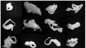
Photos of the jaw cyst clearly convince that it is easier not to get sick than to be treated. A problem that is not noticeable at first can lead to tooth loss, bone fractures, the formation of a purulent focus and sepsis, inflammation of the periosteum and bone tissue, phlegmon of the face or neck. Let's remember about cosmetic defects and painful surgical treatment, the possibility of "degeneration" into a malignant tumor. To be convincing, here are a couple more examples.
- "Lost tooth, just think ... There is still a lot left." Have you heard that? Do you know that Swedish scientists have proven that the loss of each tooth negatively affects thinking and memory, now they are calculating the connection between the number of lost teeth and Alzheimer's disease, in the common people its initial stage is called "senile dementia"; only in our time the disease has become very young, cases of the disease are described at the age of 25 - 30 years.
- Few teeth - food is poorly chewed, digestion is also difficult, a person does not receive many nutrients, problems begin - a terrible appearance, become worse eyesight and hearing, hair falls out and skin gathers in nightmarish folds, bones become thinner, a person ages much earlier than usual.
Maybe, given the above, it is worth overcoming the fear of white coats and coming to the dentist for a checkup as expected, twice a year? If you really do not want to go alone, gather the whole family or a group of like-minded people to go to the doctor, at least it will not be scary together. As a preemptive maneuver - just adhere to basic rules, they will help to detect a cyst in time or even avoid its appearance:
If a tooth hurts, be sure to take an X-ray, even if no signs of caries are visually detected. In addition to brushing your teeth, watch your mouth. Use dental floss and mouthwash. The main thing is to put the idea of self-medication out of your head forever, immediately go to a trusted specialist, and everything will be in order.
Cysts are benign growths that can form both on the surface internal organs and on mucous membranes oral cavity... At the same time, jaw cysts are most often found in young people and children. Why are they dangerous? Do they require urgent treatment? Or is a jaw cyst a completely safe formation and there is no need to fight it? Let's discuss this.
What it is?
The jaw cyst is a kind of tumor, the inner walls of which are lined with squamous epithelium. Inside it there is a pathological exudate, the amount of which is constantly increasing, as a result of which the cyst gradually grows.
The provoking factor of its formation is often the degeneration of residual bone tissues in the jaw region, which for some reason did not dissolve after eruption of a tooth or various dental procedures (for example, incorrect tooth extraction).
His appearance the cyst resembles a small vesicle that has soft but dense membranes. She is motionless and has clear boundaries. It can be formed both on the lower and upper jaw. However, as practice shows, the cyst of the upper jaw is diagnosed in patients much more often than the lower one.
Varieties
Jaw cysts have their own classification. Depending on the location and content of the formations, cysts are divided into:
- primordial (the second name is keratocysts);
- radicular;
- follicular;
- residual.
Primordial tumors are pathological formations with a thin membrane consisting of fibrous tissues. Their "favorite" place is the area where wisdom teeth erupt.
A primordial cyst can consist of one or more chambers. Inside it is a cholesteatoma (a mixture of dead cells, keratin, crystals). The peculiarity of this formation is that even after its removal in the process of restoring the tissues of the jaw, it can be formed again, while having a size much larger than before.
Most often, patients are diagnosed with a radicular cyst of the lower or upper jaw. It is formed mainly near the root of the dental tissue due to chronic periodontitis.
This tumor contains fibrous tissue inside itself. And its capsule consists of lymphocytes and plasma cells. In the case of their inflammation, the cells begin to multiply actively, gradually filling the entire cavity of the cyst, thereby provoking the onset of inflammatory processes in it, accompanied by severe pain syndrome. At the same time, the active multiplication of plasma cells can lead to the germination of a cyst in the area of the axillary cavities, provoking the development of chronic sinusitis.
The follicular cyst of the lower jaw appears against the background of a violation of teething. It is mainly formed in the area of 2 and 3 premolars. The follicular cyst of the jaw is special in that there is a follicle inside it, from which a full-fledged tooth can form.
A residual cyst can also form on both the lower and upper jaws. In this case, the place of its localization is often the area where the tooth was previously extracted. This tumor can grow rapidly, affecting more than half of the jaw.
Examining a residual cyst on an X-ray image, it can be noted that the root parts of the nearby teeth are in its cavity, but they, as a rule, diverge. Inside the formation is the root of the previously extracted tooth.
Various factors can provoke the appearance of a jaw cystic formation. Among them, the most common are:
- Violation of the integrity of the tissues of the gums or teeth in case of injury.
- Infections.
- Abnormal tooth structure.
- Dental prosthetics.
- Performing various dental procedures in violation of their technology.
Important! Most often, jaw cysts are of a kind defensive reaction the body for infections. Therefore, to prevent their appearance, it is necessary to timely treat diseases of the oral cavity, while contacting only specialized clinics.
Symptoms
Exists different kinds jaw cysts and symptoms in this case directly depend on the nature of the formation and its size. Small tumors that form on the upper or lower jaw are almost non-existent. The only thing that the patient can feel is the presence foreign body in the mouth.
When are cysts in maxillofacial area grow to large sizes, their walls can thin the tissues of the jaw, which provokes tissue inflammation and the appearance painful sensations in the field of localization of pathological processes.
Sometimes the symptoms may resemble the development of osteomyelitis and other diseases of the oral cavity, accompanied by acute purulent processes. But this happens most often in cases where the patient does not seek medical help in a timely manner and the tumor, as well as the surrounding tissues, become inflamed.
Diagnostics
Before treating a cyst, the doctor needs to establish several factors - how it appeared, that is, to identify the exact cause of its formation, and the type of formation. For this, the following diagnostic measures are carried out:
- examination of the oral cavity;
- X-ray examination of the jaw;
- MRI.
To determine the nature of the contents of the tumor, a puncture or biopsy is done. Additionally, it may be necessary to pass OAM and UAC to identify inflammatory and infectious processes in the body.
Unfortunately, cysts on the upper and lower jaws do not respond conservative treatment... They are always removed regardless of their size or the nature of their content. ChLH ( Maxillofacial Surgery) currently offers only 2 methods for removing such formations:
- cystotomy;
- cystectomy.
A cystectomy is an operation in which the entire body of the tumor is removed, followed by suturing. Applicable this method surgical intervention only when:
- The reasons for the formation of a cyst are various congenital malformations of the tissues of the oral cavity.
- This tumor covers several root sites at once and is small in size.
- If there is a tooth in the cyst.
The operation itself is performed under local anesthesia. Takes no more than 1 hour and is easily tolerated by patients. However, this method of treating jaw cysts has one significant drawback - the presence of large wounds, which can undergo infection and inflammatory processes at any time. And to prevent this, in some clinics, biocomposite materials are used with which the operated areas are treated. They contribute to the rapid regeneration of damaged tissues and help prevent infections and inflammation.
Cystotomy is a slightly different type of surgery, during which the tumor itself is not removed. Only its anterior wall is resected, after which it is connected to the oral cavity. In other words, such an operation is performed with the aim of reducing cystic formation.
Cystotomy is much easier for patients to tolerate than cystectomy. But it has a big drawback - after a short period of time, the cyst can again fill with pathological contents, after which it will be necessary to carry out a second operation. However, despite this, cystotomy is used much more often than cystectomy, for the reason that it allows you to preserve the integrity of the teeth located in the tumor zone.
If the cyst is large, two methods of surgical intervention can be used to remove it at once. Moreover, at the first stage, the tumor is reduced, and only after a few years its complete removal.
Possible complications
Despite the fact that no method surgical treatment does not give a 100% guarantee that the cyst will not reappear after a while, it cannot be left unattended. After all, any benign tumors can degenerate into cancer, which is then impossible to cure without the use of chemotherapy and radiation.
In addition, if the neoplasms in the oral cavity are not removed, they can lead to loosening of the teeth and their further loss, which will require jaw prosthetics.
Therefore, it is not worth delaying the treatment of cysts on the upper or lower jaws. The sooner they are removed, the lower the risk of future health problems.
A large cyst can disrupt the oval of the face, protruding from the side of its localization. Especially dangerous are cysts of the upper jaw, which increase towards the maxillary sinus, without manifesting outward signs... Cyst growth is always slow, the initial stage is latent without clinical signs. Cystic formation can become an accidental finding during a planned visit to the dentist, however, in 85-90% of cases, a cyst is determined during an exacerbation, when it manifests itself as suppuration and severely deforms the jaw. Dentists consider pathological fractures of the jaw provoked by thinning of bone tissue to be the most difficult cases. Also, a serious complication is the growth of a large cyst into the nasal cavity and even into the orbit area.
The jaw cyst can be of two types - odontogenic or non-odontogenic.
An odontogenic cyst is a direct consequence of a chronic advanced inflammatory process in periodontal tissues. An odontogenic cyst can cause symptoms of general intoxication, since for a long time the neoplasm releases decay products of pathogenic microorganisms into the body. Intoxication manifests itself elevated temperature body, transient dull headaches. Suppuration of the cyst is expressed in severe swelling of the jaw tissues, throbbing pain, asymmetrically swollen face. Odontogenic cysts are divided into the following types:
- Keratocyst.
- Follicular cyst.
- Radicular cyst.
- Root cyst.
Among all types, only radicular and root cysts can be considered a purely bone cyst.
- A radicular cyst is diagnosed most often, according to statistics, this type of cyst is determined in 55-60% of patients with characteristic clinical signs benign tumors of the jaw skeletal system... The cyst develops in the focus of chronic inflammation - periodontitis, often its beginning is a granuloma. The favorite localization of the radicular cyst is the bone of the upper jaw. Cysts in this zone can reach 3-4 centimeters, they tend to hyperplastic in the form of processes towards the wall of the cavity, also radical cysts quite often suppurate, while inflammatory process captures the maxillary cavity, provoking odontogenic sinusitis. A large cyst grows slowly, chronically destroying the jawbone and thinning its cortical layer. In 3-5%, radical odontogenic cysts of the jaw are capable of malignancy.
- A root odontogenic cyst is also formed as a consequence of a chronic inflammatory process. It grows very slowly, presses on the jawbone tissue, which moves compensatory, thereby disrupting the normal functions of the dentoalveolar apparatus. A root cyst is characterized by spontaneous pathological fractures of the jaw, osteomyelitis or malignant tumor jaw.
The follicular cyst of the jaw develops from the follicle of the tooth. Almost everyone admits that the cause of the formation of this pathology is the vicious development of the dental follicle, and its damage may also be the cause. The cyst occurs due to the inflammatory process at the tops of milk teeth. In most cases, follicular cysts are observed before the age of 18.
A cyst is a hollow tumor that consists of a membrane that contains liquid, semi-liquid, or mushy (cholesteatoma) contents. The membrane of the cyst consists of an outer layer, represented by dense connective tissue, and an inner layer, covered with stratified squamous epithelium, similar to the epithelium of the oral mucosa.
Jaw follicular cyst symptoms
Follicular cyst develops, as a rule, without subjective symptoms, however, clinically, a certain staging can be observed.
Stage I - latent development of the disease, which is characterized by the absence of clinical symptoms. This stage corresponds to the beginning of the pathological process around the follicle or tooth crown.
Stage II is characterized by the appearance of deformation of the alveolar process of the jaw or its body due to dense, painless swelling. With thinning of the bone wall, a parchment crunch and fluctuation are noted. Cyst infection is more common in stage II.
The diagnosis is confirmed by X-ray. The X-ray shows a bone loss of round or oval shape with clear boundaries. An impacted tooth is visible in the center.
Treatment of the follicular cyst of the jaw
Treatment is only surgical - cystectomy and removal of the impacted tooth. With extensive cysts of the lower jaw, a pathological fracture is possible, therefore, prophylactically, a fixing splint should be applied to the teeth. Fixation can be carried out both with metal bars made of aluminum or steel wire, and with aligners made of fast-hardening plastic according to the Frigof method.
The article was prepared and edited by: surgeonVideo:
Healthy:
Related articles:
- The omentum cyst occurs either as a result of blockage of the lymphatic tract, or as a result of the proliferation of a lacerated primordium of the lymphatic ...
- Fibrous dysplasia of the jaw (Braitsev-Lichtenstein disease) occurs mainly in childhood as...
- An ovarian cyst is a fluid-filled bladder ranging in size from a couple of millimeters to ten centimeters (benign ...
Among the most serious dental problems are not caries, as it might seem, but cysts. Often these neoplasms form over several years without showing any signs of life. Usually a cyst is discovered by accident, but if the patient is unlucky, it will continue to develop, sometimes triggering the oncological process. Among other types of cysts, follicular is quite rare, but this does not make it less dangerous.
Why arises
A peri-root, or follicular cyst is a direct result of improper formation or damage to the tooth-forming epithelium, as a result of which a cystic degeneration of the follicle tissue occurs.
Most often, the diagnosis of "follicular cyst" is made to young people 12-15 years old, as well as those who have reached the age of 30. Most of the patients are men. The appearance of a cyst is in one way or another associated with the presence of an unerupted tooth in the bone: both supernumerary and intact.
There is an opinion that a follicular cyst develops against the background of untreated inflammation of the root apex of a milk tooth: the inflammatory process gradually “gains momentum” and reaches the rudiment of a permanent tooth, irritating it and provoking the appearance of a cyst.
Depending on the stage at which a failure occurred in the development of the dental follicle, a cyst can either contain a dental crown or be without it.
Symptoms
The cyst is a single-chamber cavity filled with pasty, liquid and semi-liquid contents. A puncture reveals a yellow liquid with an admixture of cholesterol crystals. The cyst membrane consists of a dense connective tissue and stratified epithelium, similar to the epithelium of the oral mucosa.
In most cases, the follicular cyst is localized near the lower wisdom teeth (56% chance) and canines. Less commonly, it fills the maxillary sinus or is located in the nose or below the orbit.
The development of a cyst is a rather lengthy process that goes through two stages:
- There are no visible symptoms.
- A painless or less painful swelling of the gums appears. The pressure on it is easy, the build-up is malleable. If the cyst is large enough, a parchment crunch may be heard.
In the second stage, cyst infection usually occurs. The fluid filling it becomes cloudy and contains many white blood cells.
On average, the growth of a cyst takes from several months to several years. In many cases, due to its painlessness, the process remains unnoticed, even if the bone tissue of the jaw is significantly affected. Usually the patient goes to the doctor when the alveolar processes are already growing clearly. Among other things, in many patients there is a thickening of the jaw and a corresponding deformation of the face in this place.
On x-ray, the follicular cyst is very clearly visible: it looks like a well-defined rounded spot more than 3 mm in size. There is some difficulty in distinguishing between a normal follicular sac and a cyst: in the picture they differ only in size (normally, the enlightenment should not exceed 2.5-3 mm).
Treatment
If a person develops a follicular cyst, treatment is possible only under the supervision of a doctor: none folk remedies will not help. The only option is to trust the surgeon. Surgical intervention is planned individually for each patient, taking into account the following factors:
- where is the cyst and what size it is;
- whether there is suppuration;
- how badly the bone tissue is affected;
- are there any prospects for the eruption of an impacted tooth.
There are two main treatments for follicular cysts:
- Cystectomy - involves the complete removal of both the membrane of the cyst with its epithelial lining, and unerupted teeth.
- Cystotomy - involves the transformation of a cyst into a cavity communicating with the oral cavity. To do this, the anterior wall of the cyst is removed, the fluid is aspirated, a mucosal flap is inserted into the cyst cavity and fixed there by filling the cavity with iodoform gauze. Within a week, the mucous membrane grows with the inner epithelium of the cyst.
If the cyst is caused by inflammation, then both cystotomy and cystectomy are equally effective. When treating adult patients, the physician usually prefers the cystectomy method.
Plastic cystotomy makes it possible to preserve the tooth, near which the cyst has developed, due to its movement and further correct eruption. This method is preferred when treating children.
The "trick" of the follicular cyst in its inconspicuous development. Therefore, it is so important to monitor the condition of your oral cavity and, at the slightest symptoms of gum disease or caries, contact your dentist. This will increase the chances of early detection of the cyst and its early treatment.
Yet