The structure of the spinal cord. Short content. Spinal cord. Side rope conductors
- A rather complicated system that is responsible for many processes in the body and in which it is difficult to understand it is quite difficult. Elementary knowledge can be obtained by studying an anatomy at school, but when it comes to a deeper analysis, many incomprehensible moments arise.
Let's try to figure out what the spinal cord is as it works, what functions do, and just understand why it is needed at all.
Read also

Spinal cord as part of the nervous system
- One of the components of the human nervous system. On Latin his name looks like medulla Spinalis.
It is a thick cylindrical tube with a narrow channel located inside it. Located in the spine canal, but, speaking easier, inside the spine.
This body has a rather complicated structure and segmental structure. The main function of this organ is the transfer of various pulses and signals from the human brain to specific organs. In addition, he performs reflex activity, that is, it is responsible for human reflexes, and it is both simple and more complex reflexes.

Nervous system of man
The value of the spinal cord
There are only two main and most important features:
- Reflex. Simply speaking, a number of reflex arcs are closed on this authority. It is due to this that reflexes are carried out (the so-called spinal reflexes).
- Conductive. The body in this case acts as a conductor. It holds signals that come from various organs to the brain. It is through this body a brain receives all the information and processes it. It all works in the opposite direction.
The location of the spinal cord
The organ is located in the spinal canal (located). This channel is rather long and practically reaches the lower vertebrae. In essence, this is a special channel, which is an oblong hole in which the spinal cord is. From the parties, it is protected by vertebrae, as well as intervertebral discs.
Also, the organ is located at the lower edge of the large occipital opening, where there are connections with a brain. It is in this place that there is a huge range of roots, which is directly connected to the human brain. Such a compound is called left and right spinal nerves.

The location of the spinal cord
The bottom ends on the damage 1-11 damage. After the organ turns into a thin terminal thread. In fact, it is still a spinal cord, because the nervous tissue contains.
Topography and shape of the spinal cord
We will deal with the features of the location (topography) and forms.
To do this, consider a number of features: 
- Middle Length 42-43 centimeters. Men's length often for several centimeters more, and in women, on the contrary, less.
- Mass 33-39 grams.
- On the front there is a median gap, it is well noticeable. It can be noted that it seems to grow into an organ. In fact, it creates a peculiar partition that separates the brain into two departments.
- In the cervical and lumbar sacral departments you can
- metut two sufficiently serious thickening. This is due to the fact that the innervation of the upper and. Speaking simple language, here the nerve endings from the limbs "join" to the spinal cord, which
- sumpts them to transmit the necessary signals.
- The spinal cord is topographically practically associated with vertebrae. Various departments are disposed of not depending on the specific vertebra or several vertebrae.
Increasing the volume on these areas is due to the fact that it is here that the largest number of nerve cells are located, as well as fibers for which signals from the limbs and back are transmitted.
Despite the fact that the spine is a kind of "place for storing" the organ, the location of the nerve endings, especially at the bottom of the spine, do not correspond to the specific vertebrae. This is due to the fact that the long spinal cord is less than the length of the human spine.
That is why for physicians it is necessary to know the exact location of each of the segments, because the spine can not be focused.

Location in the spine
 The stories of our readers!
I want to tell my story, how I cured osteochondrosis and hernia. Finally, I was able to overcome this unbearable back pain. We lead an active lifestyle, I live and rejoice every moment! A few months ago I twisted me at the cottage, a sharp pain in the lower back did not move, did not even be able to go. The doctor in the hospital diagnosed osteochondrosis lumbar Department Spine, Hernia Disk L3-L4. I prescribed some kind of medicine, but they did not help, to endure this pain was unbearable. They called an ambulance, they set the blockade and hinted for the operation, all the time thought about it, that I would find a burden for the family ... Everything changed when the daughter gave me one article on the Internet. You can not imagine how much I am grateful for it. This article literally pulled me out of a wheelchair. The last months began to move more, in the spring and summer every day I go to the cottage. Who wants to live a long and energetic life without osteochondrosis,
Characteristics of the spinal cord depending on age
Consider features, depending on the age of a person:
- At the newly born child, the body length is 13.5-14.5 centimeters.
- At 2 years, the length increases to 20 centimeters.
- Approximately 10 years old can reach 29 centimeters.
- The growth ends in different ways, depending on the characteristics of the body of a particular person.
We will understand external features and changes depending on age:
- In babies, cervical and lumbar thickening are noticeable more than in adults. Also concerns the width of the central channel.
- The above features become almost imperceptible to two years.
- The volume of white substance grows at times faster than the volume of gray. This is due to the fact that the segment apparatus is formed earlier than conductive paths that connect the head and spinal cord.
In the rest age features It is practically not observed, because from the very birth of the spinal cord performs almost all the functions as in an adult person.
The features of the structure of the spinal cord
Now consider the features of the structure, alternately considering each segment individually, of which the authority consists.
Spinal cord shells
The spinal cord is in a kind of channel, but it has protection that also performs a huge number of functions.
Spinal cerebral cerebral shells, which are totaling three:
- solid sheath;
- arachnoid;
- soft shell.
All shells are interconnected, and at the bottom they grow up with terminal thread.

Spinal cord shells
In the spinal cord there is a white and gray substance.
Let's try to figure it out what it is:
- White substance - A complex system of pulp and cinema nerve fibers, as well as the supporting nervous tissue.
- Gray matter - These are nervous cells and their processes.

White and gray spinal cord substance
Spinal cord departments
Severe five main spine sections, consider them starting from above:
- cervical;
- chest;
- lumbar;
- sacral;
- copchik.
Read also

Spinal nerves
They are paired nervous trunks, which are total 31 pair: 
- 8 cervical;
- 12 chest;
- 5 lumbar;
- 5 sacral;
- couple pair.
Each nerve is responsible for a certain portion of the body. On this site there are bones, muscles, internal organs or leather. The task of a particular pair of nerves is the transfer of pulses from the area to the spinal cord and back. It is thanks to this a person can feel pain, discomfort, temperature, and so on.

Spinal nerves
Spinal cord segments
Segments as much as pairs of roots - 31. The segment is a specific portion of the human body, for which the specifically taken pair of roots.
All of them are divided into:
- cervical;
- chest;
- lumbar;
- sacral;
- copchie.
Due to the fact that the length of the spine is longer than the spinal cord length, it turns out that the roots of the nerves only in the upper part correspond to the level of intervertebral holes.
Below, to get into a special hole, the nerves of the lower departments fall below parallel to the spine. Thus, they come out at the level of the end thread.

Spinal cord segments
Vienna and the arteries of the spinal cord
The organ gets blood due to the front and pair of the rear spiral arteries. But these arteries are able to provide only 2-3 top cervical segments. The rest of the rest is the root-spiral artery, which receive blood from the branches of the vertebral and the upward cervical artery.
At the bottom of the spine gets blood from intercostal and lumbar arteries. Both of these artery are peculiar proceedings of the famous artery poses by the name of Aorta.
The functions of the spinal cord
Let us turn to the consideration of functions. For convenience, we will consider each separately.
Reflex and motor functions
This feature is responsible for human reflexes. For example, if a person touched something very hot, then reflexively he will take her hand. This is a reflex or motor function. But let's turn step by step with how it is all tripled and how the spinal cord is associated with the spinal cord.
It is best to consider everything on the example, so we will present the situation that a man has touched a very hot object:
- When touching the signal primarily receives receptors that are located throughout the human body.
- The receptor transmits the signal to the nerve fiber.
- On the nervous fiber, the signal is sent to the spinal cord.
- On the approach to the organ is a spinal assembly, where the body of the neuron is located. It is by its peripheral fiber and a pulse transmitted from receptors was obtained.
- Now the central fiber pulse is transmitted to the rear horns of the spinal cord. At this point, there is a peculiar switching of the pulse to another neuron.
- The processes of the new neuron transmit a pulse to the front horns.
- Now the return path begins, because the front horn transmits the pulse to the motor neurons. They are responsible for the movement of the upper limbs.
- According to these neurons, the pulse is transmitted directly in hand, after which a person removes it (motor function).

Reflex arc in the scheme
As a result of all this process, a person pulls out his hand from the hot object and the reflex arc is closed. The whole process takes the fraction of a second, so touching any subject, a person immediately feels its temperature, a consistency and other features.
Explore function
In this situation, the authority acts as it were. Explorer in this case it is between receptors and the brain. Receptors are obtained by a pulse, which is transmitted in the spinal cord, and after the brain. Information there is analyzed and transmitted back.
Thanks to this function, the person gets sensitivity, as well as the feeling of itself in space. It was repeatedly proven, especially the like becomes apparent with serious spinal injuries.

Generative function
About this feature is often forgotten, but it is no less important for a person than others. The integrative function is manifested in reactions that cannot be attributed to simple reflexes. In order for the body to respond, it is necessary to use other parts of the nervous system of the human body. So the spinal cord can form the bond with each other.
Here you can include reflexes of chewing, swallowing, regulation of digestion, breathing and much more. In essence, this is an inconspicuous function that provides normal life.
Spinal violation
Violation of functions can lead to serious consequences and often even to death. Violations often occur either due to injuries or because of various diseases.
For example, due to the violation of the spinal cord function, a person may lose sensitivity, in this case, for example, it can stop feeling the temperature. In the worst case, the violation may lead to uncontrolled actions of the limbs (or paralysis), violation of the work of the internal organs and the nervous system as a whole.
Diseases of the spinal cord
The list of the most common diseases that violate the full work of the body under consideration:
- Heart attack.
- Polio.
- Cross Melit.
- Tumors.
- Decompression disease.
- The defeat of nerve roots.
- Arteriovenous malformations.
If a spin hurts, neck or loin, do not tighten the treatment if you do not want to finish in a wheelchair! Chronic many pain In the back, neck or lower back - the main sign of osteochondrosis, hernia or another serious illness. Treatment must be started right now ....
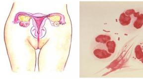
Puncture of spinal cord
Puncture spinal fluid (liquor) - a procedure that pursues diagnostic, anesthesiological and therapeutic goals. The procedure itself lies in the fact that the patient between the 3rd and 4th vertebrae is introduced the corner under a spider shell, and after a certain amount of spinal fluid for research is extracted.
In the process of procedure, the brain itself is not affected, so there is no fear of violations. But still this procedure quite serious and painful.

Puncture of spinal cord
Conclusion
Summing up, it should be said that the spinal cord is one of the most important organs in the human body. In many ways, precisely thanks to him, a person can lead normal life, as well as, thanks to this organ, almost the entire nervous system is functioning.
.Man eats, breathes, moves and carries many other functions thanks to (CNS). It consists mainly of neurons (nerve cells) and their processes (axons), for which all signals pass. It is impossible not to mark Gliya, which is auxiliary nervous fiber. Thanks to this tissue in neurons, the generation of pulses going in the head and spinal cord occurs. It is these 2 authorities that are the basis of the CNS and manage all processes in the body.
A special role is played by the spinal cord of a person and to understand where it can be located, looking at the cross section of the spine, since it is located in it. Focusing on the structure of this body, it can be understood for which it is responsible and how the relationship with most of the human systems is carried out.
The spinal cord is predominantly from the spider shell, as well as from soft and solid components. Protects the body from the damage to the fat layer localized directly under the bone tissue in the epidural space.
Most people know where the spinal cord is located, but few understand its anatomical features. This organ can be represented as thick (1 cm) the wires of the long actual half the meter, which is localized in the spine. The spinal cord is a spinal channel consisting of vertebrae, due to which it is protected from external influence.
The organ from the occipital opening begins, and ends at the level of the loin where it is represented as a cone consisting of connective tissue. It resembles a thread in shape and comes straight to the tailbone (2 vertebra). You can see the spinal cord segments in this picture:
Channels come out the roots of the spinal nerves, which serve to carry out the movements of the hands and legs. From above and in the center they have 2 thickening at the level of the neck and lower back. At the bottom of the spinal cord root resemble a tangle formed around the spinal threads.
The transverse cut of the spinal cord is as follows:

The anatomy of the spinal cord is designed to answer many questions related to the work of this body. Judging by the system of rear of the organ of localized the groove of the spinal nerve, and the special hole is located in front. It is through it that nervous rootscarrying innervation of certain systems of the body.
The inner structure of the spinal cord segment tells many details of his work. It consists of an organ mainly from the white (aggregate axon) and a gray (combination of neurons) substances. They are the beginning of many nerve paths and such segments of the spinal cord are mainly responsible for the reflexes and the transmission of signals in the brain.
The functions of the spinal cord are diverse and depend on the level of which level are nerves. For an example of white substance, nervous pathways of the front valves of the central nervous system are coming. The rear of the fibers is indicators of sensitivity. Of these, the segment of the spinal cord is formed, in which the spinal roots on both sides are collected. The main object of the white substance is the transfer of the obtained pulses in the brain for further processing.
The structure of the spinal cord of a person is not so complicated as it seems. The main thing is to remember that the spine includes 31 segments. All of them differ in size and divided into 5 departments. Each of them performs certain features of the spinal cord.
White substance
The spinal channel is the place of cluster of the white substance. It is 3 core surrounding the gray substance, and consists mainly of axons covered with myelin shell. Thanks to myelina, the signal is moving faster on them, and the substance receives its shade.
The white substance is responsible for the innervation of the lower extremities and shipping the pulses in the brain. Seeing his codes, as well as a horn of gray matter in this picture:

Gray matter
Most people do not understand what the gray substance looks like and why he has such a form, but in fact everything is quite simple. Due to the accumulation of nerve cells (motor and inserted neurons) and actually the complete absence of axons it has gray. Localized gray substance in a spinal channel and many it seems that this is a butterfly due to pillars and plates in the center.
The gray substance corresponds primarily for motor reflexes.
In his center there is a channel, which is a container of a liquid, which is a cerebrospinal fluid. Its function includes protection against damage and support for permissible pressure inside the cranial box.
The main amount of gray substance falls on the front horns. They consist mainly of motor nerve cells that perform innervation of muscle tissues at the level of this segment. Fewer substance gets back horns. They include mainly inserting neurons that serve to communicate with other nerve cells.
If you look at the spinal channel in the cut, then the intermediate zone is striking, localized in space between the front and rear horns. This area is only at 8 vertebrae of the cervical area and passes up to 2 segments of the lower back. In this area, side horns begin, which are accumulated by nerve cells.
The role of conductive paths
Conducting ways serve to communicate the spinal and brain and take their origin in the rear wire of a white substance. They are divided into 2 types:
- Rising paths (transmitting signal);
- Downlink paths (receiving signal).
To place complete information About their anatomical features need to look at this picture:

A signal is transmitted through certain beams, for example, the upper part of the body in the spinal cord represents a wedge-shaped plexus, and the lower thin. To see next to which these fibers are located in this picture:

A special role in the conductive system is performed by a renovative path. It begins from skeletal muscles and ends directly in the clogenet itself. Separate attention should be paid to the Talalamic path. He is responsible for the perception of pain and the temperature of the person. Talamus receives a signal from the front of the ceremony path, which consists mainly of inserted neurons.
Functions
A person has always had many questions regarding their body, because it is difficult to understand how all the systems are connected with each other. The spinal cord has a structure and functions are interrelated, so terrible consequences arise with any pathological changes. It is virtually impossible to eliminate them, so you need to take your spine.
Responses the spinal cord for the following functions:
- Conductive. Its essence is the transmission of the signal to certain parts of the body depending on the localization of the nervous beam. If it comes to the upper half of the body, the cervical department is responsible for it, for the authorities, the lumbar, and the sacratsone innervates the pelvis and lower limbs.
- Reflex. Such a function is performed without the participation of the brain, for example, if you touch the hot iron, the limb moves involuntarily.
Fixed spinal cosk

Many different pathologies are associated with the spinal cord, whose treatment is performed mainly in the hospital. Such diseases refers to a fixed spinal cord syndrome. This pathological process is diagnosed with extremely rarely and peculiar to the disease both children and adult people. For pathology, the fixation of the spinal cord to the spinal column is characteristic. Most often there is a problem in the lumbar department.
Fixed spinal cords are usually found in the diagnostic center with tool methods Surveys (MRI), and it arises due to such reasons:
- Neoplasms, squeezing spinal cord;
- Arising scar tissue after surgery;
- Severe injury in the field of the belt;
- Plok Kiari.
Usually, the fixed spinal cord syndrome in patients is manifested in the form of neurological symptoms and the main manifestations relate to legs and areas of damage. A person is deformed by the lower limbs, it becomes difficult to walk and failures in the work of the pelvic organs appear.
The disease occurs at any age and its course is usually consisting of an operation and a long period of recovery. Basically, after surgery, the defect is obtained and partially relieve the patient from the effects of pathology. Because of what people begin to actually walk and stop experiencing pain.

There is another pathology that some experts are associated with a spinal cord, namely hemispasm (hemifacial spasm). It is a disturbance of the facial nerve due to the reduction of muscle tissue, which is on the face. The disease occurs without pain and such spasms are called clonic. They arise due to the squeezing of the nervous tissue in the area of \u200b\u200bits exit from the brain. The diagnosis of the pathological process is carried out using MRI and electromyography. According to statistics, each year, hemicacial spasm can be diagnosed in 1 of 120,000 people and the female floor suffers from it 2 times more often.
Basically, the squeezing of the facial nerve occurs due to vessels or neoplasms, but sometimes hemispanse arises due to such reasons:
- Process of demyelination;
- Spikes;
- Bone anomalies;
- Tumors located in the brain.
Hemifacial spasm can be eliminated with medication therapy. For the treatment of facial nerve, a bitplopen, levatracy, gabapentin, carbamazepine, etc., will have to take them quite a long time, so such a course has its own minuses:
- Over time, the effect of drugs begins to end and faster and for the treatment of facial nerve will have to change drugs or increase the dosage;
- Many listed drugs have a sedative effect, therefore people who are diagnosed with hemispanis are often in sleepy state.

Despite the minuses, many cases were recorded full cure Facial nerve and removal of hemispasza. Drug therapy for the early stages of the development of pathology was particularly well influenced.
Eliminate hemicacial spasm can also be used toxin injecting botulin. It effectively eliminates the problem at any stage. Of the minuses of the procedure, you can note the high cost and contraindications in which allergic reactions The composition of the drug and eye disease.
The most efficient and rapid treatment of hemispanis is surgery. It is carried out with the aim of eliminating compression and, in the case of a patient's successfully conducted operation, written after a week. The full effect of recovery is achieved quite quickly, but in some cases it is stretched to six months.
The spinal cord is an important center of the nervous system and any deviations in its structure may affect the entire body. That is why, in the manifestation of neurological symptoms, contact a neurologist for the passage of examination and diagnosis.
From how the central nervous system is functioning, the work of all organs, as well as the overall health of a person depends. A big role is played by the spinal cord. It is located in such a way that is in relationship with each cell of the body. All motor reflexes are caused by its actions. This body transmits signals to the brain of the head - to the "Central Staff", which carries out opposite communication with the authorities.
What does the spinal cord look like
Structure of the brain
A man's spinal cord, something similar to an electrical cable, fills the spine channel. At the same time, inside this body of two halves, distributed among themselves the obligations of the right and left sides of the body.
Brain formation occurs on the most early stage Embryo development. It is he who is the basis for which all other elements of the embryo are increasing. Starting to develop at the end of the first month after conception, the spinal cord is differentiated throughout the pregnancy. At the same time, part of the departments undergoes the subsequent revision of the first childhood.
The whole spinal cord laid in the canal is biting a triple shell. In this case, the inner of them is sufficiently soft, consisting of vessels, outdoor - solid to ensure the protection of tissues. Between them is another "braid" - web. The space between this shell and the inner contains a liquid that provides elasticity. The inner space is filled with a gray shade substance, whipped white substance.

Cross-cut brain
If we consider the structure of the spinal cord in cross-section, then the structural shape of a gray substance is clearly distinguished in the section, resembling a small butterfly, laid down on a penet. Each of the part of the structure has certain features that are described below.
To the substance with gray "connected" the roots of the nerves, which, passing through the white substance, are assembled into nodes that determine the structure of the nerve of the spinal out. Bundles of nerve fibers are conductive ways that provide the links of the Central Staff with specific authorities. The spinal cord includes from 31 to 33 pairs of vertebrae formed in segments.
Cone brain
The vertebral channel is directly conjugate with the brain placed in the head, and the bottom line begins at the bottom line. In constant form, the channel passes up to the vertebrae of the lumbar and ends with a cone, which has a continuation of the terminal thread, the upper part contains the fibers of the nerves.
The cone in its structure is represented by a three-layer coupling cloth. On the vertebral in the area of \u200b\u200bthe tailbone, where he processed with the periosteum, and ends the thread indicated above. Here is the so-called "horse tail" - a bundle of the lower nerves, whining the thread.

What is the nervous system
The main assembly of nervous fibers is in 2 places - the sacroist-lumbar department and in the neck area. It is pronounced by peculiar seals responsible for the function of the limbs.
The brain spins, filling the spinal channel, has a strictly fixed position and unchanged parameters. Its length in an adult is about 41-45 cm, with this weight has no more than 38 g.
Gray substance
So, the brainstant on the cross section looks like a moth, and is inside a white tonality substance. The center for the entire length of the brain of the dorsal is the narrow channel, which is called the central one. This channel fills the liquid - a variety of liquid by the spinal surgery responsible for the operation of the system nervous.

Gray "Moth"
The brain and central dorsal canal are among themselves in relationships. Also are also compatible with spaces placed between the brain shells - they circulate the liquid of the spinal volume. It is her by means of puncture, they take research when a number of problems affect the spinal cord departments are diagnosed.
The gray substance is the similarity of pillars connected in cross-performance by plates. Spacks total 2: The back and front part constituting the central cerebral channel. They form a butterfly (Literature H).
In the sides of the substance, the horns are dismissed. Paired wide fill the front part, narrow - rear:
- In the front there are neurons of movement. Their processes (neurites) are formed in the spinal cord roots. The neurons are also created and the core of the brain of the dorsal, which in stock 5.
- The horn of the rear in the middle there is its own nucleus from neurons neurons. Each process (Akson) is located in the direction of the front horns crossing the spike. At the rear horn, an additional core has been formed from large neurons, which has the branching of dendrins in its structure.
- Between the main horns there is still an intermediate brain part. Here you can observe the branch of the horns of the side. But it does not appear on all segments, but only from the 6th cervical and to the 2nd lumbar. Nerves cells here create a lateral substance, which is responsible for the veguetative system.
White substance
Enveloping a gray substance The substance of a white shade is a set of 3 pairs of cakes. Between the furrows located the roots of the front rope. There are also rear and side, each of which is located between specific grooves.
Fibers forming a light substance pass through themselves signals emanating from nerves. Some are directed through the channel in the brain, others - in the dorsal and below the lying departments. Intersome relations are carried out by the fibers of a gray substance.
The roots of the spinal cord, located behind, are the fibers of the neurons of the ganglia of the spinal. The part is located in the horn of the rear, the rest diverge in different directions. A group of fibers included in the core is directed to the brain of the head - it is the ways of ascending type. Part of the fibers is in the back horns on the neurons of inserts, the rest goes to the Divines of the National Assembly.
Various paths
Above it has already been said that the brain receives signals emanating from neurons. On the same paths and in the opposite direction there is a movement of signals. A bundle of wedge-shaped neurons sends signals from endings located on the joints and muscles to the brain oblong.
The entire spinal cord filling the vertebral channel performs functions by bundles sending signals to the upper and lower part of the body. Each pulse group begins from the "its" site and moves according to their paths.
So, the core medial intermediate gives the beginning for the forefront. On the opposite side of the horns there is a path that is responsible for painful and thermal sensations. The signals are pre-entered into the brain intermediate, and then in the head.
Functional features
After examining the structure of the spinal cord, it is easy to conclude that this is a rather complicated system, "mounted" to the vertebral channel, and in the technical plan resembling an intricate circuit of an electronic device. In the perfect version, it should work safely and uninterrupted, performing certain functions programmed by nature.

System structure
From the described structure of the brain, it can be seen that it has 2 main responsibilities: to be conductor of impulses and provide motor reflexes:
- Under reflexes, they mean the ability to involuntarily pull the hand at risk randomly damage it with a hammer in the process of clogging the nails, or a sharp jump aside from the mouse running past. The reflex arc is caused by such actions that binds the muscles of the skeleton with the brain of the spinal. And there are relevant nerve impulses. At the same time there are reflexes congenital (laid by nature at the Gennel) and acquired, which developed in a vital process.
- The function of the conductor includes a pulse transmission on the rising paths from the scorn to the brain and in the reverse order - by descending. These pulses of the spinal cord distributes for all human bodies (according to the program). For example, the sensitivity of the fingers is developed precisely thanks to the conductor function - a person is adopted to the kitten, and the signal appears in the headquarters that form certain associations there.
The channel, which is performed by the functions of the motor, the beginning of its origin in the red core, turning gradually to the horns of the front. Here is a set of motion cells. Reflex impulses are transmitted on the front tracks, arbitrary - in lateral. The path to the brain is the front of the vestibular kernels ensures the execution of the equilibrium function.
Vascular system
The work of the brain is not possible without a normal blood supply, which is one for the whole organism. The spinal cord is constantly washed by blood passing by arteries - spinal and root-spinal. The number of such vessels is individually, because Sometimes additional arteries are observed in a number of people.

How does the blood supply for the brain
The rear roots (and therefore vessels) are always greater, but their arteries are less in diameter. Each vessel is washes his blood supply zone. But present in the system and the connection of the vessels between themselves (anastomosis), which ensures sufficient food for the brain of the dorsal.
Anastomosis is a spare channel used when functions are knocked out at the main vessel (for example, clogging thrombus). Then the spare element assumes the duty of blood transportation, immediately after the process.
Vessel plexes are formed in the shell. So each root of the nerve system is accompanied by veins and arteries that form a neuro-vascular bundle. It is his damage that leads to various pathologies manifested by pain symptoms.
To identify such a violation, you will have to go through a number of different diagnostic studies.
Each artery is accompanied by hollow veins, in which blood flows out of the spinal cord. So that the fluid has not returned back, a set of special fencing valves is located on a solid cerebral shell, which determine the correct direction of movement of the circulatory "river".
Video. Spinal cord
Without the normal reliable operation of such an important organ as a spinal cord, it is impossible not only to move, but also breathe. Any activity (digestion, measurement and urination, heartbeat, libido, etc.) is unthinkable without his participation, because Brain functions are fully guided by all these actions.
It is they who warn a person from various bruises and injuries, because Impulses carry information not only about touch, odors, movements, but also orient the body in space, and also help respond to danger. Therefore, it is so important to maintain the performance of an important component pushed into the spinal channel.
The man's spinal cord is the most important organ of the central nervous system, communicating all organs from the central nervous system and conducting reflexes. It is covered with three shells:
- solid, bow and soft
Between the web and soft (vascular) shell and in its central channel is located spinal fluid (likvor)
IN epidural space (gap between solid brain sheath and spine surface) - vessels and adipose tissue
The structure and functions of the spinal cord of a person
What is the spinal cord on the external structure?
This is a long cord in the spinal Channel, in the form of a cylindrical shape, about 45 mm long, about 1 cm wide, a flatter front and rear than on the sides. It has a conditional upper and lower boundaries. The top begins between the line of a large occipital hole and the first cervical vertebra: in this place the spinal cord is connected to the head through the intermediate oblong. Lower - at the level of 1 -2 lumbar vertebrae, after which the cord takes a conical appearance and then "degenerates" into a thin cerebrospinal thread ( terminal) with a diameter of about 1 mm, which stretches to the second vertebra of the Copshing Department. The terminal thread consists of two parts - internal and outdoor:
- internal - approximately 15 cm long, consists of nervous tissue, is intertwined with lumbar and sacrilant nerves and is located in a solid brain sheath bag
- outdoor - about 8 cm, begins below the 2nd vertebra sampling department and stretches in the form of a compound of solid, web and soft shells up to the 2nd cleaned vertebra and sprouts with periosteum
The outer, hanging to the square itself the terminal thread with the intertwing nerve fibers is very reminded by the type of horse tail. Therefore, pain and phenomena arising from pinching nerves below the 2nd sacrilate vertebra are often called horse-tail syndrome.
The spinal cord has thickening in the cervical and lumbosacral departments. This finds its explanation in the presence of a large number of nerves in these places going to the top, as well as to the lower limbs:
- The cervical thickening is distributed on the length of the 3rd - 4th cervical vertebrae to the 2nd breast, reaching a maximum in the 5th - 6th
- Lumbar-sacral - from the level of the 9th - 10th breast vertebrae to the 1st lumbar with a maximum of 12th breast
Gray and white spinal cord substance
If you consider the structure of the spinal cord in the cross-section, then in the center it can be seen a gray area in the form of a butterfly opened its wings. This is a gray spinal cord substance. It is surrounded by a white substance. The cellular structure of the gray and white substance differs among themselves, as well as their functions.

The gray substance of the spinal cord consists of motor and inserted neurons:
- motor neurons transmit motor reflexes
- inserts - provide communication between the neurons themselves
White substance consists of the so-called aksonov - Nervous processes from which descending and ascending paths are created.
Wings "Butterfly" narrower form front horns gray substance, wider - rear. In the front horns are motor neurons, in the rear - insert. Between the symmetric lateral parts there is a transverse jumper from the brain tissue, in the center of which the channel passes, communicating the upper part with the ventricle of the brain and filled with the spinal fluid. In some departments or even throughout adults in adults, the central canal can overeat.
Relative to this channel, on the left and right of it, the gray substance of the spinal cord looks like a pillars of a symmetrical shape, interconnected by the front and rear spikes:
- front and rear poles correspond to the front and rear horns on the cross section
- side protrusions form a side pillar
The side protrusions are not for the whole length, but only between the 8th cervical and 2nd lumbar segments. Therefore, the transverse cut in segments, where side protrusions are lacking, has an oval or round shape.
The connection of symmetric pillars in the front and rear parts forms two furrows on the surface of the brain: anterior, deeper, and rear. The front gap ends with a partition adjacent to the rear border of the gray substance.
Spinal nerves and segments
On the left and right on these central furrows are located respectively protectomeraland rearrelheralfurrows through which the front and rear threads come out ( axons), forming nervous roots. The front root in its structure is motor neurons Front horns. Rear responsible for sensitivity consists of inserts neuronsrear horns. Immediately at the outlet of the brain segment and the front and rear spores are combined into one nerve or nerve knot ( ganglion). Since in every segment there are two front and two rear roots, in the amount they form two spinal nerve (one on each side). Now it is not difficult to calculate how much nerves has a spinal cord of a person.

To do this, consider its segmental structure. Total there are 31 segments:
- 8 - in the cervical department
- 12 - in breast
- 5 - lumbar
- 5 - in the sacrats
- 1 - in the copier
So the spinal cord has only 62 nerves - by 31 on each side.
Departments and segments of the spinal cord and spine are not at the same level due to the difference in length (spinal cord shorter spine). This should be considered when comparing the brain segment and the vertebra number during radiology and tomography: if at the beginning of the cervical system, this level corresponds to the vertebral number, and in the lower part it lies on the vertebra above, then this difference has already been in the sacrum and sparrels of the department.
Two important spinal cord functions
The spinal cord performs two important functions - reflex and conductive. Each segment is associated with specific organs, providing their functionality. For example:
- The cervical and chest department - binds to heads, hands, organs chest, breast muscles
- Lumbar department - the gastrointestinal bodies, kidneys, muscular system Torch
- Sleepy Department - body pelvis, legs
Reflex features are simple reflexes embedded by nature. For example:
- pain reaction - take healing hand if it hurts.
- ball Reflex
Reflexes can be carried out without the participation of the brain
This is proved by simple animal experiences. Biologists carried out experiments with frogs, checking how they react to pain in the absence of a head: a reaction was noted both on weak and for severe pain irritants.
The conductive functions of the spinal cord are in conducting a pulse along the rising path in the brain, and from there - on the downward path in the form of a reverse command of some organ
Thanks to this conductor, any mental action is carried out:
stand up, go, take, throw, raise, run, cut off, draw - And many others who people, without noticing, makes in their everyday life in everyday life and at work.
Such a unique bond between the central brain, the dorsal, the entire CNS and all organs organs and its limbs, as before, remains the dream of robotics. None, even the most modern robot, is not yet able to carry out the thousandth share of those all possible movements and actions that are subject to bioorganism. As a rule, such robots are programmed for narrow specialized activities and are mainly used on conveyor automatic industries.
Functions of gray and white substance.To understand how these magnificent spinal cord functions are carried out, consider the structure of the gray and white matter of the brain at the cellular level.
The gray substance of the spinal cord in the front horns contain the nerve cells of large sizes, which are called efferent (motor) and combine five cores:
- central
- frontocel
- rearverteral
- promotional and rearbed
The sensitive roots of small cells of the horses are specific cell processes from sensitive spinal cord assemblies. In the back horns, the structure of the gray substance is heterogeneously. Most of the cells form their own nuclei (central and chest). The border zone of the white substance, located near the rear horns, is adjacent to the spongy and pupil zone of the gray substance, whose cells, together with the proceedings of small diffuse scattered cells of the rear horns, form synapses (contacts) with the neurons of the front horns and between adjacent segments. These neurites got the name of the front, side and rear eating beams. The connection of them with the brain is carried out using the conductor paths of the white substance. On the edge of the horns, these bundles form a white cut.
The side horns of the gray substance perform the following important functions:
- In the intermediate zone of the gray substance (lateral horns) are located sympathetic Cells Vegetative nervous system, it is by means of them with internal organs. The processes of these cells are connected to the front roots
- Here is formed sneakerszhachy way:
At the level of the cervical and upper breast segments is Reticular Zone - a beam from a large number of nerves associated with the activation zones of the cerebral cortex and reflex activity.

The segmental activity of the gray matter of the brain, the rear and front roots of the nerves, its own beams of white substance, barking gray, is called the reflex function of the spinal cord. Reflexes themselves are called unconditional, By definition of Academician Pavlov.
The conductive functions of the white substance are carried out by three cakes - the outer areas bounded by furrows:
- Front rope - a plot between the front median and lateral furrows
- Rear rope - between the rear median and lateral furrows
- Side rope - between frontoolel and rear agent furrows
The axons of the white substance form three conductivity systems:
- short bundles called associative fibers that bind various spinal cord segments
- ascending sensitive (afferent) Bundles aimed at brain departments
- descending motor (efferent) Bundles directed from the brain to the neurons of the gray substance of the front horns
Ascending and descending pathways.Consider for example Some functions of white substance rope paths:
Front ropes:
- Front pyramid (cortical-spinal) path - Transmission of motor pulses from the cortex of the brain to the cerebrospinal ones (front horns)
- Spinolamic front track - transfer of impulses of touching the skin surface (tactile sensitivity)
- Covenno-cerebral pathway - binding visual centers under the cortex of the brain with the nuclei of the front horns, creates a protective reflex caused by sound or visual stimuli
- Held and Leventhal bundle (predver-spinal path) - fibers of white substance bind vestibular nuclei eight pairs of cranial brain nerves with engine neurons of the front horns
- Stretch rear beam - Combining the upper segments of the spine with the brain barrel, coordinates the work of the eye muscles with the cervical and others.
The rising paths of the side cords are carried out impulses of deep sensitivity (sensations of their body) on cortical-spinal, spinatelamic and cooked-cerebrospinal routes.
Downlink ways of side cordics:
- Lateral cortical-spinal (pyramid) - transfers the pulse of movement from the cortex of the brain to the gray substance of the front horns
- Red-cerebral pathway (It is ahead of the lateral pyramidal), the neighboring rear and spinnotelamic side path are adjacent to it.
The red-cerebrospinal output path automatic control of movements and muscle tone at the subconscious level.

In different spinal cord departments, various ratios of gray and white brainstatons. This is explained by different ascending and descending paths. In the lower spinal segments larger than the gray substance. As you move upwards it becomes less, and white substance On the contrary, it is added, as new rising paths are added, and at the level of the upper cervical segments and the middle part of the chest white - most. But in the area of \u200b\u200bboth cervical and lumbar thickening, the gray substance prevails.
As you can see, the spinal cord has a very complex structure. The connection of the nerve beams and fibers is vulnerable, and serious injury or disease are able to break this structure and lead to a violation of conductive paths, which is why the point of "cliff" conductivity can be a complete paralysis and loss of sensitivity. Therefore, at the Malley hazardous signs The spinal cord must be examined and treated on time.
Puncture of spinal cord
To diagnose infectious diseases (encephalitis, meningitis, etc. Diseases), the puncture of the spinal cord is used (lumbar puncture) - the needle in the cerebrospinal channel. It is carried out in this way:
IN subarachnoidal The spinal cord space at the level below the second lumbar vertebra is introduced by a needle and a fence is carried out. spinal fluid (likvora).
This procedure is safe, since below the second vertebra in an adult, the spinal cord is absent, and therefore there is no threat to his damage.
However, it requires special care, so as not to be taken under the shell of the spinal cord infection or epithelial cells.
The puncture of the spinal cord is carried out not only for diagnosis, but also for treatment, in such cases:
- introduction of chemotherapeutic medicines or antibiotics under the brain shell
- for epidural anesthesia during operations
- for the treatment of hydrocephalus and reducing intracranial pressure (removal of excess liquor)
The puncture of the spinal cord has such contraindications:
- stenosis of the spinal canal
- displacement (dislocation) of the brain
- dehydration (dehydration)
Take care of this important organ, engage in elementary prevention:
- Take antivirus drugs during the epidemic of viral meningitis
- Try not to arrange picnics in the forest park zone in May-early June (the period of activity of the encephalitic tick)
- After each hike in the forest, inspect the whole body, and at the very first signs of the disease, go to the doctor. Signs such: headache, high temperature, neck rigidity (movement difficultness), nausea.