The consequences of the laser coagulation of the retina. Retinal strengthening operation with laser: restrictions Repeat procedure
The laser of the retina of the eye is an integral part of modern ophthalmic microsurgery. Such operations are carried out by many patients with the degenerative pathologies of the retina, as well as to prevent complications.
Causes and symptoms of the retina
Laser coagulation is most often carried out during thinning and. Each person should be able to distinguish the symptoms of pathology from eye fatigue to seek help.
Causes of retinal pathologies:
- disorders of refraction (myopia, hyperopiasis);
- circulatory defects;
- age violations;
- cataract;
- negative impact (injury, excessive load);
- diseases of other organism systems (stress, neurological disorders, sharp increase in blood pressure).
The risk of retinal rupture is that pathology is poorly expressed, and sometimes it does not appear at all. The patient simply cannot recognize the presence of a problem and ask for help. Therefore, it is very important to regularly undergo prophylactic inspections at the oculist.
Symptoms of retinal rupture:
- the appearance of light outbreaks and glare in front of the eyes (the symptom is enhanced in the dark);
- reduction of visual acuity;
- image insanity;
- narrowing of fields of view;
- distorted perception of objects.
The retinal breaks often end with detachment. Only an experienced doctor who has special equipment is capable of identifying violation. When the retinal breaks are detected, it is recommended to undergo a laser coagulation procedure. In these cases, preventive or limiting operations are prescribed.
If not to treat the retinal gap, it will definitely provoke heavy complications. The most common - retinal detachment - can deprive a person of vision forever. With sudden I. harsh deterioration Vision to help the patient will be very difficult. Not always doctors manage to return to the place the peelled retina, but even after successful operations, vision is often completely restored.

Features of laser coagulation in retinal pathologies
Most often degenerative retina processes develop on high and middle degrees myopia when the form changes eyeballAs well as when stretching the shell and cellular disorders. Strengthening the retina with a laser is one of the few procedures to eliminate such violations.
Purpose laser operation When retinal detachment is the creation of spike (merge) between the retina and the adjacent vascular sheath. It is possible to achieve such an effect with the help of a laser coagulator, which increases the temperature in the tissues and forms local micro-rigid retina.
Laser coagulation allows you to limit the flat detachment in people who have contraindications to radical surgical intervention. The operation is also shown as an additional measure after surgical correction of detachment.
Types of laser coagulation
- Restrictive prophylactic. This procedure is considered therapeutic and prophylactic. The laser makes dosage burns, creating a barrier around the retinal dystrophy, which helps to stop the process and promotes the prevention of complications.
- Peripheral prophylactic. The procedure consists in preventive strengthening the retinal periphery to avoid the detachment. The laser processes the fattened areas, soldering them to the vascular shell and creating spikes around the existing breaks.
- Papretina. The method involves applying microscopic burns to the entire retina area (with the exception of the center). Typically, the procedure is carried out in several steps and with an interval of 2-4 months to reduce the load on the eyeball. The number of these stages will depend on the stage of pathologists (3-5 sessions). At each stage, 500-800 point burns are applied, which takes an hour of time.
Laser coagulation is common for the reason that it makes it possible to improve vision, restore blood supply, effectively prevents separation and appropriate complications, up to full blindness. Laser coagulation is considered one of the best ways to treat pathologies of the retina.
Advantages of laser coagulation:
- efficiency (the procedure takes 10-20 minutes without the need to put the patient in the hospital);
- bloodless;
- seamlessness;
- high efficiency (studies show that the procedure gives good results in 70% of cases).
It is noteworthy that laser coagulation is well tolerated by patients from different ages. The operation can be assigned even to a child, since the process uses light and safe local anesthesia.

Indications and contraindications
Retinal LaserGugulation is the best and almost only method of treating angomatosis, diabetic retinopathy, thrombosis central Vienna retina, age-related changes in Makula and other pathologies of this part of the eye.
Indications for laser retinal coagulation:
- dystrophy of this element;
- vascular changes, including angiomatosis (the growth of vessels in the eye);
- gap and retinal detachment;
- central veins thrombosis;
- obstruction of the central artery;
- diabetic retinopathy;
- benign I. malignant tumors retina.
Preventive laseroagulation recommend men before conducting laser correction vision. Laser coagulation is also used to eliminate vascular retinal defects and certain types of tumors. The operation is recommended for retinal pathologies to patients with diabetes and arterial hypertension.
Contraindications for laser coagulation retina:
- pathological growth of vessels in the field of iris (iriscimization of iris);
- expressed hemorrhagic syndrome (hemorrhage in the eyeball or high predisposition to it);
- pathological clouding of the environment of the eye (or to laser coagulation should be carried out cryoplexia through the conjunctival);
- epirtytal gyosis accompanied by traction syndrome (detachment fiscame body 3-4 degrees that provokes retinal detachment);
- severe retinal detachment (risk of maculopathy, choroid debris and pathologies in the macular region increases);
- retinal thrombosis (in certain cases);
- central serous chorioretinopathy (sometimes);
- rough Range Range;
- pronounced changes in the elements of the eye dna;
- heavy mental and somatic diseases.

Preoperative examination
When breaking and detaching the retina, laseroagulation makes it possible to limit the focus of dystrophy. It is possible to identify the defect only with a thorough examination of the periphery of the eye bottom at the maximum expansion of the pupil. With the increased risk of delaying the delay, it is necessary to pass such a survey at least twice a year. Patients with myopia, the presence of retinal detachment in a family history and those who have suffered surgical intervention in the visual system should be registered with an ophthalmologist.
The laser coagulation survey should include such procedures:
- tonometry (inspection of intraocular pressure);
- visometry (determination of visual acuity);
- (inspection of the eye dna);
- tomography retina.
If there are related pathologies, other studies can be assigned. It is important before the operation to visit the therapist, otolaryngologist and the dentist to eliminate the presence of contraindications from other organism systems. Immediately before surgical intervention, you need to hand over blood and urine on analyzes, to check for HIV, syphilis and hepatitis, to make an x-ray of the chest and face.
Conducting laser retina
The advantage of the laser coagulation of the retina is its simplicity. The operation is carried out in outpatient conditions using local drip anesthesia. Since the retina is operated on with a laser, there is no blood loss and minimizes the traumatic effect on the eyes. This reduces the risks of infection to zero.
Before the patient's eye operation, special drops are buried: some expand the pupil, while others are anesthesia. During the procedure, the patient may be in a sitting position. With laser coagulation, a three-meters of a goldman lens is installed, which allows you to focus the laser radiation in certain areas of the eye dame. The low-frequency laser affects the retina to 20 minutes. The patient can see outbreaks of light and feel a lens, but discomfort or painfulness Do not arise.
The surgeon controls the course of the procedure through the stereomicroscope (provides surround perception). Depending on the peculiarities of pathology, the laser may affect limited sections or pass through the periphery. When the retina is broken, the laser is directed along the edge to bore the defect. Bonding with subjectable shells guarantees the deceleration of the degenerative process and prevent detachment in this place.
Laser compound causes a sharp rise in temperature in the operated area. This phenomenon stops bleeding and forms a spike in the field of pathology. When the retina is broken, the laser helps glue damaged areas. Laser coagulation occurs without cut the shells of the eye.
After the operation, the patient must be in the clinic for several hours. When the doctor is convinced of the successful outcome of the procedure, the patient is sent home.
Specificity of laser coagulation:
- The zone of breaks or the local detachment is limited to several dummy rows.
- For the formation of durable adhesions takes up to two weeks.
- The lack of progression of the defect (distribution abroad) is considered a sign of a successful outcome of the operation.
When an aggressive impact of a laser on a large retinal area, an exudative detachment is possible, as well as the defects of the vascular shell and changes in the macular area. For timely treatment Complications can be stopped in a matter of days.

Laser budget retina during pregnancy
Laser coagulation of the retina eye during pregnancy is admissible. Typically, the operation is prescribed in cases where, on the background of myopia, pronounced changes of the retina and the high risk of retinal detachment during the natural genera are revealed.
Peripheral retinal dystrophy is included in the list of reasons for banning natural labor. Such patients are strongly recommended to conduct a cesarean section, since during the birth, the strongest load on the visual system occurs. Therefore, in the presence of pathologies of the eyes, before conception, it is necessary to undergo a survey and, if necessary, to strengthen the retina. Conduct preventive laser coagulation by up to 35 weeks of pregnancy.
Negative effects of laser coagulation
In most cases, the laser coagulation retina is successful. Complications, as a rule, testify to the inexperience or negligence of the surgeon, or on insufficient preoperative diagnostics. Before the operation, it is important to check all contraindications and appreciate the opportunity allergic reaction on the medications. However, even when these conditions are fulfilled, there are chances of the development of complications, since laser coagulation is surgical intervention. And the operation is always a risk.
Possible consequences of retina laseroagulation:
- the development of inflammation in conjunctiva (conjunctivitis is an unpleasant complication, but it can always be cured with drops for 4-5 days);
- lounge the shells of the eyeball.
It is also very important after the operation to fulfill all the recommendations of the doctor and protect your eyes. So patients after laser coagulation can not be transferred to strong physical exertion, otherwise the eye membranes can occur. It is noteworthy that moderate activity should be limited after the surgery throughout the life.
After the laseroagulation, it is necessary to attend an ophthalmologist every six months (regardless of the availability of discomfort or symptoms). If necessary, the operation can be repeated.
The cost of a laser operation
You can strengthen the retina with a laser at different prices. Depending on the clinic, the experience of doctors and technical equipment, the digit varies from 3 to 50 thousand rubles. The cost of pyretinal laseroagulation is 6-15 thousand rubles per session. When calculating the cost, the doctor will also take into account the complexity of the procedure and the retina area that needs to be operated. The price can also take it on whether a full survey and annual postoperative observation is included in the service package.
Modern microsurgery of the eye reached such a development that with the help of laseroagulation, it is possible to cure not only the ruptures and thinning of the retina, but even complex cases of detachment. To avoid retinal pathologies, it is necessary to carry out early diagnosis and treatment of the pathologies of the visual system, to pass surveys twice a year, especially in the presence of retinal pathologies and risk factors (myopia), as well as check the status of the retina at the beginning and at the end of pregnancy.
More than eighty percent of the incoming information, a person perceives with the help of organs of vision. That is why it is so important to keep eye health. However, problems with ecology, incorrect meals, failures in day, stress and bad habits, as well as the constant use of electronic gadgets leads to the fact that the load on the organs of vision is constantly increasing. As a result, the main element of the structure of the eye is the retina.
What is a retina
The retina is the main element of the structure of the organs of the vision, the sensitive shell, which is covered with the entire inner cavity of the eyes. It is she who makes it possible to perceive light pulses. Moreover, the retina is a kind of intermediary, which ensures the interaction of our optical system and located in the brain of visual departments. In other words, it is the retina that receives, processes and transfers visual information.
The possible problems with the retina should be thought if the vision deteriorates significantly in a short period of time. The reason for an immediate visit to the doctor should be "double-minded" image, blur and fuzziness pictures, the appearance of strange "sparks", "midges" or "sand", as well as dizziness and migraine.
One of the most common patterns of retina is its dystrophy. This is the destruction of fabrics mesh shell. In the risk group there are not only people of old age, but also those who have suffered an eye injury or head, and also suffer from vascular diseases of the systemic nature.
Why the retina dystrophy develops and what it is fraught
Various factors can lead to dystrophic changes in the mesh shell. Thus, due to violations in the vascular system of the organs of vision or general immunite, the process of scarring tissues may begin. Many specialists bind the development of dystrophic phenomena with the imbalance of the diet and with bad habits, in particular, smoking and alcohol passion. The retina may suffer as a result viral diseasesIf the patient was not provided timely and qualified medical care.
Also to dystrophic changes in the mesh shell can lead diabetes, increased blood pressure, heart disease and metabolic disorders.
In the event that the patient was diagnosed with retina dystrophy, it needs immediate medical care. Otherwise, serious complications are possible, including the retinal detachment (detachment).
 The start of the retinal detachment may indicate the so-called "photopsy" - the appearance before the eyes of "outbreaks", "spark" and "lightning". Also, pay attention to the "flies" before the eyes in combination with the so-called "Weiss Ring" - a turbid zone of rounded outlines. The beginning of the detachment may indicate the permanent "Pelon" before the eyes, rapid deterioration, distortion of the outlines of objects and their size.
The retinal detachment usually begins with the periphery, but over time it also affects its center, which is fraught with complete loss of vision. Therefore, measures must be taken already when the first signs of retinal dystrophy is manifest. Most often doctors recommend holding laser coagulation.
Recently, laser coagulation is becoming more and more common operational intervention. This procedure has, first of all, preventive value, because it helps to prevent the gap or retinal detachment during its dystrophy, and also helps to improve blood flow and increase visual acuity. At the moment, coagulation is recognized as one of the most effective procedures in the treatment of retinal diseases.
Indications for the procedure
Coagulation with a laser is shown in many diseases of the organ of vision.
- In the hereditary or acquired retinal dystrophy, as a result of which photoreceptor cells suffer. In order to prevent the development of the peripheral retinal detachment, which is often developing due to dystrophy, the problem areas are "searched" with a laser.
- With vascular diseases of the organ. The procedure, for example, will help to prevent the so-called neovascularization - in other words, the growth of blood vessels.
- During retinopathy - the appearance of local areas of mesh layer thinners. This phenomenon may arise due to different reasons, but it usually does not exhibit in any way in adult patients. Nevertheless, Retinopathy may further lead to ruptures, and therefore specialists in such cases are often prescribed by laser coagulation.
- With retinal detachment. It is usually developing as a result of other diseases, but it is considered as a separate serious problem.
Contraindications to the operation
As with any surgical intervention, laser coagulation exists their contraindications in which the procedure is categorically not recommended or should be transferred to later.
When cataract and other diseases that lead to a violation of the transparency of the media, it may simply not be able to clearly consider the affected areas. Therefore, before carrying out laser coagulation, you must first cure the main disease.
When the retinal detachment is running, the procedure is simply useless, so the laser coagulation is not carried out. Hemorrhage (hemorrhages) may also be a serious interference to carry out coagulation, because they interfere with the ophthalmosurgeon to consider the problem areas. If hemorrhage is one, the procedure is carried out after the blood dispels.
 Finally, with glyose III degree and higher, when the photosensitive cells of the retina are replaced connective tissue, coagulation simply does not help solve the problem, so it does not make much sense.
It is noteworthy that pregnancy is not an absolute contraindication for laser coagulation. It is possible to carry out the procedure to patients to almost the birth. Moreover, some women are recommended to hold it, if they decided to give birth naturally, without resorting to cesarean section. In this case, when the risk of retinal breaks increases, and therefore coagulation is effective prophylaxis. Of course, before conducting the procedure, the patient should consult from.
Advantages of laser coagulation
Laser coagulation is carried out in an outpatient basis. No special preparation is required. The procedure is non-contact, and therefore bloodless, and also does not require a long time (one session continues maximum to twenty minutes). Because local drip anesthesia is used, the anesthesia is not a load on internal organs and patient system. Already a few hours after the operation, the patient may return to a conventional lifestyle, observing only minor time limitations.
How is the operation going
The basis of the laser coagulation is the effect of argon laser rays on a limited retinal area. Under their influence in this zone there is a local increase in temperature, as a result of which the coagulation process (coagulation) of tissues occurs. Since this process is very rapid, hemorrhage does not occur, and the processed areas in the literal sense of the word "solder" to the vascular sheath of the eye.
The procedure under local drip anesthesia is in the sitting position. In the patient's eye, annesthetic instant acting and drops, expanding the pupil, which makes it possible to provide sufficient access even to the most hard-to-reach sections of the retina.
After that, a special lens is installed on the patient's eye, due to which the laser radiation can penetrate the eye apple and will be focused on the problem area. At this stage, the patient may feel insignificant discomfort. It should be noted that the lens also reliably fixes the eye so that the laser beam does not hit a healthy retina.
Controls the process of performing intervention through stereomicroscope. Coagulation can be performed on both limited areas of the mesh shell and circularly, by its periphery - in each case, it all depends on the testimony. In the process of procedure, the argon laser warms up the retinal surface, "gluing" it with a vascular bottom. Coagulation points act as "nails", which hold tissue from breaking and detachment. Total doctor can make up to three hundred cavities.
The laser action itself will see as bright flashes of light. As a rule, no unpleasant sensations in the patients arise, although some complain about tingling, bouts of dizziness and nausea. After the operation is completed, the doctor removes the lens. Patient must sit calmly for a few minutes to come to himself.
Features of the postoperative period
 During a certain time, after the operation, the effect of expanding pupil drops will be saved. In the event that coagulation was carried out only on one eye, it practically will not affect vision. Nevertheless, even in this case, before the pupil comes to normal, care should be taken and not engaged in activities that require the attention and speed of the reaction.
Depending on what kind of hospital, private or state, the operation was carried out, the patient may leave the medical institution or immediately or in a few days. As a rule, the second option is more preferable, because the doctor gets the opportunity to check the retinal state daily by controlling the healing process. Also, a nurse will be bought out all the necessary drugs.
Most private clinics permits patients to leave the walls of the institution within a few hours after the laser coagulation. Experts recommend going home in two or three hours, when the usual visual acuity is finally restored. In addition, in any case, it is better to ask you to get familiar or relatives from the clinic.
As a rule, in most cases, a small swelling and redness develops during the first hours after the operation. These symptoms are considered quite normal and pass independently.
Fully retina is restored in approximately two weeks. During this period, it is desirable to observe a number of restrictions. In particular, give up activities related to vibrations, shaking or incidence of falling. Reduce the visual loads to a minimum, spend as little time as possible at the computer. Do not drink alcohol and do not smoke, and also give up other stimulants. Do not attend the bathhouse, solarium, sauna. It is not recommended to raise gravity and expose yourself to serious physical exertion. Finally, do not lean and do not sleep in a position in which the legs are above the head.
It is imperative to avoid cold illnessbecause the operated eye is in high degree vulnerable to any inflammatory processes. During this period, it is better to refuse to visit the places of mass accumulation of people.
Possible complications and consequences
Like any operational intervention, the coagulation of the retina can cause complications. One of the most common negative consequences is the inflammation of conjunctiva. It is easier to prevent it to be treated, and therefore immediately after coagulation is recommended to use certain drops. If the inflammatory process has nevertheless begins, antibiotics are applied.
Also often occurs repeated retinal detachment. This occurs in the event that the procedure did not help eliminate the main cause of the disease. During relapse, all the same method is used - re-laser coagulation.
Sometimes the operation can provoke a variety of violations. They can develop immediately - and in this case pass after swelling subsides - either begin after a while. The patient may complain about the narrowing of the field of view, the appearance of stains or points before the eyes. In this case, immediate medical care is needed.
Part of the patients complain about the "dry eye syndrome", which occurs after the operation due to the insufficient generation of the tear fluid. As a result, the patient feels burning and discomfort. You can buy them with the help of special drops.
Other types of complications are extremely rare. As a rule, they are not provoked by the act of laser rays, but by the fact that the main disease progresses. Therefore, at least once a year is recommended to visit an ophthalmologist. Also shows a periodic study of the eye dna.
All materials on the site are prepared by experts in the field of surgery, anatomy and profile disciplines.
All recommendations are approximate and without advising the attending physician not applicable.
Laser coagulation of the retina eye - operation carried out under local anesthesia and easily tolerated by patients. Modern equipment allows you to direct the beam exactly in the path of pathological change. As a result of the impact of the laser, coagulation (partial destruction) of retinal proteins occurs. This causes the "sealing" of the problem area and stops the progression of the disease, and in some cases it leads to its cure.
Indications for the operation
Laser coagulation is carried out with the following eye diseases:
- Retina dystrophy. The disease can be hereditary or acquired. It is expressed in the defeat of photoreceptor cells. One of the variants of the disease is retinocyisis - peripheral retinal detachment. It is under this pathology that the most promising "searches" of problem areas is the most promising.
- Vascular diseases organ. Laser coagulation can only be used in some cases, for example, to prevent non-disconnection (vascular growth).
- Retinopathy - Local areas of refinement of the retinal layer. They arise for various reasons and, as a rule, in adults do not show themselves. However, such thinnesses can subsequently cause breaks, therefore, in some cases, retinal strengthening is prescribed by laser coagulation.
- Retinal disinsertion. Although it is often a consequence of certain processes, it is usually considered as a separate disease. One of frequent manifestations The detachment is a lattice dystrophy, in which the retinal breaks and thinning are between the interbedded oblique (in advance) vessels. It gives it a network type, lattice.
Contraindications for laser coagulation retina
The operation is not carried out at the following pathologies:

Important!Pregnancy is not a contraindication for the operation. Laser coagulation can be carried out up to 35-36 weeks. Some women need to be carried out for the possibility of natural childbirth. The risk of retinal ruptures increases at bills, so if there is such a probability, the doctor recommends resorting to Caesarean cross section. For coagulation, not only the direction from the ophthalmologist is necessary, but also the resolution of the gynecologist.
Course of operation
Intervention is carried out under local anesthesia. In the eyes of the patient, anesthetic is buried, which begins to act instantly. The procedure itself is painless, but putting on a special lenses can cause discomfort.
 The course of operation rarely exceeds 15 minutes. At first, the doctor will once again examine the patient, having previously expanding the pupils atropine. After that, he drives anesthetic and puts on an eye lens. It looks like an eyepiece of a microscope. This will prevent involuntary movements of the eye and will allow you to accurately focus a laser beam on a problem area.
The patient will see the laser action as flashes of bright light. As a rule, they do not cause any unpleasant sensations, but some patients note in their lung tingling, dizziness or bouts of nausea. The operation passes in the sitting position. The doctor is located opposite the patient and directs the laser on the problem areas. The eye itself is reliably fixed, and the beam hit on a healthy retina is excluded.
After the end of the operation, the doctor removes the lens. Patient is offered to sit for a few minutes to come to themselves. After that, it is carried out in the ward.
Postoperative period
After the operation, the effect of expanding pupil drops will be saved. If the procedure was carried out only on one eye, then on the patient's vision, it practically will not affect. However, it's not recommended to sit behind the steering wheel in such a state.
Depending on whether an operation will be carried out in a private or state medical institution, the patient may leave the hospital on her day or after 3-5 days. The last option, although it takes a certain time, is preferable, since during the postoperative period, the doctor will check daily how the retina is healing, and the medical sister is to bury all the necessary drugs.
If in a private clinic it is allowed to leave it immediately after the operation, still do not hurry. It is worth waiting for 2-3 hours until it is to end the action of the organized drugs and the usual clarity of vision will return. And in any case, it is better to ask someone from relatives or friends to pick you up from the clinic and hold home.
During the first hours after the operation, a small swelling may develop and redness.These symptoms usually pass on their own. The retina recovery itself takes about 2 weeks. At this time, it is advisable to refuse:
- Activities related to falls, shaking, vibrations;
- Strong visual loads, work at the computer;
- Alcohol, smoking;
- Lifting weights physical Loads;
- Slopes or sleep in a position in which the head is below the legs.
It is important to avoid colds, since the operated eye is susceptible to inflammation. It is better to refuse to visit large clusters of people, especially children's teams in the recovery period.
Possible complications after laser coagulation
The most frequent consequence of the operation is the inflammation of the conjunctiva. For prevention, the doctor prescribes drops (for example, Tobrax), if the patient is in the hospital, to control their reception and the condition of the eye will be specialists. If inflammation still began, it is possible to use special baths, antibiotics inside.

Repearly occurs also quite often. This happens when the main cause of the disease is not eliminated or in principle cannot be eliminated. Recurrement is treated with the same method - the laser coagulation of the retina of the eye.
Sometimes after the operation there are violations of vision. They can develop immediately and pass after the swelling of the edema or begin after a while. The patient may disturb the narrowing of the fields of view, the occurrence of bright stains or points in front of the eyes. Each case is examined by the doctor separately, the recommendations are appointed individual. Important! If you find such symptoms, you need to immediately consult with a specialist.
Some patients face after surgery with such a phenomenon like a "dry eye".It is associated with the fact that not a sufficient amount of tear fluid is produced, as a result there are sensations of burning, discomfort that pass, if the yawn. This symptom is easy to fix special drops, in particular, the SYSTEAN BALANCE that help restore the protective lipid layer.
Other changes are rare and most often a consequence of non-laser actions, but progressing the underlying disease. For their warning, it is recommended at least once a year to visit an ophthalmologist and conduct research of the eye bottom.
Price, OPM Policy Operation
 The cost of laser coagulation is an average of 7,000 - 10,000 rubles for strengthening the retina on one eye. The price does not include stay in hospital and medicines. The price is affected by the location of the clinic used equipment.
P rIPS TO THE STATE MEDICAL INSTITUTION The operation is possible for free. To obtain such a kind of assistance, the patient must come to inspect their ophthalmologist and get a direction to the hospital that conducts laser coagulation. After that, you can sign up for the reception to the surgeon. He will hold another inspection and assign a date of operation.
Perhaps you have to wait 1-2 months, the provision of services is carried out in a queue. During this time, the patient will need to pass all the necessary analyzes and appear with their results on the appointed day. In private organizations, as a rule, the time chooses the patient, and no surveys are required.
The recovery period after LCS is held quite quickly and easily, but it plays an important role in the aspect of the effectiveness of laser treatment. There are a number of factors capable of affecting the wound surface healing process. This can not only minimize the efficiency of retinal laser coagulation, but also lead to the development of a variety of exacerbations.
In connection with the foregoing, it is very important to adhere to all the recommendations of the Doctor and in a timely manner to apply for qualified help in the emergence of negative phenomena.
The flow of the postoperative period after
Based on type medical institutionIn which the procedure under consideration was carried out, the patient can leave in the clinic for a couple of days - or after a couple of hours after the operation, let go home. The second option is practiced more often. In a similar situation, after a complete recovery of the visual function (about 2 hours), the doctor conducts a control examination of the eye bottom under the slit lamp.
The first day after the laser exposure, the operating plot swells and blushes. In physical activity, as well as at eye straining, eye and headaches occur. Often, these states are expected after 2-3 days, but in terms of non-compliance with the recommendations of the Doctor can delay for a longer period.
On the complete recovery of the retina, as a rule, leaves 2 weeks.
The specified period may increase, depending on the individual characteristics of the patient's body.
Video: Laser coagulation retina
Early recovery period after LCS - What is it possible that it is impossible?
Due to the fact that the first few days after the patient's manipulation under consideration will be disturbed by pain, they are prescribed packaging eye drops.
In order to prevent the infection of the operating plot also apply anti-inflammatory local means.
To minimize the risk of developing complications, before the formation of high-quality spike after laser exposure (which goes, on average, 14 days) must adhere to the following recommendations:
- Do not rub your eyes with your hands and try not to touch them.
- We take yourself from colds: first of all, the wound surface will suffer. In this regard, public events, as well as places where many children are better not to visit.
- Do not overvolt the eyes reading, watching TV, needlework, as well as driving vehicle.
- When exiting the street to wear sunglasses. On the day of operation, it is better not to go out into the street, because There is a pronounced light.
- Do not lift anything heavy. Even wearing bags weighing 1 kg can provoke eye / head pain. After the expiration of the rehabilitation period, it is not recommended to raise gravity over 5 kg.
- Refuse to occupy sports, in which the high risk of injury that provoke vibrations, shaking the body (running, jumping, etc.). The first day after the specified procedure is better to organize bedding, and at work to take leave until complete recovery.
- Limit the amount of salt, spices, fluid in the diet. The same applies to drinking alcohol and caffener-containing drinks. There are no prohibitions in the aspect of tobacocuria, but smoke getting into the eye is discomfort.
- Refusal of decorative cosmetics: mascara, shadows, etc. It is forbidden to also wipe the eyelids with alcohol-containing tonic.
- As much as possible to protect yourself from stressful situations and experiences.
- Avoid slopes of the body forward (including when washing the head). It follows to sleep in such a way that the legs are not above head. The first night after the laser coagulation is better to sleep on the back.
If the manipulation under consideration was carried out by a person with diabetesshould follow sugar levels in its blood.
Those who suffer regular increase arterial pressure You need to control this indicator and in time to take the necessary drugs.
Late recovery after the laser coagulation of the retina - prevention of complications and lifestyle
A couple of weeks After laser treatment, the patient needs to appear on the reception to the oculist for the test inspection.
With sugar diabetes Similar visits should be more frequent: monthly during the first 6 months. In the next six months, preventive inspections must be held every 3 months, and in the future (in the absence of any exacerbations) the number of visits is reduced to 2 per year. During such techniques, the doctor checks the eye bottom to thinning the retina.
With a sharp deterioration in the visual abilities, the appearance of "flies" in the eyes, outbreaks, other uncomfortable sensations in the field of laser exposure, To the doctor must be treated immediately!
Vision correction after the laser coagulation of the retina - when can I wear glasses or lenses?
In the absence of any exacerbations after the procedure under consideration, lenses / glasses can be worn already the day after the operation.
In the presence of discomfort in the field of laser exposure, apply optical devices for vision correction is better for 2-3 days.
In the event that the patient has a cornea swelling, inflammatory processes in the region of the iris, or in the field of view appear dark spots - Wearing contact lenses should be postponed before the elimination of these phenomena.

What to do if the eye hurts, there were other complications after the laser coagulation of the retina?
In the first days after the procedure under consideration, it is extremely rare, but the following negative states may occur:
- Inflammatory phenomena in the zone of conjunctiva. This is due to education small rascal With laser exposure, which under the influence of various factors can be inflated. To prevent this exacerbation, anti-inflammatory drops are prescribed. If infection occurred - resort to antibiotics.
- In which patients complain about discomfort and burning in the eyes. A similar phenomenon is a consequence of insufficient production of tear fluid. When yawning, the patient feels a significant relief. Eliminate the complication under consolidation can be eliminated by the use of special eye drops.
- Repeated retinal detachment. It may occur if the main aless was not installed either cured, as well as with an extensive initial retinal detachment. With this state, repeated laser coagulation is required.
- Disruption of visual functions. An edema may be provoked in the first days after manipulation. In such a situation, no action should be taken - the situation stabilizes after the decline in the swelling. If the vision field is noticeably narrowed, and flies or dark spots appear before your eyes - you need to immediately seek help.
Laser therapy is actively used for treatment wide spectrum Ophthalmological pathology is already several decades. For the first time, the xenon laser was developed in the Carl Zeiss laboratory in the 1950s. Argon laser was opened in 1964 William Bridges. Since then, this area is actively developing, new installations are being introduced, work techniques, which allows you to cope with various diseases retina. Photocoagulation implies the use of high-energy light rays to form thermal burns on the retinal surface. The energy of light rays is absorbed by a retinal pigment epithelium and transformed into thermal. At the same time, the zone of coagulation necrosis is formed, when the temperature indicator is exceeded with a temperature of 65 ° C, denaturation of cell proteins occurs. Effective laser photocoyaging requires a complete view of a retinal tissue for accurate target effects. The absorption of light rays occurs with pigments of melanin, xanthophyll and hemoglobin. At the same time, Melanin absorbs green, yellow, red and infrared spectrum, xanthofill (mainly the Macula region) - mostly blue spectrum, minimally - yellow and red. Hemoglobin, in turn, preferably absorbs the waves of blue, green, yellow and minimally red range.
Indications for the operation

Laser coagulation of the retina eye is most often used in modern ophthalmological practice For the treatment of retinal ischemia and non-disclaiming of any etiology. There are other indications for the use of this comfortable and painless technique:
- Phartetinal laser effect is relevant with any proliferative states, such as diabetic retinopathy, occlusion of retinal veins, pathological changes in sickle celluliac anemia.
- Focal coagulation is shown in the macular edema of diabetic genesis and thrombosis of the branches of the veins of the retina.
- Treatment of retinopathy premature.
- Closure of retinal microvascular anomalies - micronevarisms, teleangectasis and perivascular progesting.
- Focal ablation of the extractive choroidal neovascular membrane.
- The formation of chorioretinal adhesions surrounding gaps and detachment zones.
- Treatment of pigment anomalies, for example, the progestion due to central serous chorioretinopathy.
- Treatment of retinal neoplasms.
- The impact on the ciliary body to reduce the products of the liquid during glaucoma.
Laser treatment can be both a separate operation and part of a complex surgical impact. It can complement vitrectomy or episcileral sealing.
Contraindications for laser coagulation retina
The method described, as a rule, is applicable from the overwhelming majority of patients due to its small-acting, efficiency and speed. However, there are clinical situations in which with laser influence it is necessary to postpone or abandon such interference. Consider contraindications for retina photocoagulation: 
- The most important limitation is a violation of transparency of any of the Eye Apple environments to such an extent that the laser surgeon cannot adequately visualize the retina. It concerns this vitreous body, lens and cornea. A similar situation is often developing with hemophthalm - blood getting into the cavity of the vitreous body. If the source of bleeding is not detected and the inspection of the Eye DNA is necessary, Vitrectomy is carried out - removal of the vitreous body, followed by laser influence, if necessary.
- The remaining contraindications are relative and are determined by the clinical situation. For example, a massive retinal detachment requires more radical exposure than the laser coagulation of the retina.
The general somatic state of the patient and the concomitant diseases rarely impede the execution of the described operation. If the patient is not able to hold his head in a fixed position for a long time (for example, Parkinson's disease), if there are appropriate indications, the procedure under general anesthesia is permissible. Such cases are considered individually by a specialist or consultation of doctors.
This operation, despite the speed, requires anesthesia. Most patients carry it under local anesthesia in the form of eye drops with an appropriate drug. Some are subject to subconjunctive, peribulbar or retrobulbar injections of local anesthetic. Common anesthesia with cardiorespiratory monitoring is used by premature newborns, children and adult contingent according to the testimony. 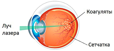 Laser exposure is carried out using a slit lamp or an indirect ophthalmoscope. Consider both methods in Read more:
- When using a slit lamp, the laser is attached to it. The patient is in the sitting position, the chin is placed on a special stand. Contact lenseswhich contributes to the focusing of the laser on the retina, is placed on the cornea of \u200b\u200bthe operated eye.
- In the second case, the patient may be in the sitting position and lying. The doctor puts up a standard illuminator with a laser attached to it. Holded in the hands of the lens is used to review the retina and focusing the laser beam in the required area.
Regardless of the methodology, the operation implies the implementation of 1500-5000 laser "burns" during 1-4 therapeutic approaches. It all depends on the protocol adopted in a specific clinic. In the process of vitrectomy, the energy of the laser can be delivered directly to the cavity of the eye. For this purpose, a special endolar needle is used, which is introduced into the gland body cavity during the operation, and the photocoagulation beam is directed directly to the retina. Procedure control is carried out by a surgeon through an operating microscope. For the prevention of complications, the patient must be convenient for the surgeon is positioned sitting or lying. It is necessary to explain that the work of a specialist requires high accuracy and concentration, the movement of the head or eye is unacceptable. Local anesthesia is usually sufficient to create comfortable for the operated conditions and relief of pain syndrome. The duration of such an impact is 15-20 minutes. The procedure is an outpatient, that is, after its end and a short observation, a person can go home.
Postoperative period
The recovery period as a whole depends on the underlying disease, on which the retina was treated with a laser. Browse of view, the feeling of burning and moderate pain syndrome is the norm during the first postoperative days. Then the unpleasant symptoms should gradually disappear. If the described features persist or the state worsens, there is no need to wait for the next scheduled visit. You need to go to the doctor on the same day. For complete recovery after the intervention takes about 14 days. During this period, it is recommended to avoid heavy physical exercise and sports, limit reading and working at the computer. You can manage the car only after inspection in the clinic and with the permission of the attending physician. Persons with diabetes must carefully monitor blood glucose levels and prevent its excessive increase. In the case of diabetic retinopathy, the laser coagulation of the retina cannot return the visual angiopathy, however, the treatment suspends the progression of the underlying disease.
Possible complications after laser coagulation
Scientists have proven that this procedure is safe for patients. However, as with any operation, there is a certain risk of developing complications. With such a patient, a must acquaint the attending physician before signing informed consent. Consider the most common complications:
- Development or progression of macular edema.
- The loss of visual fields is proved the negative effect of pyretinal photocoagulation on peripheral vision.
- Loss of color vision.
- Reduction of contrast sensitivity.
- Hemorrhagic complications.
- Problems from the front segment - clouding of the cornea or lens.
- Problems with night vision.
- Transient vision loss.
An experienced specialist is always trying to perform the operation as clearly and quickly to minimize the above-described risks. Many of the listed states are transit. Others are well treated. It is important to notice the deterioration in time. From the patient's part, strict compliance with all postoperative recommendations and timely appeal professional help At the first signs of deterioration.
Price of operation
The cost of laser impact on the retina depends on the required amount of coagulation and the underlying disease. Often laser therapy is part comprehensive treatment severe disease, for example, retinal detachment. In this case, the price will be higher through the implementation of several procedures. Isolated retinal photocoagulation costs from 7000 to 10,000 rubles depending on the volume. Should not forget about preoperative surveys, laboratory and instrumental diagnostics. In this case, we are talking about private ophthalmologic clinics. When contacting B. state Institution Perhaps the free execution of the procedure in the presence of readings and subject to the time limits, if there is a queue in the clinic.


