The structure of the human spinal cord. The structure and functions of the spinal cord of the brain and its function
The spinal cord is part of the central nervous system. It is difficult to overestimate the work of this body in the human body. After all, with any of its defects it becomes impossible to carry out the fulfillment of the body with the world from the outside. It is no wonderful of its innate vices, which can be found using ultrasound diagnostics already in the first trimester of the child tooling, are most often indicated to interrupt pregnancy. The importance of spinal cord functions in the human body causes the complexity and uniqueness of its structure.
Located in the spinal Channel, being a direct continuation of the oblong brain. The conditionally upper anatomical border of the spinal cord is considered to be the line of connecting the top edge of the first cervical vertebra with the lower edge of the occipital opening.
The spinal cord ends approximately two first lumbar vertebrae, where it is gradually a narrowing: first to a cerebral cone, then to a brain or terminal thread, which, passing through the channel of the sacrum of the spine, is attached to its end.
It is interesting that the spinal brain embryo in length is equal to the spine, but then they grow unevenly - the growth of the spine goes much more intense. As a result, already in an adult, a spinal cord for several tens of centimeters shorter than a spinal column.
This fact is important in clinical practiceSince the spinal cord wide known at the lumbar level is absolutely outside the danger from mechanical damage.
Spinal shells
The spinal cord is reliably protected not only by the spine bone tissue, but also in three own shells:
- Solid - from the outside includes fabrics of the vertebral canal tissue, then the epidural space and the inner layer of the solid shell.
- Cowital - thin, colorless plate, fragile with a hard shell in the area of \u200b\u200binterghangers. Where there are no fights, there is a subdural space.
- Soft or vascular - separated from the previous shell with a subpaste with a spinal fluid. The soft shell itself is adjacent to the spinal cord, consists most of the vessels.

The entire body is fully immersed in the spinal fluid of the subpasic space and "floats" in it. The fixed position gives it special ligaments (gear and intermediate cervical partition), with which the inside of the inside with the shells is carried out.
External characteristics
- The shape of the spinal cord is a long cylinder, slightly flattened in front-back.
- The length is average of about 42-44 cm, depending on
From human growth. - Weight about 48-50 times less brain weight,
is 34-38 g
Repeating the outlines of the spine, the spinal structures possess the same physiological bends. At the level of the neck and lower part of the chest, the beginning of the lumbar departments allocate two thickening - these are the places of the exit of the roots of the spinal nerves, which are responsible for the innervation of hands and legs, respectively.
There are 2 furrows, which share it into two absolutely symmetrical halves at the back of the spinal cord. All over the body, in the middle there is a hole - the central channel, which is connected to one of the ventricles of the brain. At the bottom of the brain cone area, the central channel is expanding, forming the so-called terminal ventricle.
It consists of neurons (nervous tissue cells), the bodies of which focused in the center form a cereal gray matter. According to calculations of scientists in the spinal cord, only about 13 million neurons - less than in the head, thousands of times. The location of the gray substance inside the white is by no means somewhat different in shape, that on the cross section remotely resembles a butterfly.

The specific type of transverse cut can be isolated in the spinal-gray substance such anatomical structures:
- Front horns are rounded and wide. Consist of motor neurons that transmit pulses to muscles. From here begins the front roots of the spinal nerves - motor roots.
- The rear horns are long, non-screens, consist of intermediate neurons. They take signals from the sensitive roots of the spinal nerves - rear roots. Here are neurons that, through nerve fibers, carry out the relationship of different areas of the spinal cord.
- Side horns are found only in the lower segments of the spinal cord. Contain so-called vegetative nuclei (for example, the centers of the expansion of the pupil, innervation of sweat glands).
The gray substance from the outside is surrounded by a white substance - it is essentially its neuron processes from a gray substance or nerve fibers. The diameter of the nerve fibers is not more than 0.1 mm, but their length reaches a one and a half meters.
Functional purposes of nerve fibers can be different:
- ensuring the relationship of the multi-level areas of the spinal cord;
- transmission of data from the head to the spinal cord;
- ensuring the delivery of information from the scorn to the head.
Nervous fibers, integrating in bundles, are located in the form of conductive cerebrospinal tracks along the entire length of the spinal cord.
 Narrowing (stenosis) of the spinal canal in most cases requires operational treatment. The causes and symptoms of stenosis are described in.
Modern effective method Treating back pain - Pharmacopuncture. The minimum doses of drugs introduced into active points work better than pills and ordinary injections :.
What is better for the diagnosis of spinal pathologies: MRI or computed tomography? We tell.
Spinal nerve roots
The spinal nerve in nature is neither sensitive nor motor - it presents the nerve fibers of both types, as it combines the front (motor) and rear (sensitive) roots.

The area of \u200b\u200bthe spinal cord, which is the "starting platform" for one pair of nerves, is called segment or neuromer. Respectively, the spinal cord consists of everything
Of 31-33 segments.
It is interesting and it is important to know that the spinal segment is not always in the spine department with the same name due to the difference in the length of the spine and the spinal cord. But the spinal roots still come out of the corresponding intervertebral holes.
For example, the lumbar spinal segment is in the thoracic spinal column, and the corresponding spinal nerves come out of the intervertebral holes in lumbar Department spine.
Spinal nerve roots overcome some distance to achieve a "their" intervertebral opening - this fact is based on the appearance of a structure in the spine channel, called the "horse-tail" and is a beam from the spinal roots.
The functions of the spinal cord
And now let's talk about the physiology of the spinal cord, about what "duties" is assigned to it.
In the spinal cord, segmental or working nerve centers are localized, which are directly related to the human body and manage it. It is through these spinal work centers that the human body is subject to control from the brain.
At the same time, certain spinal segments control well-defined parts of the body by obtaining nerve pulses from them from sensitive fibers and transmission of response pulses by motor fibers:
| Spinal segments (location, sequence number) | Inneveloped plots |
| Cervical: 3-5 | Diaphragm | |
| Cash: 6-8 | Hand joints | |
| Breast: 1,2, 5-8 | Muscles and leather hands | |
| Breast: 2-12. | Muscles and skin of the body | |
| Breast: 1-11 | Intercostal muscles | |
| Breasts: 1-5 | Muscles and leather head and neck, heart and lungs | |
| Breast: 5-6 | Lower of the esophagus | |
| Breast: 6-10. | Digestive organs | |
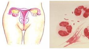
| Lumbar: 1-2 | Groove bunch, adrenal glands, kidneys and ureters, bladder, prostate, uterus | |
| Lumbar: 3-5 | Muscles and leather legs | |
| Sleeps: 1-2 | Muscles and leather legs | |
| Sleeps: 3-5 | External genital organs, crotch (reflex centers of urination, erection and defecation) |
Some vegetative or complex motor reflexes of the spinal cord makes it at all without the intervention of the brain, thanks to its bilateral connection with all parts of the human body - so the spinal cord performs its reflex features. For example, urinary reflector centers or erection are in 3-5 sacral segments and during the spinal damage in this place these reflexes may be lost.
Conductive spinal function It is ensured by the fact that all conductive paths connecting the areas of the nervous system between themselves are localized in the white substance. On ascending ways, information from tactile, temperature, pain receptors and receptors of movement from muscles (proprigororeceptors) is transferred first in the spinal cord, and then to the corresponding heads of the head. The descending paths are associated with the head and spinal cord in the reverse order: with their help the brain is monitored over the human muscles.Danger of damage and injuries
Any spinal cord injury threatens human life.
The most dangerous are damage to the cervical spinal segments - in the absolute majority of cases, this leads to an immediate stopping of breathing and death.
Serious damage to other spinal segments located below, the cause of death may not become, but to partial or complete loss of disability will lead almost 100% of cases. Therefore, nature and intended so that the spinal cord is under reliable protection of the spine.
The expression "healthy spine" in most cases is equivalent to the expression "healthy spinal cord", What is one of the necessary conditions for the high-quality human life.
Offer interesting videowhich will help to understand the anatomy of the spinal structures and their functioning.
Central nervous system (CNS) in human organism represented by two brain elements: head and spinal. In the skeleton of man there is a vertebral channel, where the spinal cord is localized. What functions does he carry out?
It exercises two vitality functions:
- conductive (transmission paths of impulse signals);
- reflex-segmental.
The conductor function is carried out by the transmission of the pulse along the ascending brainstamps to the brain and back to the executing executors on the downstream brainways. The long transmission paths of pulse signals allow them to be transmitted from the spinal cord into different functional departments of the head, and short - provide a link between adjacent segments of the spinal chute.

The reflex function is reproduced by activating a simple reflex arc (knee reflex, extension and bending of hands and legs). Complex reflexes are reproduced with the participation of the brain. The spinal seas is also responsible for performing vegetative reflexes that control the work of the internal environment of a person - digestive, urinary, cardiovascular, sexual systems. The diagram illustrates the functions of the autonomic system in the body. The management of vegetative and motor reflexes is carried out due to proproporeceptors in the thickness of the cerebrospide. The structure and functions of the spinal cord have a number of features in humans.
Consider the structure of the cerebrospinal tightness for a better understanding of which functions it performs.
Anatomical features
The structure of the cerebrospinal tightness of man is not such a simple, as it may seem at the beginning. Externally, the brain resembles a cord with a diameter of up to 1 cm, length in the range of 40-45 cm. Its beginning it takes from the oblong brain department and ends with a horse tail to the end of the spinal column. The vertebrae protect the spinal cord from damage.
The spinal cord is litter, it is formed by a cerebral cloth. On all its length, it has a rounded form in section, exceptions are only the thickening zones, where it is sealing. The cervical thickening is located from the third vertebra of the neck to the first thoracic. Lumbar-sacral flattening is localized in the region of 10-12 vertebra of the chest department.

The front and rear of the spinal cord on its surface has a groove, which divide the organ into two halves. Brain gravity has three shells:
- solid - represents a white shiny dense fibrous tissue rich in elastic fibers;
- cowital - made of connective tissue-coated endothelium;
- vascular - shell from loose connective tissue with rich vessels to ensure the nutrition of the spinal cord.
Between the two lower layers there is a liquid (spinal fluid).
The central deposits of the spinal aliment are made with a gray substance. On the drug cutting of the organ, this substance in the outline has similarity with a butterfly. This component of the brain from the bodies of nerve cells (inserted and motor-type). This section of the nervous system is divided into functional zones: front and rear horns. The first contains the neurons of the motor type, the second have insert nerve cells. Throughout the segment of the spinal aliment from the 7th cervical segment to the 2nd lumbar there are additional side horns. Here are the centers responsible for the functioning of the vegetative NA (nervous system).

The rear horns are characterized by the heterogeneity of its structure. As part of these spinal cord areas there are special kernels performed inserted neurons.
The outer part of the spinal aliment is formed by a white substance made by the butterfly neurons axon. The spinal grooves are conditionally crushed white substance for 3 pairs of cakes, known as: side, rear and front. Axons are combined into several conductive paths:
- associative fibers (short) - provide the relationship of various spinal segments;
- ascending fibers, or sensitive, - transmit nerve signals to the head unit of the CNS;
- downward fibers, or motor, - transmit pulse signals from the bark of the hemispheres to the front horns controlling the executive officers.
Rear channels contain conductors only ascending, and the remaining two pairs are characterized by the presence of descending and upward routes. The number of conductive paths in the corders is different. The table showers the location of the conducting paths in the spinal part of the CNS.
Side rope of conductors:
- the spinal cerebellar path (rear) - transmits in the cerebellum pulse signals of proprioceptive nature;
- the spinal cerebelling path (front) is responsible for contacting the cerebellum bark, where and translates pulse signals;
- the spinal thalamic tract (outdoor side) is responsible for transmitting pulse signals to the brain from receptors that react to pain and temperature change;
- the pyramid tract (outer side) - conducts from the bark of the large hemispheres of motor pulse signals to the cerebrospinal seal;
- the red-cerebral-spinal tract - controls the maintenance of the tone of the skeleton muscles and adjusts the execution of subconscious (automatic) motor functions.
Front channel conductors:
- the pyramid tract (front) - broadcasts the motor signal from the cortex of the UPS TSNS to the bottom;
- the spinal thalax tract (front) - transfers the transfer of pulse signals from tactile receptors;
- the predver-spindy - coordination of conscious movements and equilibrium, and is also characterized by the presence of communication with the oblong brain.
Rear wire conductors:
- the slim bundle of the Gully fibers - is responsible for the transfer of pulse signals from proprigororeceptors, interior beams and skin receptors of the lower body and legs to the brain;
- the wedge-shaped bundle of Burdach fibers - is responsible for the transfer of the same receptors in the brain from the hands and upper parts of the body.

A man's spinal cord in its structure refers to segmental authorities. What number of segments does he have in the human body? Total brain litter contains 31 segments, respectively, spinal departments:
- in cervical - eight segments;
- in breast - twelve;
- in the lumbar - five;
- in the sacrum - five;
- in the smoking - one.
Cerage segments have four roots forming spinal nerves. Rear flares are formed from axons of sensitive neurons, they are included in the rear horns. Rear roots have sensitive ganglia (one at each). Then, in this place, synaps is formed between the sensitive and motor cells of the NA. Axons of the latter form the front roots. The shown scheme demonstrates the structure of the spinal cord and its roots.

In the center of the cerebrospide, the channel is localized on all its length, it is filled with liquor. To the head, hand, light and heart muscle conducting fibers stretch from cervical and uppergrate segments. The belt segments and the pectoral section of the brain give the nerve ending to the muscles of the body and abdominal cavity with its contents. The lower-friendly and sacral segments of a person give the nerve fibers of the legs and muscles of the lower press.
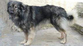
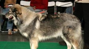
The spinal cord of a person or an animal is the most important part of the CNS. Through it, the brain is associated with muscles, leather, internal organs, vegetative nervous system. This ensures the vital activity of the human body, dogs, cats or another mammal. The structure of the spinal cord is characterized by a complex organization and a narrow specialization of each region. Biology is so arranged that any serious violation is manifested in problems with motor functions, somatic anomalies.
Externally, this body very much resembles a cord stretched out in a special spinal channel. He has the right side and left. In length, it does not exceed half the meter, and the diameter is around a centimeter.

We will consider in detail the structure of the spinal cord, the features of his organization, the principles of work. Knowing what the structure of the spinal cord can be easily understood how our movements are born, how neurons can appear. We also describe what functions do the spinal cord.
In the spinal cord there is from 31 to 33 pairs of nerves, so it is divided into 31-32 segments. Everyone relates to part of our body and continuously performs its functions. The mass of such an important organ, without which no movement is impossible, is only 35 grams.
Location area - vertebral channel. At the top it immediately goes into medullaAnd at the bottom it is completed by the spine cleaner.
Division into segments
The role of the spinal cord is to organize any human movements. To ensure maximum efficiency of its work, during evolution segments were allocated, each of which ensures the functioning of a certain area of \u200b\u200bthe body.
This part of the nervous system begins to form already at the 4th week of embryonic development, but the basic functions of the spinal cord can not be performed immediately.
The spinal cord departments and their functions are now well studied. He seglessly divides:
- cervical segments (8 pieces);
- chest (12 pieces);
- lumbar (5 pieces);
- sacral (5 pieces);
- copter (from 1 to 3 pieces).
A man's back ends with a small smoking. He is Rudiment, that is, part of the fact that during the evolution lost its significance. This is actually the rest of the tail. Because a person has very little cleaned segments. The tail he is just no longer needed.
What is it needed for
The spinal cord is a center that collects all the information coming from the periphery. Then he sends the teams to the muscles and fabrics, leading them to the tone. So all movements are born. This is a complex and painstaking work, because a person makes hundreds of thousands of smallest movements per day. His physiology is characterized by a complex organization and interaction of all parts of the CNS.
The spinal cord reliably protect three shells at once:
- firm;
- soft;
- powered.
Inside there is a spinal fluid. The center of the brain fills the gray substance. In the context, this area is similar to the butterfly, which deployed wings. The gray substance is the concentrate of neurons, it is they can transmit a bioelectric signal.

Each segment consists of dozens and even hundreds of thousands of neurons. They provide a full operation of the engine device.
In the gray substance there are protrusions of three types (horns):
- front;
- rear;
- side.
Between zones are distributed different types Neurons. This is a complex and well-organized system that has its own characteristics. In the front horns area there is a huge number of large motor neurons. In the back of the horns there are small inserts neurons, and in side - visceral (sensitive and motor).
It is nervous fibers that form the paths by which the signal is carried out.
In total, in the spinal cord of a person, scientists counted more than thirteen million nerve fibers. The protective function for them is performed by external vertebrae, which form the spine. It is in them that there is an internal gentle and vulnerable spinal cord.
The gray substance from all sides is surrounded by many nerve fibers. The transmission of bioelectric signals is carried out through the thinnest process of neurons. Each may take from one to a variety of such processes. Neurons themselves are extremely small. Their diameter is not more than 0.1 mm, but the processes are affected by their length - it can reach one and a half meters.
In gray matter there different types cells. The front departments consist of motor cells, they are very large. As can be seen from the very name, they are responsible for motor functions. These are thin, but very long fibers, which from the spinal cord go directly to the muscles and lead them into motion. Such fibers form large bundles and leave the spinal cord. This is the front roots. One of them goes to the right, and the other is to the left.

In each department there are such sensitive fibers, of which a pair of roots is formed. Part of the sensitive fibers is suitable with a brain. The second part is directed directly into the gray substance. In it, fibers ends. The ending for them becomes different types of cells - motor, intermediate, insert. Through them are continuous regulation of movements and organs.
Organization of conductive paths
Conducting the entire body is customary to divide on:
- associative;
- afferent;
- efferent.
The task of associative tract is to connect neurons between all segments. These connections are considered short.
Afferent provide sensitivity. These are ascending paths that receive information from all receptors and send it to the brain. Efferent paths transmit signals from the brain to the neurons of the whole organism. They relate to downward paths.
Functions
The activity of the spinal cord is continuous. It provides the motor activity of the body. There are two main functions of the spinal cord of a person - reflex and conductor.
Each department ensures the operation of a completely defined zone of the body. Segments (cervical, chest, for example) provide the functions of the sternum organs, hands. For the full work of the muscles and the digestive system corresponds to the resurrector segment. For the functions of the pelvis organs, the legs are responsible for the sacral segment.
Reflex
The reflex brain function lies in the organization of reflexes. This allows the body, for example, to instantly respond to a signal about pain. Reflexes affects its efficiency. Man pulls out his hand from a hot object for a split second. During this time, information from receptors to the brain and back managed to do a considerable path on the reflex arc.
When the sensitive nervous end of the skin, muscle fibers, tendons, joints get irritation, it means that a nervous impulse was sent to them. Such signals apply to the rear roots of nerve fibers and come in the spinal cord. Receiving a signal, motor and insert cells are excited. Then the motor fibers of the front roots pulses are sent to the muscles. After receiving such a signal, muscle fibers are reduced. Simple reflexes occur in this mechanism.
Reflex is the reaction of the body in response to the resulting irritation. All reflexes are provided by the work of the CNS. One of the spinal cord functions is reflex. It provides the so-called reflex arc. This is a complex path that nerve impulses pass from the peripheral components of the body to its spinal cord, and from it directly to the muscles. It is not an easy, but vital process.

The simplest reflexes can save a person life and health. By pulling into the hand that touched hot, we do not even suspect that the signal from the skin lightningly passed through the nerve fibers in the head, and then in the spinal cord. In response, the impulse was sent to him, which reduced the muscles of the hand to avoid burns. This is a bright manifestation of the reflex function.
Neurophysiologists studied in detail almost all reflexes and nervous arcs that ensure their execution. These data allow for effective rehabilitation after injury and a number of diseases, and also help with their diagnosis.
It is on such a reflex that the diagnosis of a neuropathologist is founded, in which the doctor will easily hit the hammer on the tendon knee Cashechka patient. This is how the knee reflex is studied, according to which one can judge the state of a certain spinal cord department.
However, the spinal cord is not an independent reflex system. Its functions tirelessly controls the brain. They are closely linked by special beams of nerve fibers. Fibers are very long, thin, consist of white substance. Signals according to one are transmitted to the brain upwards, and in others - in the dorsal.
All CNS participates in the formation of coordinated complex movements. Each movement is a continuous flow of pulses from the brain to the dorsal, from it - to muscle fibers.
Conductor
This is the second important feature. It is that nerve signals are transmitted above from the spinal cord. There, in the subcortical and cortical area, all information is instantly processed, and in response, the corresponding signals are sent to it.
The conductor function works in those moments when we decide to take something, get up, go. This happens instantly, without time spent on thought.
This feature is mostly provided with intermediate or insert neurons. They send a signal to motor neurons, and also process information that comes from leather and muscles. There are peripheral signals and pulses from the brain.

The excitation impulse is sent using plug-in cells to different groups of motor cells. In this case, the activity of other groups is braked. Exactly like this difficult process Provides coherence and high coordination of human movements. So the depotless movements of the pianist, ballerina appear.
Possible diseases
In the human body there is a unique department that is called the "horse tail". It does not have a spinal brain itself, but only the spinal fluid and beams of nerves remained. If they are squeezed, the body begins to experience pain, there are disorders of the musculoskeletal system. This disease at the location of the localization of the main reason is called the "horse tail".
If a "horse tail" is developing, a person is worried about a number of symptoms. There is pain in the lower back, the muscles are weakness, the body begins to react much more slowly into external stimuli. Inflammation may occur, even the temperature rises. If these alarming symptoms ignore, the state is aggravated. It becomes difficult for a person to move or sit for a long time.
- A rather complicated system that is responsible for many processes in the body and in which it is difficult to understand it is quite difficult. Elementary knowledge can be obtained by studying an anatomy at school, but when it comes to a deeper analysis, many incomprehensible moments arise.
Let's try to figure out what the spinal cord is as it works, what functions do, and just understand why it is needed at all.
Read also

Spinal cord as part of the nervous system
- One of the components of the human nervous system. On Latin his name looks like medulla Spinalis.
It is a thick cylindrical tube with a narrow channel located inside it. Located in the spine canal, but, speaking easier, inside the spine.
This body has a rather complicated structure and segmental structure. The main function of this organ is the transfer of various pulses and signals from the human brain to specific organs. In addition, he performs reflex activity, that is, it is responsible for human reflexes, and it is both simple and more complex reflexes.

Nervous system of man
The value of the spinal cord
There are only two main and most important features:
- Reflex. Simply speaking, a number of reflex arcs are closed on this authority. It is due to this that reflexes are carried out (the so-called spinal reflexes).
- Conductive. The body in this case acts as a conductor. It holds signals that come from various organs to the brain. It is through this body a brain receives all the information and processes it. It all works in the opposite direction.
The location of the spinal cord
The organ is located in the spinal canal (located). This channel is rather long and practically reaches the lower vertebrae. In essence, this is a special channel, which is an oblong hole in which the spinal cord is. From the parties, it is protected by vertebrae, as well as intervertebral discs.
Also, the organ is located at the lower edge of the large occipital opening, where there are connections with a brain. It is in this place that there is a huge range of roots, which is directly connected to the human brain. Such a compound is called left and right spinal nerves.

The location of the spinal cord
The bottom ends on the damage 1-11 damage. After the organ turns into a thin terminal thread. In fact, it is still a spinal cord, because the nervous tissue contains.
Topography and shape of the spinal cord
We will deal with the features of the location (topography) and forms.
To do this, consider a number of features: 
- Middle Length 42-43 centimeters. Men's length often for several centimeters more, and in women, on the contrary, less.
- Mass 33-39 grams.
- On the front there is a median gap, it is well noticeable. It can be noted that it seems to grow into an organ. In fact, it creates a peculiar partition that separates the brain into two departments.
- In the cervical and lumbar sacral departments you can
- metut two sufficiently serious thickening. This is due to the fact that the innervation of the upper and. Speaking simple language, here the nerve endings from the limbs "join" to the spinal cord, which
- sumpts them to transmit the necessary signals.
- The spinal cord is topographically practically associated with vertebrae. Various departments are disposed of not depending on the specific vertebra or several vertebrae.
Increasing the volume on these areas is due to the fact that it is here that the largest number of nerve cells are located, as well as fibers for which signals from the limbs and back are transmitted.
Despite the fact that the spine is a kind of "place for storing" the organ, the location of the nerve endings, especially at the bottom of the spine, do not correspond to the specific vertebrae. This is due to the fact that the long spinal cord is less than the length of the human spine.
That is why for physicians it is necessary to know the exact location of each of the segments, because the spine can not be focused.

Location in the spine
 The stories of our readers!
I want to tell my story, how I cured osteochondrosis and hernia. Finally, I was able to overcome this unbearable back pain. We lead an active lifestyle, I live and rejoice every moment! A few months ago I twisted me at the cottage, a sharp pain in the lower back did not move, did not even be able to go. The doctor in the hospital was diagnosed with osteochondrosis of the lumbar spine, the hernia of the L3-L4 discs. I prescribed some kind of medicine, but they did not help, to endure this pain was unbearable. They called an ambulance, they set the blockade and hinted for the operation, all the time thought about it, that I would find a burden for the family ... Everything changed when the daughter gave me one article on the Internet. You can not imagine how much I am grateful for it. This article literally pulled me out of a wheelchair. The last months began to move more, in the spring and summer every day I go to the cottage. Who wants to live a long and energetic life without osteochondrosis,
Characteristics of the spinal cord depending on age
Consider features, depending on the age of a person:
- At the newly born child, the body length is 13.5-14.5 centimeters.
- At 2 years, the length increases to 20 centimeters.
- Approximately 10 years old can reach 29 centimeters.
- The growth ends in different ways, depending on the characteristics of the body of a particular person.
We will understand external features and changes depending on age:
- In babies, cervical and lumbar thickening are noticeable more than in adults. Also concerns the width of the central channel.
- The above features become almost imperceptible to two years.
- The volume of white substance grows at times faster than the volume of gray. This is due to the fact that the segment apparatus is formed earlier than conductive paths that connect the head and spinal cord.
In the rest of the age characteristics, it is practically not observed, because from the very birth of the spinal cord performs almost all the functions, as in an adult person.
The features of the structure of the spinal cord
Now consider the features of the structure, alternately considering each segment individually, of which the authority consists.
Spinal cord shells
The spinal cord is in a kind of channel, but it has protection that also performs a huge number of functions.
Spinal cerebral cerebral shells, which are totaling three:
- solid sheath;
- arachnoid;
- soft shell.
All shells are interconnected, and at the bottom they grow up with terminal thread.

Spinal cord shells
In the spinal cord there is a white and gray substance.
Let's try to figure it out what it is:
- White substance - A complex system of pulp and cinema nerve fibers, as well as the supporting nervous tissue.
- Gray matter - These are nervous cells and their processes.

White and gray spinal cord substance
Spinal cord departments
Severe five main spine sections, consider them starting from above:
- cervical;
- chest;
- lumbar;
- sacral;
- copchik.
Read also

Spinal nerves
They are paired nervous trunks, which are total 31 pair: 
- 8 cervical;
- 12 chest;
- 5 lumbar;
- 5 sacral;
- couple pair.
Each nerve is responsible for a certain portion of the body. On this site there are bones, muscles, internal organs or leather. The task of a particular pair of nerves is the transfer of pulses from the area to the spinal cord and back. It is thanks to this a person can feel pain, discomfort, temperature, and so on.

Spinal nerves
Spinal cord segments
Segments as much as pairs of roots - 31. The segment is a specific portion of the human body, for which the specifically taken pair of roots.
All of them are divided into:
- cervical;
- chest;
- lumbar;
- sacral;
- copchie.
Due to the fact that the length of the spine is longer than the spinal cord length, it turns out that the roots of the nerves only in the upper part correspond to the level of intervertebral holes.
Below, to get into a special hole, the nerves of the lower departments fall below parallel to the spine. Thus, they come out at the level of the end thread.

Spinal cord segments
Vienna and the arteries of the spinal cord
The organ gets blood due to the front and pair of the rear spiral arteries. But these arteries are able to provide only 2-3 top cervical segments. The rest of the rest is the root-spiral artery, which receive blood from the branches of the vertebral and the upward cervical artery.
At the bottom of the spine gets blood from intercostal and lumbar arteries. Both of these artery are peculiar proceedings of the famous artery poses by the name of Aorta.
The functions of the spinal cord
Let us turn to the consideration of functions. For convenience, we will consider each separately.
Reflex and motor functions
This feature is responsible for human reflexes. For example, if a person touched something very hot, then reflexively he will take her hand. This is a reflex or motor function. But let's turn step by step with how it is all tripled and how the spinal cord is associated with the spinal cord.
It is best to consider everything on the example, so we will present the situation that a man has touched a very hot object:
- When touching the signal primarily receives receptors that are located throughout the human body.
- The receptor transmits the signal to the nerve fiber.
- On the nervous fiber, the signal is sent to the spinal cord.
- On the approach to the organ is a spinal assembly, where the body of the neuron is located. It is by its peripheral fiber and a pulse transmitted from receptors was obtained.
- Now the central fiber pulse is transmitted to the rear horns of the spinal cord. At this point, there is a peculiar switching of the pulse to another neuron.
- The processes of the new neuron transmit a pulse to the front horns.
- Now the return path begins, because the front horn transmits the pulse to the motor neurons. They are responsible for the movement of the upper limbs.
- According to these neurons, the pulse is transmitted directly in hand, after which a person removes it (motor function).

Reflex arc in the scheme
As a result of all this process, a person pulls out his hand from the hot object and the reflex arc is closed. The whole process takes the fraction of a second, so touching any subject, a person immediately feels its temperature, a consistency and other features.
Explore function
In this situation, the authority acts as it were. Explorer in this case it is between receptors and the brain. Receptors are obtained by a pulse, which is transmitted in the spinal cord, and after the brain. Information there is analyzed and transmitted back.
Thanks to this function, the person gets sensitivity, as well as the feeling of itself in space. It was repeatedly proven, especially the like becomes apparent with serious spinal injuries.

Generative function
About this feature is often forgotten, but it is no less important for a person than others. The integrative function is manifested in reactions that cannot be attributed to simple reflexes. In order for the body to respond, it is necessary to use other parts of the nervous system of the human body. So the spinal cord can form the bond with each other.
Here you can include reflexes of chewing, swallowing, regulation of digestion, breathing and much more. In essence, this is an inconspicuous function that provides normal life.
Spinal violation
Violation of functions can lead to serious consequences and often even to death. Violations often occur either due to injuries or because of various diseases.
For example, due to the violation of the spinal cord function, a person may lose sensitivity, in this case, for example, it can stop feeling the temperature. In the worst case, the violation may lead to uncontrolled actions of the limbs (or paralysis), violation of the work of the internal organs and the nervous system as a whole.
Diseases of the spinal cord
The list of the most common diseases that violate the full work of the body under consideration:
- Heart attack.
- Polio.
- Cross Melit.
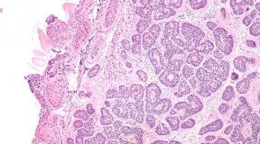
- Tumors.
- Decompression disease.
- The defeat of nerve roots.
- Arteriovenous malformations.
If a spin hurts, neck or loin, do not tighten the treatment if you do not want to finish in a wheelchair! Chronic many pain In the back, neck or lower back - the main sign of osteochondrosis, hernia or another serious illness. Treatment must be started right now ....
Puncture of spinal cord
Puncture spinal fluid (liquor) - A procedure that pursues diagnostic, anesthesiological and therapeutic purposes. The procedure itself lies in the fact that the patient between the 3rd and 4th vertebrae is introduced the corner under a spider shell, and after a certain amount of spinal fluid for research is extracted.
In the process of procedure, the brain itself is not affected, so there is no fear of violations. But still this procedure quite serious and painful.

Puncture of spinal cord
Conclusion
Summing up, it should be said that the spinal cord is one of the most important organs in the human body. In many ways, precisely thanks to him, a person can lead normal life, as well as, thanks to this organ, almost the entire nervous system is functioning.
.The spinal cord is the most important internal body relating to the structure of the central nervous system. The surface of the spinal cord has 3 shells - web, solid and soft. The anatomy of the spinal cord is designed in such a way that the internal body is a dominant system that supports the vital activity of the whole organism.
The structure of the spinal cord
The spinal cord is located in the cavity of the spinal canal, which is formed using vertebral processes and their bodies. The beginning of the spinal cord structure is the head of the brain's occipital hole. Next, the spinal cord is located in the channel, representing 40 - centimeter "cord", surrounded by three shells.
The internal body ends with the accumulation of nerve fibers at the level of the first vertebrae in the lumbar department, called the horse-tail. It also begins a narrowing, and then the internal body is "pulls out" into the terminal (terminal, finite) thread, the diameter of which is 1 mm. The terminal thread stretches to the cockerel department, where it grows with the periosteum.
The lower part of the nithe filament is tightly wrapped in the "horse-tail" fibers. In the occurrence of pain in the field of the cocoon, doctors talk about the syndrome with the same name. The structure of a spinal cord of a person is such that the actual brainstant is under constant protection - this is provided by the shell and the vertebral pole itself.

The external structure is the shell and space between them.
Brain shell
- Solid shell. It is immediately behind the vertebral periosteum, but it does not fit close to it. Epidural space is located between the periosteum and solid sheath. The tissue of the solid shell is connective, there are vessels, lymphatic and blood vessels. Epidural space is filled with fatty tissue. Here are venous plexuses.
- Arachnoid - The network of thin plates from the connective tissue, in structure resembling a web. The plates are composed of collagen and elastic fibers. Between the web and soft shell there is a subarohnoid space with a liquor, providing the exchange and nutrition of neurons.
- Soft shell.This is a vascular environment that has gear ligaments for fixing and provides communication and nutrition between the liquor and the brain.
Terminal thread
Terminal thread has 2 parts:- Internal, the length of which is approximately 15 cm. The inner part of the terminal thread consists of nervous tissue, intertwined with lumbar and sacral nerves and is located in a peculiar bag of hard shell.
- The outdoor, the length of which is 8 cm. The outdoor portion of the end thread begins below the second vertebra of the sacrilate department, it stretches to the second cleaner vertebra, where it splits with the perception.
Features
The inner structure of the spinal cord has thickening in lumbar sacrings, as well as cervical departments. This structure is formed because in the corresponding parts of the spine there are a large number of emerging nerves that are directed to the lower or upper limbs.
- The cervical thickening is located at the level of the third and fourth cervical vertebra and lasts to the second breast vertebra.
- Lumbar-sacrive thickening is located on the level of 9-10 breast vertebra and lasts to the 1st lumbar.
White and gray spinal cord substances
The scheme of the spinal cord structure in the context is a similarity of the butterfly wings, it is this part of the internal organ called gray substance. Outside, the gray substance is surrounded by white substance, but the cellular structure and functions of such substances differ significantly.
The gray substance consists of insert and motor neurons:
As a composition of the white substance, axons are present - these are nervous processes that create fibers of descending and ascending wired pathways.
Opinion expert
Pain and crunch in the back and joints with time can lead to terrible consequences - a local or complete limitation of movements in the joint and the spine down to disability. People who are scaled with bitter experience to cure the joints enjoy a natural means that the orthopedist Bubnovsky recommends ... Read more »
Spinal nerves and segments
On the right and left of the central grooves of the spinal cord are located oppositely, as well as rear agents, and the rear axons pass through them, which form nerve roots.
- front root - these are motor neurons;
- rear root is sensitive neurons.
The front and rear roots at the outlet of the brain department are connected to a single nervous node (gangliya). Since each segment has 2 front, as well as 2 rear nerve root, in the aggregate, they form 2 cerebrospinal nerves - one on each side. 

Total spinal cord has 64 nerves - that is, 31 nerves on each side.
The location of the nerve endings as follows:- in the cervical department - 8;
- in the thoracic department - 12;
- in the lumbar department - 5;
- in sressy Department — 5;
- in the pulp section - 1.
Segments and spinal cord departments are located not at one level in the spine due to different lengths (the spinal cord is much shorter than the spine).
A little about secrets
Have you ever experienced constant back pain and joints? Judging by what you read this article - with osteochondrosis, arthrosis and arthritis you are already familiar personally. Surely you tried a bunch of drugs, creams, ointments, injections, doctors and, apparently - nothing of the above did not help you ... And this is an explanation: pharmacists simply not profitably sell a working remedy, as they lose customers! However chinese medicine Millenniums knows the recipe for the relief from these diseases, and it is simple and understandable. Read more »
Functions organ
The structure and functions of the spinal cord is the most important system that supports the normal vital activity of the human body.
The functionality of the spinal cord is divided into 2 parts:- the reflex - these are the simplest motor reflexes of the body, for example, with a hand burn, a man begins to pull his hands from the focus of injury or with a strike of a hammer over the knee there is a reflex extension of the knees;
- the conductor function is the transmission of nerve pulses from the brain area to the inside of the dorsal, as well as the transfer of nerve pulses from the brain and to the internal organs of the human body.
With the help of the conductor, almost every mental action is carried out - it is to get up, go, lie down, sit, draw, convey, cut off, etc. On the most part of the actions, the person does not even think, performing them on the reflex level in everyday life.
It is important to note that reflex features can be executed without the participation of brain functions. This feature of a living organism has been proven by scientific experiments carried out on the frogs. For example, the researchers were determined how frogs were reacted into pain of different character without the participation of the brain - reflexes were manifested both on weak pain and on severe pain. 
If you describe the structure and functions briefly, the human body is a unique system where all internal organs and systems interact harmoniously.
It is important to note that each spine segment is directly related to specific internal bodies, providing them with the necessary functionality:- the cervical and chest departments are associated with heads, chest muscles, chest organs;
- the lumbar department is associated with the internal gastrointestinal bodies, kidneys and muscular system human bodies;
- the sacral division "replies" for functionality lower extremities and pelvis organs.
The spinal cord is a complex and vulnerable system, from the state of which does not depend not only overall well-being, but also many reflexes. Diseases or injuries affecting the spinal cord are unusually dangerous, because it is planned, and at best, lead to disabilities.