Diagram of the structure of the human gastrointestinal tract. The structure of the stomach: divisions, layers Divisions of the human stomach
Nutrition is a complexly coordinated process aimed at replenishing the energy of a living organism through processing, digestion, splitting, and absorption of nutrients. All these and some other functions are performed by the gastrointestinal tract, which consists of many important elements combined into a single system. Each of its mechanisms is capable of performing various actions, but when one element suffers, the work of the entire structure is disrupted.
This is due to the fact that food, entering our body, undergoes multi-stage processing, this is not only the familiar processes of digestion in the stomach and absorption in the intestines. Digestion also includes the assimilation of those very substances by the body. Thus, the scheme digestive system a person takes on a big picture. Pictures with captions will help to visualize the topic of the article. 
In the digestive system, it is customary to allocate organs gastrointestinal tract and additional organs called glands. The organs of the digestive tract include:
A visual arrangement of the organs of the gastrointestinal tract is shown in the figure below. Having familiarized yourself with the basics, it is worth considering the structure of the organs of the human digestive system in more detail. 
The initial section of the gastrointestinal tract is oral cavity... Here, under the influence of teeth, mechanical processing of the received food is performed. Human teeth have a varied shape, which means that their functions are also different: incisors are cut, canines are torn, premolars and molars are crushed.
In addition to mechanical treatment, chemical treatment begins in the oral cavity too. This happens under the influence of saliva, or rather, its enzymes that break down some carbohydrates. Of course, the full breakdown of carbohydrates cannot occur here due to the short stay of the food lump in the mouth. But enzymes saturate the lump, and the astringent components of saliva hold it together, allowing it to easily move to the pharynx.
Pharynx- This tube, consisting of several cartilages, performs the function of carrying the food bolus to the esophagus. In addition to carrying food, the pharynx is also a respiratory organ; 3 sections are located here: the oropharynx, the nasopharynx and the hypopharynx - the last two belong to the upper respiratory tract.
More on the topic: Gastric lavage in case of poisoning
From the throat, food enters esophagus- a long muscular tube, which also performs the function of carrying food already to the stomach. A feature of the structure of the esophagus is 3 physiological narrowing. The esophagus is characterized by peristaltic movements.
With its lower end, the esophagus opens into the stomach cavity. The stomach has a rather complex structure, since its mucous membrane is rich in a large number of tissue glands, a variety of cells that produce gastric juice. Food stays in the stomach from 3 to 10 hours, it depends on the nature of the food taken. The stomach digests it, impregnates it with enzymes, turns into chyme, then "food gruel" enters the duodenum in portions.
The duodenum belongs to the small intestine, but it is worth focusing on it, since it is here that some of the most important elements of the digestion process come - these are intestinal and pancreatic juices and bile. Bile is a liquid produced by the liver that is rich in special enzymes. Distinguish between gallbladder and hepatic bile, they differ somewhat in composition, but perform the same functions. Pancreatic juice, together with bile, intestinal juice, constitute the most important enzymatic factor in digestion, which consists in the almost complete breakdown of substances. Mucous duodenum have special villi capable of capturing large lipid molecules, which, due to their size, are not able to be absorbed by the blood vessels. 
Further, the chyme passes into the jejunum, then into the ileum. The small intestine is followed by the large intestine, it begins with the cecum with a vermiform appendix, best known as the appendix. The appendix does not carry any special properties during digestion, since it is a rudimentary organ, that is, an organ that has lost its functions. The large intestine is represented by the blind, colon and rectum. It performs functions such as absorption of water, the secretion of specific substances, the formation of feces and, finally, the excretory function. A feature of the large intestine is the presence of microflora, which determines the normal functioning of the entire human body as a whole.
More on the topic: Stomach atony: during treatment we focus on diet
Digestive glands are organs capable of producing enzymes that enter the digestive tract and digest nutrients.
Large salivary glands. These are paired glands, they are distinguished:
- Parotid salivary glands (located in front and below the auricle)
- Submandibular and sublingual (located under the diaphragm of the mouth)
Produce saliva - a mixture of secretions from all salivary glands. It is a viscous transparent liquid, consisting of water (98.5%) and dry residue (1.5%). The dry residue includes mucin, lysozyme, enzymes that break down carbohydrates, salts, etc. Saliva enters the oral cavity through the excretory ducts of the glands during meals or during visual, olfactory and auditory stimulation. ![]()
Liver... This unpaired parenchymal organ, located in the right hypochondrium, is the largest gland in the human body, its weight in an adult can be approximately 1.5-2 kg. In shape, the liver resembles an irregular wedge, with the help of ligaments it is divided into 2 lobes. The liver produces golden bile. It consists of water (97.5%) and dry residue (2.5%). The dry residue is represented by bile acids (cholic acid), pigments (bilirubin, biliverdin) and cholesterol, as well as enzymes, vitamins, inorganic salts. In addition to digestive activity, bile also performs an excretory function, that is, it is able to remove metabolic products from the body, for example, the already mentioned bilirubin (a breakdown product of hemoglobin).
Hepatocytes are specific cells of the liver lobules; it is from them that the organ tissue consists. They serve as filters for toxins coming from the blood, therefore, the liver has the ability to protect the body from poisons that poison it.
Gall bladder located under the liver and adjacent to it. It is a kind of reservoir for the hepatic bile, which enters it through the excretory ducts. Here bile accumulates and enters the intestines through the bile ducts. This bile is now called gallbladder and has a dark olive color.
When discussing anatomy, the phrase "form determines function" comes to mind. This means that the structure of an organ largely explains what it does. The stomach is a muscle sac that provides a favorable environment for breaking down and digesting food. He also sends to the next stage of processing the material that a person or any other mammal takes for food. The stomach is located in the upper abdomen. Human anatomy reliably hides the organ under the cover of the lower ribs and thus protects it from mechanical damage. In front, it adjoins the abdominal wall, left hypochondrium, left lung, diaphragm and liver, and behind it - with the lesser omentum, diaphragm, spleen, left adrenal gland, upper part of the left kidney, splenic artery, pancreas and transverse colon. The stomach is fixed at both its ends, but is mobile between them, constantly changing shape depending on the filling. The organ can be called a part of the digestive tract chain and, undoubtedly, its most important link. It is located in front of the duodenum and, in fact, is a continuation of the esophagus. The stomach and the anatomy of the tissues lining it walls are: mucosa, submucosa, muscular and serous membranes. The mucous membrane is where acid is produced and secreted. The submucosa is a layer of connective tissue that separates the mucous from the muscle outer surface. Muscular - consists of fibers, which are divided into several types, named for their location in the organ. This is the inner oblique layer, the middle circulation layer and the outer longitudinal layer. All of them are involved in uniform mixing and grinding of food, as well as its further movement along the tract. The final layer, serosa, is a connective tissue that lines the outer walls of the stomach and prevents it from sticking to adjacent organs. Behind the organ is the pancreas and the greater omentum. The main areas of the stomach structure and anatomy consist of: the esophageal sphincter (cardiac sphincter), fundus, body, antrum (pyloric) section and pylorus. In addition, it has a large curvature (rear convex part) and a small curvature (anterior concave), which are located on the left and right sides, respectively. The esophageal sphincter is contained in the cardiac region and controls the flow of material into the stomach. The bottom is its upper section, the wall of which is formed by the upper curvature, and the body represents the main region of the organ. The end part - antrum, serves as an exit and entrance to the small intestine and ends with the pyloric sphincter (gatekeeper). Cardiac hole. It lies near the heart, where the esophagus enters the gastric mass. This hole does not have anatomical constipation, but contains a special mechanism through which food is not thrown back. In this system, the lower circular smooth muscle fibers of the esophagus serve as a physiological sphincter. The pyloric hole. Formed by the pyloric canal that joins the first part small intestine- Duodenum (duodenum) - and creates an exit route for the chyme. It differs from cardiac in that it has a pyloric sphincter with a valve. It consists of a circular muscular layer that thickens around it. The gatekeeper controls the rate at which stomach contents are excreted into the duodenum. Less curvature. It is also part of the anatomy of the stomach, or rather, its right outer border, and stretches from the cardiac opening to the pylorus. It faces the liver and comes into contact with it and other organs. Large curvature. It is much longer than the smaller one and runs to the left of the cardiac foramen, along the bottom and left border of the stomach. Its anatomy extends to the gatekeeper; the hepato-gastric ligament of the lesser omentum diverges from the upper part, and the greater omentum diverges from the lower part. The walls are composed of muscle tissue and contain three layers: longitudinal, round and oblique. Longitudinal. The most superficial fibers of the muscle wall, concentrated along the curvature. Circular. Lies under the longitudinal and surrounds the body of the stomach. It thickens significantly at the pylorus in order to form the sphincter. Only a few circular fibers are found in the bottom area. Oblique. Forms the innermost lining of the stomach. The anatomy of this muscle tissue is structured as follows: it loops along the bottom and runs along its front and back walls, which run almost parallel to the lesser curvature. The stomach receives an extensive blood supply. Left artery. It arises directly from the celiac trunk, supplies blood, according to the name, to the left side of the stomach, partly to its right side, as well as the esophagus. Right artery. It is a continuation of the hepatic artery and extends from the superior border of the pylorus to the lesser curvature. Further, it diverges along the bottom of the right side of the stomach and at the end merges with the left gastric artery and anatomy. You can see a photo of the blood supply to the entire organ below. Short arteries. These are small branches that radiate from the large splenic artery, supply the lower part of the organ, and connect to the left and gastroepiploic arteries. Left gastroepiploic artery. It also continues the splenic artery, runs along the greater curvature and between the layers of the greater omentum. Right gastroepiploic artery. A branch of the gastroduodenal artery that moves to the left and connects to the left gastric artery. It diverges along the right side of the organ and the upper part of the duodenum. There are exactly as many veins in the stomach as there are arteries, and they are called exactly the same. The right and left flow directly into the portal vein. The short and left gastroepiploic veins flow into the splenic vein, and the right gastroepiploic vein flows into the superior mesenteric vein. The stomach receives signals from the sympathetic and parasympathetic nervous system... The sympathetic fibers are derived from the celiac plexus, and the parasympathetic fibers are derived from the right and left vagus nerves. Vagus nerves in chest form the anterior and posterior vagal trunks. The anterior trunk is formed mainly by the left nerve. It gets into abdominal cavity along the outer surface of the esophagus and stretches along the anterior edge of the stomach. The posterior nerve, on the other hand, is located along the posterior walls of the organs. The pyloric sphincter receives motor fibers from sympathetic system and inhibitory fibers from parasympathetic. The main tasks in the anatomy of the stomach include killing bacteria, processing food, and then pushing it further into the small intestine while maintaining a constant rate of material release. The pH inside the organ is maintained at a very high acidic level, which helps digestive enzymes such as pepsin break down food to move it further along the tract. Finally, the stomach, along with the small intestine, takes part in the absorption of vitamins. After chewing and swallowing food, it moves down the esophagus, then entering the stomach. There it stays for a certain amount of time (depending on the nature of the food) until it takes on the appropriate consistency for digestion and absorption in the small intestine. The organ mixes food with its secretions, forming a semi-liquid gruel. Thus, after the chemical and mechanical breakdown of food, the stomach controls the amount of mass that passes on. This is to ensure that food is not skipped forward faster than it is being processed. They are circular muscles associated with the stomach, structure and function. The anatomy of these organs opens and closes passages for food entry and exit. Thus, the first check valve (cardiac) is located between the esophagus and stomach, allowing food to enter and helping to prevent food retention in the esophagus. If the sphincter is not working properly, the acid spills back and causes what is commonly known as heartburn. Another valve (gatekeeper) allows food to pass from the stomach to the small intestine. As mentioned above, this sphincter helps the stomach control how much food is sent to the duodenum at one time. Since everything we eat ends up in the stomach, the anatomy and function of this organ cannot be imagined without chemicals to help break it down. Some of them include enzymes like pepsin. It helps break down proteins that enter the body when eating food. It also contains stomach acid, sometimes called stomach acid, which is produced by some of the cells in the body. This increment is a liquid composed of of hydrochloric acid, mucus, enzymes, water, and other substances that help break down food and kill germs. Since such an impact may not always be sufficient, in addition to chemical destruction, there is also a mechanical effect. It is carried out with the help of muscle contraction. As they shrink, they rub all the food inside the organ and help break it down into a pasty mass. Chyme is a pasty substance that forms when the muscles of the stomach contract and are exposed to gastric juice. They mix the incoming ingredients and break them down into smaller fractions. During the meal, chyme is mixed with gastric juice and enzymes. The organ will begin to contract, as if kneading all the substances together, and produce this pasty substance. Further, peristalsis, which is these wave-like contractions, pushes food towards the pyloric sphincter. It opens and allows a small amount of mass to pass from the stomach to the intestines. The anatomy of this organ allows you to take all the nutrients from the substance and gradually bring it out. Now you have learned about the structure and functions of the stomach, all the essentials in order to properly take care of it. Monitor your health, and this body will repay you with a long and uninterrupted service. The gastrointestinal tract (GIT) contains organs responsible for the mechanical and chemical processing of food. The unique structure of the gastrointestinal tract and the well-coordinated functioning of all its departments allow the body to extract useful components from food, to absorb the necessary substances into the lymph and blood, and remove the remains through the anus. It has a complex structure. Each organ in a healthy body functions in a specific sequence, without any disruptions, which guarantees high-quality food processing and a person's well-being. This is due to the characteristic structure of the elements and the functions performed. The digestive system is represented by the following organs: The salivary glands are located in oral cavity... Their structure allows the production of a certain amount of secretion necessary for the normal formation of a food lump and its further movement. The liver is a kind of filter, it helps to release nutrients and eliminate toxins from the body. The gallbladder produces bile, which is directly involved in the digestion process. The stomach is responsible for the processing of incoming food and its further movement to the intestines. The pancreas secretes special enzymes involved in the cleavage process. Each of the presented elements of the digestive structure performs its own specific work and is responsible for the normal movement, splitting and processing of incoming products. It is difficult to imagine human life without the normal functioning of the digestive system.
The role of each section of the gastrointestinal structure is important. A disruption in the performance of one of the organs affects the entire process of digestion. Its failures, in turn, worsen the general well-being of a person. Functions of the gastrointestinal tract The gastrointestinal tract is divided into eight main parts, each with a unique structure. The passage of food is carried out in the following sections. All organs of the gastrointestinal tract are hollow. Consistently connecting with each other, they form a single alimentary canal. Functions of the organs of the gastrointestinal tract Let's consider the organs of the gastrointestinal tract in detail. The mouth is the highest and most starting point of the gastrointestinal tract. Its structure is represented by lips, hard and soft palate, tongue and cheeks. The oral cavity is responsible for the production of the required amount of saliva, which will allow the food to be mechanically mixed and easily move it to the pharynx and esophagus. The oral cavity, due to its structure, is in close contact with the pharynx through the throat isthmus. Its inner part is covered with a mucous membrane, the surface of which is dotted with multiple ducts of the salivary glands. The soft palate is distinguished by the muscles involved in the swallowing process. The tongue is a movable organ based on muscle tissue. Its main tasks are chewing food, the process of swallowing and sucking. The tongue is characterized by the following sections: body, apex, root and back. Its upper part is represented by a mucous membrane dotted with nerve endings. Collectively, these receptors are responsible for recognizing the taste of food. The tip of the tongue determines the sweet taste, the root is bitter, the middle and sides are sour. Top part tongue adjoins the gum by means of a special frenum. The salivary glands are located on its surface. The pharynx is represented by a 15 cm long tube that connects the oral cavity to the esophagus. Consists of three main divisions: the nasopharynx, the oropharynx and the laryngeal part. Due to its structure, it is responsible for the process of swallowing and further movement of food. This section is the main transport route for food from the mouth to the stomach. It is a soft elastic tube, the length of which is 25 cm. A distinctive feature of the esophagus is the ability to stretch and adapt to the size of the passing lump of food. Then the organ contracts and returns to its original position.
Thanks to thorough chewing and a sufficient amount of saliva, the food lump quickly moves from the esophagus to the stomach. Food movement time does not exceed 7 seconds. The structure of the lower end of the organ is represented by the sphincter, or constrictor. It "closes" after food is swallowed, thereby preventing the acidic stomach contents from being thrown back into the esophagus. The stomach is located in the upper part of the peritoneum. Its volume is 500 ml. Under the influence of excessive food intake, the stomach is able to stretch. In the normal state, the volume increases to one liter. It is an important organ of the gastrointestinal tract that takes in all food coming from the pharynx. The special structure of the stomach allows it to produce gastric juice and additional components that are actively involved in the processing of products. It is noteworthy that all food comes in a weak alkaline environment, and after a short period of time it adapts to an acidic one. This is due to the acidic environment of the stomach itself and its unique structure. The organ contains many enzymes, including gelatinase, amylase, and lipase. They are responsible for the breakdown of collagen, gelatin and oil tributarins. It takes about two hours to break down food in the stomach. Absorption of nutrients is carried out exclusively here, in this part of the gastrointestinal tract. Small intestine is responsible for the main process of digestion. It is represented by several departments: the duodenum, the jejunum and the ileum. All parts are arranged in a sequential manner. The special structure allows you to freely move the remains of food further along the gastrointestinal tract. Intestines The anatomy of the colonic gastrointestinal tract is complex. It contains: blind, colic, ascending colonic, transverse colonic, descending colonic and sigmoid colon... They are responsible for the absorption of fluids and beneficial components. The main function is the formation of feces from the remnants of the incoming food, which is provided for by the structure of the organ. The length of this intestine is 18 cm. It is a complex closure apparatus. Its structure: the muscles of the pelvic diaphragm and the sphincter anus... Above the specified part of the gastrointestinal tract is an ampoule, it contains feces, under the weight of which the walls of the department expand. This process gives the urge to emptying. In the absence of pathologies and diseases of the gastrointestinal tract, the ampoule should be empty. Under the influence of provoking factors, namely unhealthy nutrition, it is constantly clogged, which provokes poisoning with poisons and toxins. With the correct activity of the gastrointestinal tract feces are regularly excreted from the body through the anus. Disturbances in the work of the human gastrointestinal tract lead to improper processing of food and poisoning with toxins. A moderate pace of life and proper nutrition will help to normalize the functioning of all departments. Comments: What is the structure of the stomach and what does this organ look like? The stomach is a sac-like expansion of the digestive tract. In this organ, food accumulates after it moves through the esophagus, the initial stages of digestion go through, when the solid components of the food must turn into a liquid composition or porridge. The food that has entered the organ undergoes further digestion, which begins in the oral cavity. The sections of the human stomach are presented in the following form: This body is located in the epigastrium. Most of the organ is located to the left of the middle of the plane. The greater curvature of the organ, if filled, will be located in the regio umbilicalis. The fornix of the stomach can reach the lower part of the V rib. Ostium cardiacum is located on the left side of the spine, at a distance of 2-3 cm from the extreme part of the sternum. The syntopy of the stomach is as follows: the pylorus in the case of an empty organ will lie along the midline or to the right of it. In the case of a full state, the abdomen at the top will be in contact with the lower base of the left side of the liver. In the back, the organ is in contact with the upper pole of the left kidney and the adrenal gland, with the anterior base of the pancreas. When the abdomen is not full, due to the contraction of the walls, the organ will go deep, and the freed space will be occupied by the transverse colon. The latter can be located in front of the stomach, under the diaphragm. The size of the organ can vary. In the case of an average level of stretching, the length of the element is approximately 20-25 cm. The dimensions of the stomach of a newborn are small (length is 5 cm). The capacity of the organ will largely depend on the dietary habits of the subject; the value is most often in the range of 1-3 liters. Back to the table of contents Back to the table of contents In case of contraction of the pylorus contractor, the inverter will completely separate the abdominal cavity from the duodenal cavity. There is also a device that regulates the entry of food from the stomach into the intestine and prevents its return. Otherwise, neutralization of the acidic environment of the stomach could occur. Classification of glands: In some places, single follicles are scattered in the mucous membrane. Impregnation of food with stomach juice can be achieved due to the ability of the mucous membrane to join into folds. This can be achieved by the presence of a loose submucosal base, which contains blood vessels and nerves and allows the mucous membrane to join in a variety of folds. The blood supply to the stomach occurs thanks to the vessels that surround it. Along a small curvature, the folds of the stomach, the structure of which is being considered, will have a longitudinal direction and form a path, which, in the event of muscle contraction, will become a channel through which food fluids will pass from the esophagus to the pylorus, bypassing the fundic element. The peritoneal ligaments of the stomach from the side of the small curvature refer to the small omentum. In addition to folds, the mucous membrane may have rounded elevations called margins. Small pits can be found on their basis. The glands will open into these pits. At the entrance of the esophagus under a microscope, you can see a clear border between the epithelium of the stomach and esophagus. A circular fold is located in the area of the pylorus opening, which delimits an acidic medium from an alkaline one. Back to the table of contents The structure of the ruminant stomach is characterized by a complex digestive system. The outer layer of the wall will be formed by the serous film, which is an element of the peritoneum. The serous film will connect to the stomach in all places except two curvatures. Vessels will be located between several sheets of the peritoneum. At the base of the stomach, on the left side of the ostium cardiacum, there is a small area that is not covered by the peritoneum. At this point, the organ is in contact with the diaphragm. Despite its relatively simple shape, the human stomach, controlled by an innervation device, is a perfect organ that allows a person to easily adapt to various eating patterns. Back to the table of contents To prepare the stomach for diagnosis, contrast with a suspension of barium sulfate should be used. In the contrast image, you can see that the cardiac sphincter, the fornix and the body of the organ will form the descending part of the shadow. The pyloric section of the stomach forms the ascending part of the shadow. The ratio of these parts can be individual in each case. Most often, the following types and positions of the organ are observed:
The stomach is located on the left side of the midline and slightly extends beyond it in some places. The organ is placed vertically. A correlation can be noted between the shape and placement of the stomach: the horn-shaped organ in most cases has a transverse position, the hook-shaped organ is oblique, the elongated organ is vertical. The shape of the organ is more associated with the body type. In patients with a dolichomorphic physique and an elongated body of a small width, an elongated organ with a vertical arrangement can often be found. Almost the entire stomach is located to the left of the spinal column. The gatekeeper will be projected onto the spine, the lower line of the organ in question will fall below the linea biiliaca. In patients with a transitional constitution, the shape of the organ in the form of a hook can be found. The stomach is placed obliquely. This form and position is most common. The shape is also influenced by the tone of the muscles. On an empty stomach, the organ is in a dormant state. If food gets into it, then the stomach will begin to stretch to cover its contents. The stomach meridian starts from the wing of the nose and rises to inner corner the eyes, where it connects with the bladder meridian. The glands of the mucous membrane will secrete sap, which contains digestive pigments, as well as hydrochloric acid. This juice will have a bactericidal effect. Nutrition is a process necessary to ensure the vital activity of the human body. The stomach plays one of the main roles in this process. The functions of the stomach are the accumulation of food mass, its partial processing and further advancement into the intestines, where the absorption of nutrients takes place. All these processes take place in the gastrointestinal tract. It is a muscular hollow organ located between the esophagus and duodenum. It consists of the following conditional departments: The shape of this organ looks like a hook. This is especially noticeable on X-rays. The stomach has a lesser curvature that faces the liver and a large curvature that faces the spleen. The wall of the organ consists of four layers, one of which is external, it is the serous membrane. The other three layers are internal: Due to the hard muscle layer and the submucosa lying on it, the mucous membrane has numerous folds. In the area of the body and the fundus of the stomach, these folds have an oblique, longitudinal and transverse direction, and in the area of the lesser curvature - only longitudinal. Due to this structure, the surface of the gastric mucosa is significantly increased. This makes the food bolus easier to digest. What is the function of the stomach? A lot of them. Let's list the main ones. Each of these functions plays an important role in the digestion process. Next, we will consider the functions of the stomach in more detail. It is known that the digestion process begins in the oral cavity, from there food enters the stomach through the esophagus. Further in the stomach the motor function of the stomach consists in the accumulation of food mass, its mechanical processing and further advancement into the intestine. During a meal and in the first minutes after that, the stomach is relaxed, which contributes to the accumulation of food in it and ensures the secretion of secretions. Further, contractile movements begin, which the muscle layer provides. In this case, the food mass is mixed with gastric juice. The musculature of the organ is characterized by the following types of movements: After a meal, peristaltic waves are initially weak. By the end of the first hour after a meal, they intensify, which contributes to the movement of the food lump to the exit from the stomach. The pressure in the pyloric stomach increases. The pyloric sphincter opens and part of the food mass enters the duodenum. The remaining most of this mass returns to the pyloric section. The evacuation function of the stomach is inseparable from the motor function. They provide grinding and homogenization of the food mass and thereby promote better absorption of nutrients in the intestines. The secretory function of the stomach consists in the chemical processing of the food lump with the help of the secretion produced. An adult produces from one to one and a half liters of gastric juice per day. It contains hydrochloric acid and a number of lipase and chymosin. Glands are located on the entire surface of the mucous membrane. They are external secretion glands that produce gastric juice. The functions of the stomach are directly related to this secret. The glands are divided into several types: The secretory function of the stomach is provided by three types of cells: cardiac, fundic, or main, and pyloric. This activity of the organ, rather, has a secondary role, since the main absorption of the processed nutrients occurs in the intestine, where the food mass is brought to a state in which the body can easily use all the substances necessary for life that come with food from the outside. It lies in the fact that from the lymph and blood into the cavity of the stomach through its wall some substances enter, namely: If the concentration of these substances in the blood increases, then their entry into the stomach increases. The excretory function of the stomach is especially important during fasting. The protein in the blood cannot be used by the cells of the body. They are only able to assimilate the final product - amino acids. Getting from the blood into the stomach, the protein undergoes further processing under the action of enzymes and breaks down into amino acids, which are further utilized by the tissues of the body and its vital organs. This function is provided by the secret that the organ produces. Trapped pathogens die from exposure to gastric juice, more precisely, from the hydrochloric acid in its composition. In addition, the stomach is designed in such a way that when poor-quality food enters it, it is able to ensure its return and prevent ingestion hazardous substances into the intestines. Thus, this process will prevent poisoning. This function is carried out by the endocrine cells of the stomach, which are located in its mucous layer. These cells produce more than 10 hormones that are capable of regulating the functioning of the stomach itself and the digestive system, as well as the entire body as a whole. These hormones include: We found out that the stomach plays an important role in the process of digestion and maintenance of vital functions of the body. Its structure and functions were also designated. Diseases of the gastrointestinal tract, as a rule, are associated with a violation of any of its structure. In this case, dysfunction of the stomach is observed quite often. We can talk about such pathologies only if the patient does not have any organic lesions of this body. Violations of the secretory or motor function of the stomach can occur with pain and dyspepsia. But with correct treatment these changes are often reversible.Location
Structure
Holes

Two curvatures
Stomach sections

The walls of the stomach

Innervation

Functions
Sphincters

Stomach substances

How the digestive system works
General functions of the gastrointestinal tract and its departments

Oral cavity and pharynx
Esophagus and stomach
Small and large intestine

Rectum and anus
Skeletopia of the stomach
 The abdomen has anterior and posterior walls. The bent, directed upward and to the right, the extreme part of the organ is called the lesser curvature. The convex, downward and leftward-oriented extreme part of the organ is called the greater curvature. On the slight curvature, near the exit end, you will notice a notch where several sections of slight curvature join at an acute angle.
 Syntopy, holotopy, skeletotopy, the structure of its walls - all of this develops the topographic anatomy of the stomach.
The structure and function of the stomach
The structure of the mucous organ
 The wall consists of several shells:
 The pancreas is located behind the organ in question.
What you need to know about the structural features of the stomach?
 The oblique muscle fibers are connected into bundles that fit the left side of the ostium cardiacum and create a loop for support.
X-ray anatomy and physiology of the stomach
 Such a diagnosis of this organ in sick people makes it possible to reveal the dimensions, shape, location of the stomach and the image of the folds of its mucous membrane. In this case, the tone of the muscle shell matters. A person's stomach will not contain the X-ray rays and therefore will not produce shadows in the X-ray image. You can only find enlightenment, which corresponds to a gas bubble: air and gases penetrating along with food, rising to the fornix of the stomach.
 In patients with a brachymorphic body type and a small torso, a horn-shaped stomach can often be found. The organ is located transversely, the lowest part is 3-5 cm above the line that connects the iliac crests.
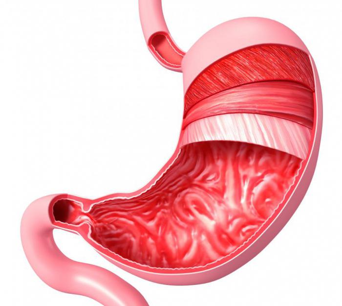
Functions

Motor function

Secretory function. Stomach glands

Suction function
Excretory function
Protective function

Endocrine function
Functional disorders