Pancreatic contours. Pancreatic pancreatitis. The flow of chronic pancreatitis
Press on the pictures to enlarge.
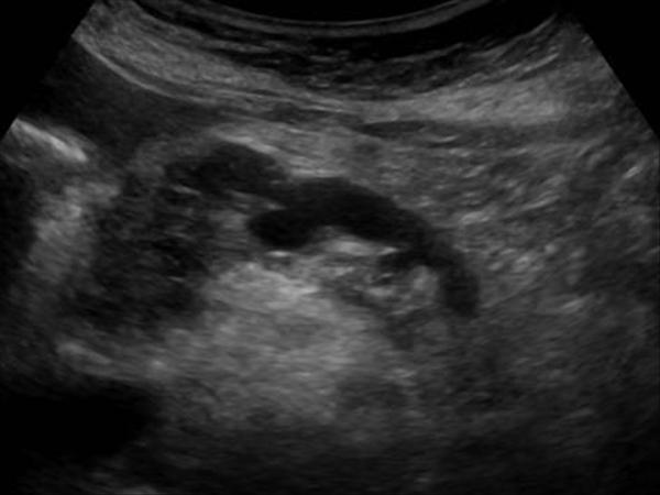
Pancreatic cysts on ultrasound
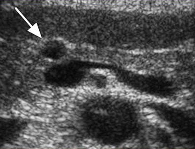
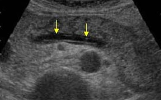
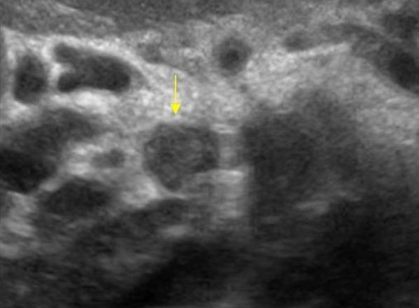
Single small simple cysts are found as random finds in a healthy pancreas. In chronic pancreatitis, small simple cysts are very common. When suspected of a cyst, pay attention to the gain of the outline of the far wall and the effect of the signal amplification in the tissues behind. Simple cysts are isolated from the parenchyma with a smooth thin wall. Inside there should be no partitions or irregularities of the wall, the contents of the cysts anechogenic. Simple cysts are always benign. But, if the cyst is not obvious "simple", further research is required.
Sasha and Vasily's life has not changed since her mother left her. There is guaranteed neglect. In these institutions, children with congenital deformities are even mentioned in official documentation as fools and backward. Despite their number, they are not considered part of the general population.
Stories about sexual violence in orphanages are widespread, even in the most serious cases. Since she was born ten years after the disaster, it is impossible not to doubt that the state of Sasha is associated with the consequences. In addition, it cannot be clearly demonstrated that the congenital disorder of Denis and George - our next contact is not just because of the failure. They affect the cochin syndrome, a disease that is rarely found outside of Russia, Ukraine and Belarus. Condition also affects growth.
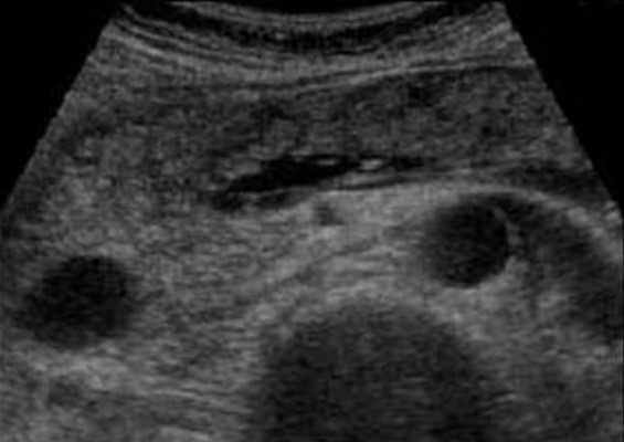
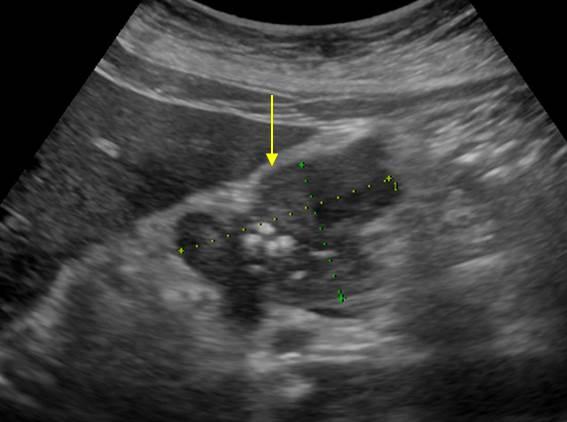
| Photo. Simple pancreatic cysts on ultrasound. A, b - single simple cysts in the field of body (a) and neck (b) pancreas with a thin smooth wall and anechogenic content. In - classic signs of chronic pancreatitis: the main pancreatic duct is expanded against the background of the parenchyma atrophy, an outline of the gland is uneven with jar, in the parenchyma of ordinary and small cysts. | ||
 |
 |
|
Important!!! Often there are simple pancreatic cysts, but do not forget about cystic tumors. Cancer is most dangerous disease pancreas.
If you look from the back, you can easily save them for babies. Like Sasha, they rarely, if at all ever, felt fresh air on the skin. Unlike Sasha, they never saw sunlight. Acute sensitivity of them nervous system Makes them to be in a darkened room - in this boiling hot apartment. Your bed, folding futon.
They weighed their boys, carry them on their shoulders, sing something to them and stroke them on his back. Wedding photography hanging on the wall, shot five years ago, when Mishe was twenty-three years old, and Olga twenty. She is in a sapphire blue dress, in a black suit, in a black shirt, without a tie. They rarely visit, so they are glad to see us. Like Vasily, they do not get help from the state.
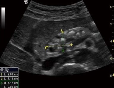
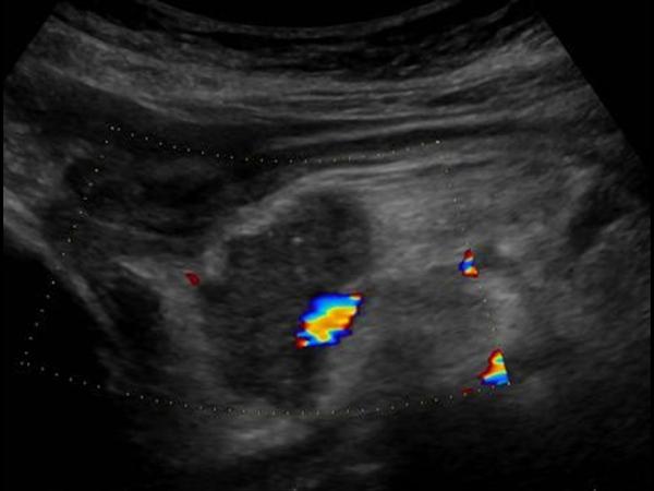
There are two types of cystic pancreatic tumors: a benign microciste adenoma and malignant macrocyse adenoma. The microciste adenoma consists of a plurality of small cyst and looks like a dense education on ultrasound. Macrocyse adenoma, as a rule, includes less than five cyst of more than 20 mm. Sometimes in such cysts you can see polypoid formations.
We sit down and watch George when he carefully trying to make a few steps, having heard how he uttered a few words. A year ago, Denis was still able to develop encephalitis, the edema of the brain, which made it stupid and almost immobile. Young parents George smile and clap in approval sign.
Pancreatic tumors on ultrasound
These cases are all except exceptions in this country. While President of Belarus Alexander Lukashenko lays the foundation for a new nuclear power plant, the proportion of children with chronic diseases It is much higher in his country than during the years, immediately after the Chernobyl catastrophe. According to experts, only ten percent of the expected common damage from the point of view of congenital deformations in the first generation after a disaster can be observed.
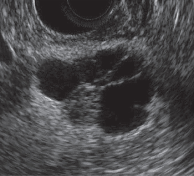
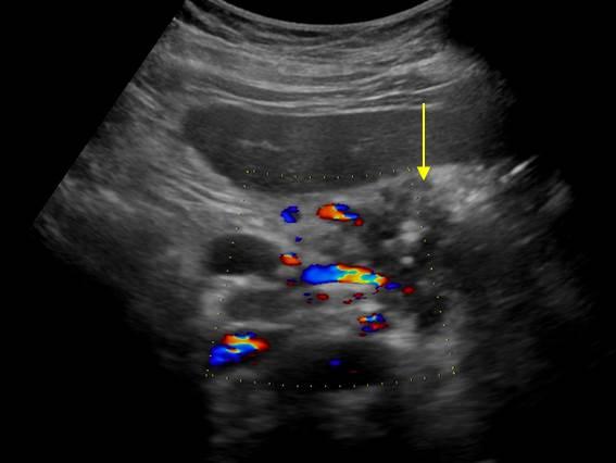
| Photo. A, b - benign microciste pancreas adenoma: large cystic education In the pancreas head. B - pancreatic adenoma with the macro and microciste component. | ||
 |
 |
 |
With pancreatitis, the secretion of the pancreas digesors the surrounding tissues and pseudokists are formed. Pseudokists out abdominal cavity can go in chest and mediastinum. Often, pseudokists are found in patients who have undergone acute pancreatitis (see below).
"Our sufferings will make peace with heart." The people he described, he called the ill disgust that literally means "poor souls." Platonov used this expression rather descriptively than compassion. He explained to him that if someone is removed from life in life, only the soul remains, the ability to feel and suffer. "From our suffering," he writes, "the world will become one heart."
Three years later, it was 74 percent. In less contaminated areas, this number has increased from 40 to 53 percent. For several short years, Gomel was at the forefront of medical research of nuclear pollution. Moving a couple to the city was not associated with career intentions. Rather, they considered their duty to provide their knowledge of those who had no choice but to live with chronic radiation impact. Having received the post of rector of the Gomel Medical Institute, Bandazhevsky faced the anxious nature of the problems with the heart, strokes and rare innate defects among local children.
As a result of a pronounced expansion of pancreatic duct, a distal place of obstruction can form retention pseudokists.
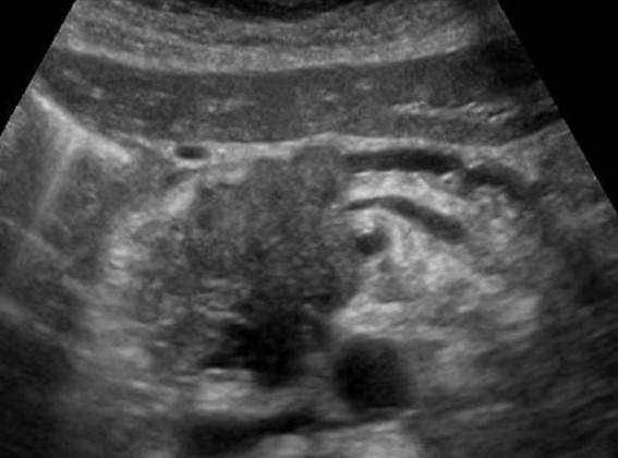
Acute pancreatitis on ultrasound
Acute pancreatitis is a severe complication of a gall-eyed illness or a consequence of toxic effects, such as alcohol.
Light pancreatitis is not visible on the ultrasound (CT more sensitive method). Heavy pancreatitis is easily determined by ultrasound. When extraordinarily clear and contrasting pancreas stands out against the background of surrounding tissues, you can assume the edema of parenchyma and the surrounding fatty fiber. If around the pancreas, along the stomach, at the gate of the liver and spleen, a thin layer of free fluid is viewed, you can confidently diagnose pancreatitis.
After his speech, Lukashenko arrested him
During this, he began a series of long-term biological research by sample victims. After nine years of systematic collection and analysis of data, including the development and production of advanced dosimetric devices, Bandazhevsky presented the results of its research by the Belarusian Parliament and President Alexander Lukashenko. Following the lecture of Bandayzhevsky, Lukashenko arrested him. In anticipation of the court, Bandazhevsky summed up the research "Radioactive Cesium and Heart". He was sentenced to eight years of compulsory work and repeatedly subjected to torture in the first months of his detention.
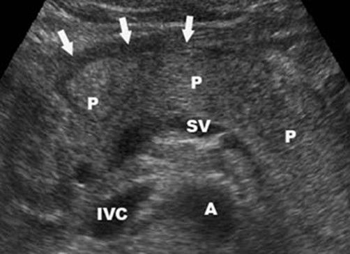
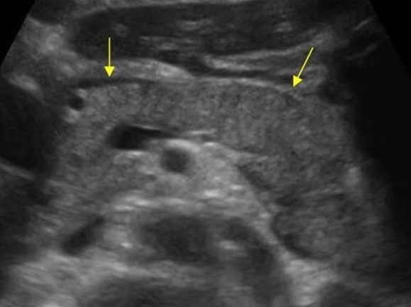
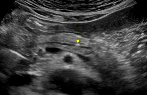
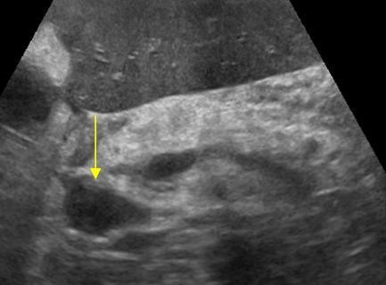
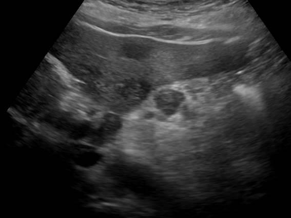
| Photo. Acute pancreatitis on the ultrasound: a - edema of pancreatic parenchyma (P), the edge of the gland is extremely clear, a small cluster of the liquid along the boundary (arrows). B, B is a cluster of the liquid along the contour of the pancreatic body, the thin rim of the liquid in the course of the spleen vein (arrows), the parenchyma is inhomogeneous, the surrounding fiber of hyperhekin - swelling and inflammation, expanded the overall bile duct (B). In this case, it is necessary to exclude a gall-eyed disease. | ||
 |
 |
 |
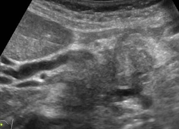
Almost all tumors of the pancreas are hypo echogenic compared to the normal pancreas. Only by ultrasound it is impossible to distinguish focal pancreatitis and pancreatic tumor. Tumor and pancreatitis can be combined.
The Belarusian Secretary Police also quickly made a raid on his offices at the Gomel Medical Institute and destroyed their archival slides and samples. Most of Pupils of Bandyezhevsky were dismissed, many of them were further attracted to criminal liability. They appointed a new director who killed the work of Bandazhevsky and closed his research clinics. A few years later, these vile actions were distributed to the destruction of all medical files containing information about the victims of the Chernobyl catastrophe.
Acute pancreatitis on ultrasound
When four years later, Bandyezhevsky was released, many of those who were evacuated after the collapse were returned to highly polluted areas. Bandazhsky is currently in exile. His most important discovery was that regular consumption of radioactively contaminated food immediately leads to cardiac arrhythmias and irreversible damage to cardiac tissue and other vital organs. These findings are important in themselves, but even more significant is the discovery of Bandyezhevsky that Cesium-137 is one of the most common radionuclides allocated to the atmosphere from Chernobyl concentrates in organs, and not evenly distribute throughout the body.
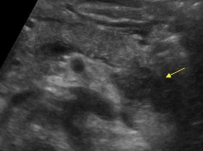
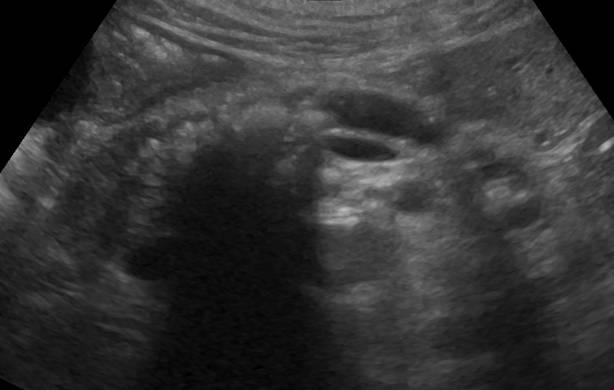
| Photo. Acute pancreatitis on the ultrasound: pancreas is unusually contrasted against the background of hyperheogenic surrounding tissues, a thin band of liquid along the contour (a), in the tail of a hypooehogenic focus (b), in the gate of the spleen liquid (B). A hypo echogenic tail can be mistaken for a tumor. | ||
 |
 |
 |
In severe cases of pancreatitis, pancreatic fluid digest the surrounding tissues, forming pseudocysts. Such cysts may be single or multiple. They can increase in size and break.
This reveals the idea of \u200b\u200b"justified dose" as a misleading. Just as the radioactive substance is distributed randomly on the land plot and forms radioactive foci, the body also absorbs radioactivity unevenly and processes it through the pancreas, the brain, thyroid gland, adrenal glands, the heart, the intestinal walls, and, beyond any doubt, in many other ways that We still need to open.
Some people can absorb significant doses without visible damage, while others can absorb a small amount to cause cancer or serious damage to organs. Most victims from long-term damage are born and unborn children. Their immune system has not yet mature, and their cells are developing much faster, so any change in the cell structure more and has a much greater influence than in adults.
At ultrasound, pseudokists are defined as oval or rounded hypo echogenic formations with clear contours. In the early phases of the formation of a cyst, it represents a semi-liquid education and has a complex echostructure with internal reflections and fuzzy contours. Later, due to the autolytic processes and precipitates the suspension of blood and pus, clear signs of liquid content appear and a false capsule is formed with even walls. Often there is an infection of the pseudocyst, then internal echoes or thin tender partitions can be determined. When cyst is found, it is important to trace the connection of cysts with a duct, as it is important to determine therapeutic tactics. When a pseudokist is more than 10 cm in size, difficulties arise in determining its source.
Each nuclear reactor regularly explodes radioactive gases to the atmosphere.
Even leaving aside the results of Bandayzhevsky, a sample of evidence in most of the regions affected by Chernobyl showed that a chronic low dose of stress leads to diseases of the circulatory system, endocrine system, immune system and respiratory tract; In addition to reproductive disorders, changes in the structure of the bone, damage to the brain, blindness, congenital defects and anomalies, to cancer thyroid gland, leukemia, to increased infections, organ insufficiency, premature aging, genetic mutations, to "Chernobyl AIDS", "Chernobyl" "Heart", "Chernobyl limbs" and "Vegetative circulatory dystonia".
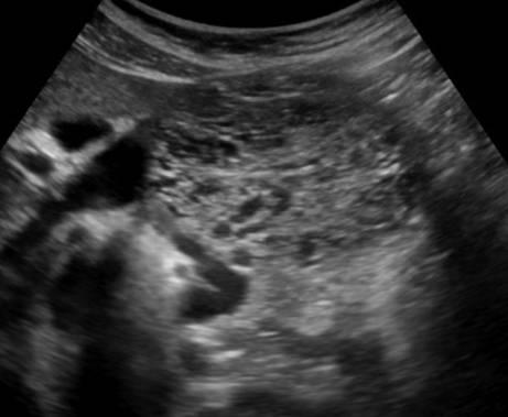
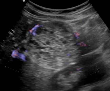
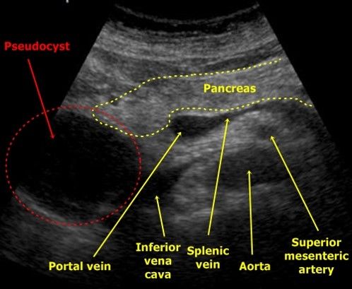
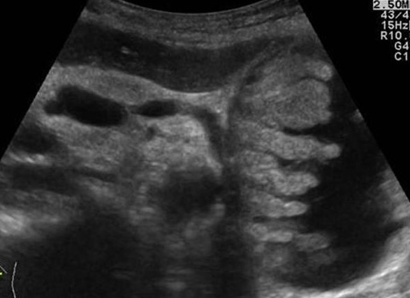
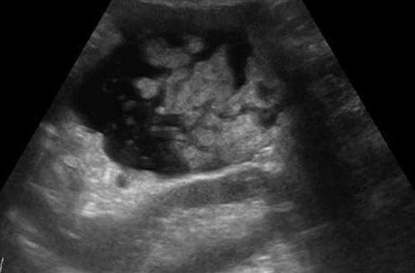
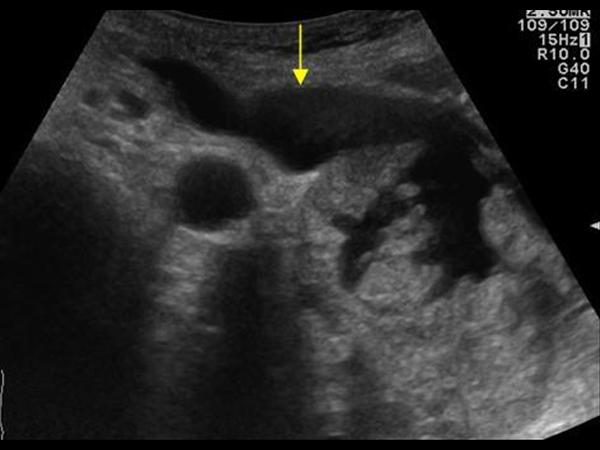
| Photo. A is a large pseudocyst between the head of the pancreas and the liver after the transferred pancreatitis. B, B - heavy necrotic pancreatitis longitudinal (b) and transverse (c) cuts: extensive necrosis, melting surrounding in the area of \u200b\u200bthe tail, accumulation of fluid around the gland. | ||
 |
 |
 |
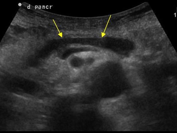
Chronic pancreatitis on ultrasound
Chronic pancreatitis can have different manifestations, from almost normal gland to pronounced atrophy and occurrence of parenchyma. Pancreas becomes thinner, pancreatic duct sometimes seems slightly expanded, the contour of the gland is often uneven with jar. Often there are simple cysts, and they can become quite large. Often, stones are formed in the pancreatic duct.
The last terms in this list are common terms for many new syndromes that were discovered by medical professionals only in the years after the Chernobyl disaster. The symptoms are so diverse and diverse that the doctors are forced to group them under relatively general names.
The opening of Bandazhevsky is a serious threat to the nuclear lobby, because each nuclear reactor regularly produces radioactive gases into the atmosphere. These "ventilation holes" are not exceptions; They are planned, approved and systematically immanent.
Calcifications in the pancreas on ultrasound
Important!!! If there is a dilatation of pancreatic duct, stones in the pancreatic duct and in the overall bile database should be searched.
Calcifications inside the pancreas can give an acoustic shadow, however, if they have small dimensions, they look like a separate bright echo essential shadow. In chronic pancreatitis, calcifications are distributed diffuse throughout the pancreas. Stones in the duct are located in the course of the duct. The bile stones in the distal choleret can be enrolled in the calcifications in the pancreas. Calcifications are clearly visible on the CT, and for unused stones, preferably MRI or ultrasound.
In most cases, about hundreds of cubic meters of radioactive gases are sent hourly from the capacitors of each reactor. If the reactor is temporarily disabled due to the mechanical failure, these ventilation holes increase in frequency and length.
Although radiation as a phenomenon also takes place in nature, long-lived radionuclides emitted by nuclear reactors, are new to us as species. They did not exist in significant quantities on Earth during the entire evolutionary history of complex forms of life and in millions of times more toxic than natural radionuclides.
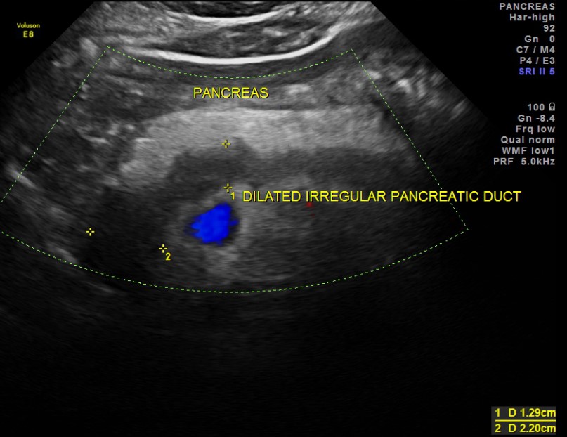
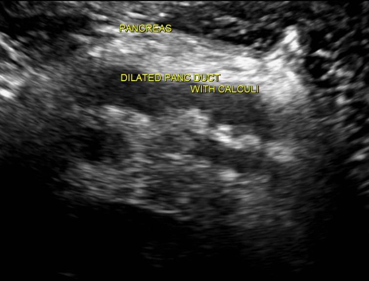
| Photo. A - in the extended dummy a small stone. B - in the extended pancreatic duct, a number of several stones with shading behind. B - in a patient with chronic pancreatitis huge stones in an extended dash. Pay attention to the intensive shading behind. | ||
 |
 |
|
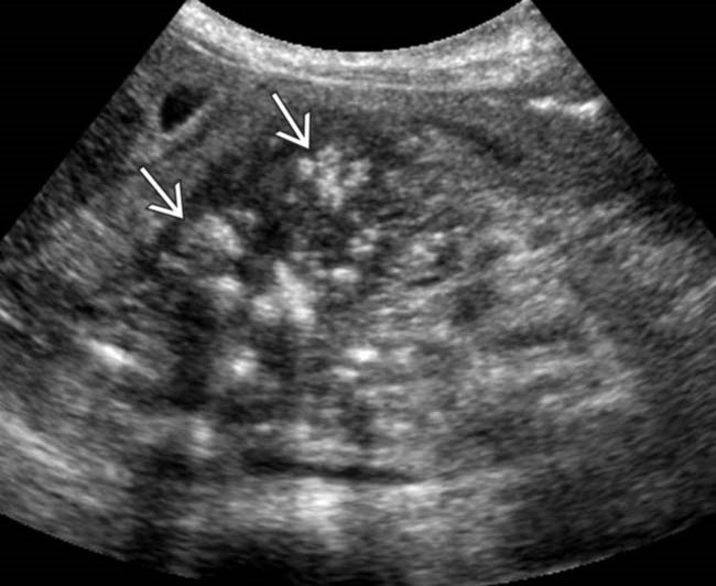
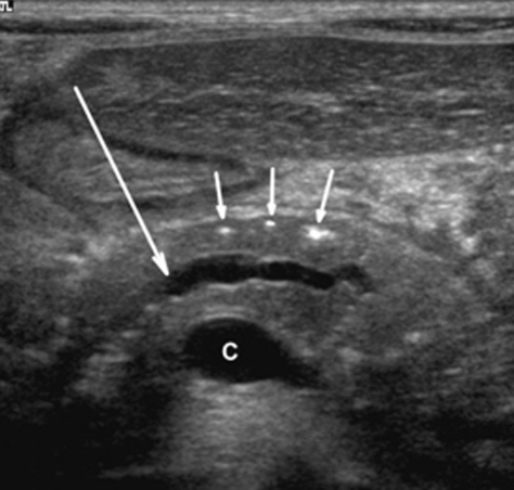
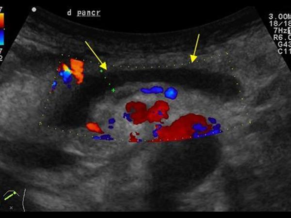
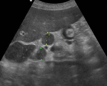
| Photo. A, b - calcifications in the pancreas parenchyma in patients with chronic pancreatitis. Some calcifications have a shadow. In - a boy of 5 years with chronic hereditary pancreatitis: Outbreaks (small arrows) and dilatation of pancreatic duct (big arrow). C is the merger of the upper mesenteric and spleen veins. | ||
 |
 |
 |
Advanced pancreatic duct on ultrasound
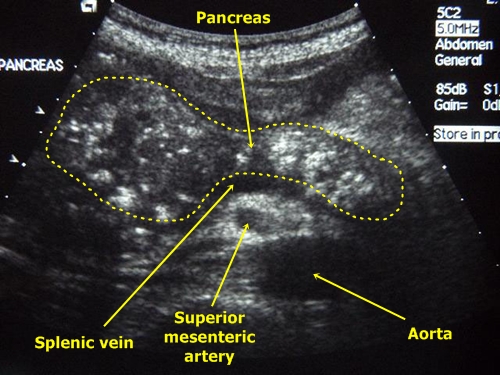
The internal diameter of the normal pancreatic duct is less than 3 mm. The duct is better visualized with transverse scanning in the middle third of the pancreas body. In order to make sure that you discovered the duct, you need to see the pancreas fabric on both sides of it. The spleen vein is behind or the stomach wall in front can be falsely interpreted as pancreatic duct.
Even in the distant future, the deterioration of the situation is most likely
Nikolai Omelyan: We found that child mortality increased by 20-30% due to chronic irradiation after the accident. In the province of Sumy, it is 96, 5 percent, in the Donetsk region - 96 percent. Even in the distant future, the situation will most likely escalate, since the genetic consequences of the catastrophe still have to deploy. Animal studies show that after twenty generations, radioactivity resistance is noticeably reduced among those who are exposed to, which is likely to lead to even more diverse and malignant diseases through four hundred years.
The walls of the pancreatic duct should be smooth, and lumen clean. When the duct is expanded, the walls become uneven; Scan not only the head of the pancreas, but also the entire biliary tract.
The main reasons for the expansion of the pancreatic duct: the tumor of the head of the pancreas or the ampoule of the nipple of the nipple (combined with the jaundice and dilatation of the biliary tract); Stones of general gall or pancreatic duct; chronic pancreatitis; Postoperative spikes.
At the same time, even problems associated with the storage of nuclear waste are taken into account. Wallpaper around me shows a brick pattern with areas where the stones seem to have fallen and repeatedly show views of the rural house. On the edge of this scene, a woman throws chickens on a peak for a picket. On TV in the corner, the cook prepares Spaghetti Carbonara with a label of a culinary show. Then he suddenly breaks out of his role and conscientiously cleaned with a spray bottle and soft fabric. He smiles, puts the bottle of cleaning agent into the chamber and praises her quality.
"Did you talk about it with him?" - "Are you kidding?"
Alexy returned me to the airport in his gray truck. He hesitates with one hand, and to another holds a cigarette between big and index fingers. Landscape was shrouded in fog. "My dad was on cleaning." You had to mention it. He could not talk.
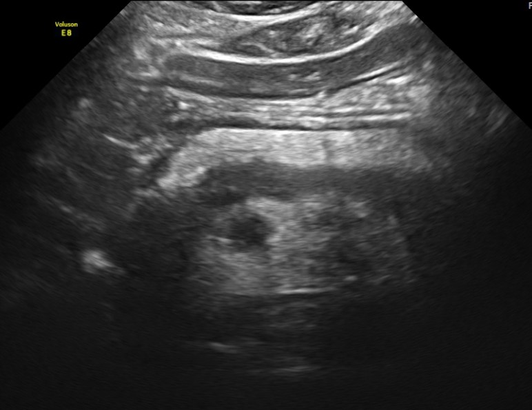
| Photo. A man with insulin dependent diabetes Complaints for weight loss and abdominal pain for a few months. On the ultrasound, the extensive overall dump of the pancreas with an uneven wall. With a further inspection, it is clearly visible in the calcification duct with the shadow behind (B). | ||
 |
 |
 |
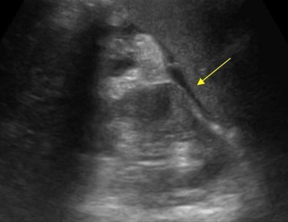
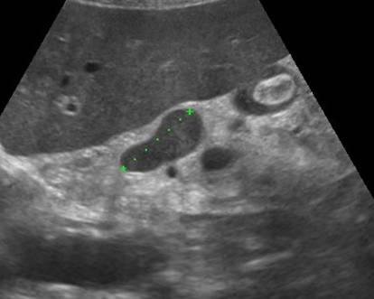
| Photo. Patient with sharp pancreatitis: a large pseudokist was formed at the tail level (see above), the extended pancreatic duct opens in the pseudokist. | ||
 |
 |
 |
Pancreatic tumors on ultrasound
In most (50-80%) cases, the tumor affects the head of the pancreas. Head tumors squeeze the overall bull duct. When cancer contour pancreas, fuzzy, characterized by a local increase or esteem, sometimes embedded in the surrounding tissue in the form of languages \u200b\u200bor pseudopodies.
"How long did he hurt?". He had two heart attacks and strokes. His friends are also sick. They can not leave their homes. They never encounter each other. "Do you have memories about this time?". He pulls out a cigarette from the window. "Did you talk about it with him?".
His pallor strikes his behavior as if the last bit of life was washed out of him. What about your friends? If you drink together, do you talk about what you know? He continues to look outside. Then he folds his head and ironizes. You just do not understand this.
In most cases, the pancreas tumor is a hypo echogenic formation, almost deprived of internal echoes. However, there are tumors with diffuse scattered echoes and high-intensity echoes in the center in their absence on the periphery. Despite the fact that the border between the tumor and the rest of the parenchy gland is fuzzy, it can always be approximately carried out due to the difference in the echogenicity of normal tissue and tumor focus.
Although the hypo echogenic structure of the tumor, especially in the absence of small sections of increased density in it, resembles such at cysts, the absence of the effect of distal amplification makes it possible to eliminate the liquid nature of education. For a cyst, moreover, a much more flattened and clear boundary is characterized.
| Photo. The carcinoma of the pancreas head (arrow): the overall bull duct (A) and the pancreatic duct (b), hypo echogenic tumor surrounds the upper mesenteric vein (B). | ||
 |
 |
 |
In tumors of the pancreatic head, the overall biliary and pancreatic duct is very often expanded, unlike chronic wall pancreatitis, it is smooth and infertable.
Important!!! The visualization of the main pancreatic duct within the hypo echogenic zone indicates the benefit of local edema and against the tumor.
Sometimes in the pancreatic cancer, typical signs of chronic pancreatitis are revealed, as well as pseudokists distal than tumor obstruction site. This is a consequence of obstruction. Intrahranny metastases, enlarged currency, periportal and retroperitoneal lymph nodes indicate cancer.
| Photo. The carcinoma of the pancreas head: the outline of the head is uneven due to volumetric hypochogenic education, the body parenchyma is very thin (atrophy), expanded pancreatic (a) and common bull (b) ducts, in the gate of the liver, a large rounded lymph node (B). | ||
 |
 |
 |
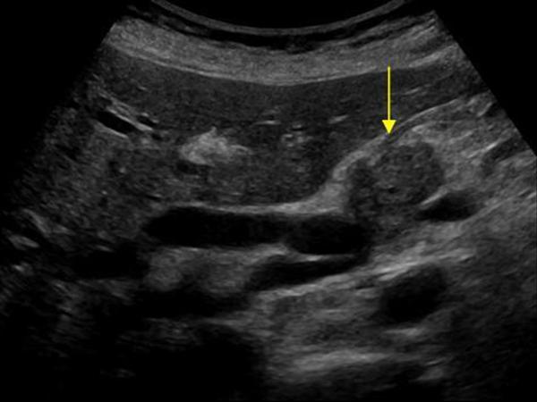
| Photo. Large lymph node (arrow) near the pancreas can be mistaken for a head tumor. Increased mesenteric lymph nodes of the rounded shape, hypo echogenic and without a central rutter, which indicates their malignancy. |
||
 |
 |
 |
| Photo. Big neuroendocrine tumor (arrows) of the pancreas with observation and metastases in the liver (B). | ||
 |
 |
 |
Take care of yourself, Your diagnostic!
Problems with pancreas are familiar with a rather wide circle of people. Pancreatitis does not bypass or adults or children. And the sooner the disease is detected, the more effective will be the treatment.
Clearly see clinical picture Helps an ultrasound study. The picture on the monitor screen will show how the pancreas looks like and how far the disease is rooted in the body.
What does healthy pancreas look like
There is an organ between the stomach and duodenalist, on average by 5 - 10 cm above the place where the navel is. Consists of head, body, duct and tail. The length is 78 - 87 mm, the diameter duct fluctuates in such limits: 1.5 - 2 mm. The boundaries have a clear and even outline.
S-shaped
Typically, the pancreas has an S-shaped form. But the science is also known to the anomalies, which may be due to a number of diseases of the gastrointestinal tract, as well as as a result of the narrowing of the ducts or the appearance of additional. Most often you can meet such deviations from the norm:
- spiral;
- split;
- ring-shaped;
Types of pancreatitis
The inflammation of the pancreas has three varieties: acute, chronic and reactive. For acute form The disease is characterized by changes in tissue (it disintegrates), possibly hemorrhage and accumulation of purulent substance.
For chronic form The disease flows slowly. Requires a constant diet and medication treatment.
The reactive is called the disease, if the attack of acute pancreatitis is manifested with the beginning of the liver disease, stomach, duodenal gut, gallbladder.
Symptoms of the disease
In chronic form, the patient detects painful sensations at the top of the abdomen. Painful feelings May be felt in the left, less often the right hypochondrium. Appear on the background of improper nutrition, alcohol consumption. The pain can be like stupid and sharp. And the feeling of nausea and attacks of vomiting appear.
For acute form, the following picture is characteristic:
- sudden, severe pain at the top of the abdomen, adjusted to shock, can give under the left blade;
- exhausting vomiting;
- diarrhea.
With reactive pancreatitis, the patient appears pain, cutting and stupid, wear a look. In his mouth, the taste of bile is also characteristic of vomiting.
What can be determined by ultrasound
All patients with inflammation of the pancreas are prescribed an ultrasound study. It is carried out in acute, and in chronic pancreatitis, and if suspected a disease.
Ultrasound will show, in what condition the contours and fabrics are located:
- if the contour is vague, then inflammatory changes occur in the pancreas, swelling began. But he may be evidence of the illness of the stomach or duodenum;
- the contour is convex, but at the same time smooth - a cyst was formed on the gland;
- uneven outlines are characteristic of pancreatitis and neoplasms;
- bug, blurred edges talk about cancer formations.
Uz studies also demonstrates the state of the pancreatic tissue. W. healthy man It is medium density. If the density is increased, it means that connective tissue. This condition is characteristic of chronic pancreatitis, but may be evidence of age-related changes. On the screen, such sections of white.
With a reduced tissue density, the picture on the screen will be black. This condition is characteristic of the acute form of pancreatitis.
At each stage of the disease, the pancreatic contours and its structure are modified. At the first stage of the acute form of the disease of the contour railway gland, blurred and uneven, the density is lowered, the duct is expanded.
In the second stage, the appearance of cyst or abscess. The outline will be rounded, the tissue density is reduced.
In chronic pancreatitis, the contours become fuzzy. If stones appeared in the gland, the contour will acquire a rounded outline, the density in this place will be increased.
Cyst and tumor
If the pancreas during the ultrasound is visible to black neoplasm with even well-defined edges - this is evidence that liquid has gathered in these places (cyst rises) or rose gathered.
Cancer formations on the gland look black or white spots. It depends on what type of cancer is developing in the body. Mixed cases are possible.
With the anomalies of the pancreas, two pancreatic ducts are visible and a blurry structure, unevenly transmitting bonds.
How to prepare for ultrasound
First of all, it is necessary to organize proper nutrition. Do not use products that contribute to gas formation: soda, sweets, legumes, mayonnaise, cabbage, etc. Gaza is inflated intestinal loops, and those in turn close the pancreas. Research can break.
To reduce gas formation, it is appropriate to take adsorbents, pancreas enzymes, wind turbines.
For the period of preparation for ultrasound Research, It is necessary to reduce the consumption of meat and meat products, dairy products, fish. And still follows:
- eliminate alcoholic beverages;
- forget about smoking;
- can take medicinal productswhich reduce gas formation;
- the last meal must be held for 6 to 8 hours (in children for 3 hours) before the study began.
When the ultrasound is prohibited
The doctor will not appoint an ultrasound if there is an allergy to the gel used or if the patient's life is under threat. When the patient's condition stabilizes, ultrasound research can be carried out. Patients whose obesity in the third stage is not carried out, as there is no good review.
In the presence of Absadin and the Russian Academy of Sciences, ultrasound will not hold. This is explained by the fact that it is impossible to get good contact. As a result, a bad review.
During problems with the pancreas, it is necessary to organize treatment correctly and on time, to go through the research prescribed doctor. Rely on your own strength and grandmother's money - a dangerous health game.
Read also ...
- The work of "Alice in Wonderland" in a brief retelling
- That transformation. "Transformation. Attitude towards the hero from the sister
- Tragedy Shakespeare "King Lear": the plot and the history of the creation
- Gargantua and Pantagruel (Gargantua et Pantagruel) Francois Rabl Gargantua and Pantagruel Brief