Methods of preparation of microscopy microscopy. Microscopic laboratory. a) formed the arch
Not everything that is at hand, it makes sense to put on the subject table and try to consider in the eyepiece of the microscope. For example, opaque objects (finger, coin) will not consider at all, and there is quite a strong magnifying glass to consider.
Usually, specially prepared microscopes are used for microscopes. When preparing a microdaparation, the subject glass is taken (size 25 × 75 mm), which is placed the object in question; Usually, for better safety and ease of use, the object is covered with a thin coating glass (size 18 × 18 mm).
By the method of cooking and storage time distinguish:
- permanent drugs - the object is placed in a transparent hardening environment (usually canadian balm) and covers with coating glass; Such drugs can be stored for years and decades, but their preparation is quite laborious (the object must be carefully prepared: dehydrated, painted, etc.);
- temporary drugs - the object is placed in a liquid medium (water, saline, glycerin-gelatin, etc.), such drugs are suitable for use for several hours, however, experiments can be carried out with them (for example, to replace water with a salt solution of a certain concentration, add dye, etc. .).
Permanent drug |
The table shows the types of preparations depending on the nature of the object under study.
Description |
Permanent / temporary |
Example |
||
Total preparation |
Whole organism or its small parts (limbs, oral apparatus). Usually require processing (clarification) |
Permanent, temporary |
 |
|
The object is poured into the plastic material (paraffin, acrylic) and after its solidification it is cut into thin plates with a microtoma (devices for producing thin sections) |
Usually standing |
 |
||
The current-layer preparation without coating glass (usually the blood product): a drop with a polished glass is smeared over the surface of the slide glass thin layer, dried |
Temporary, less frequent |
 |
||
Thin (thickness 0.03-0.02 mm) The plate of rock or other solid sample (bone, fossil), glued to the covering glass |
Constant |
 |
In some cases, the temporary preparation can be viewed directly without covering the covering glass. This drug will be almost impossible to consider dramatically all - some part will be in focus, and the rest is not. But if we consider such a drug with a small increase in the microscope, you can consider some interesting objects.
Also, in the form of open time, it is possible to grow crystals of different salts (acetylsalicylic acid, or aspirin; copper sulfate, or copper sulfate; red blood salts, or potassium hexationanoferrate). A few drops of aqueous or alcohol salt solution are applied to the finger-purified dust and fingerprints (for better quality crystals, the solution is pre-high, because it will reduce the number of crystallization points and get larger and neat crystals). Drops are allowed to dry (it is possible to carry out an experiment with one of the glasses, heating it, in order to accelerate evaporation, and another to evaporate the solvent, and the crystals are dropped independently). You can also consider the semi-prepared micro-process at the crystallization process: you can see how the crystal increases directly in the eyes or how the fluid flow occurs around the embryo of the solid due to the difference in the concentrations of ions.
A simple and spectacular way of making microfrets without cuts, allowing to consider the relief of the surface of the object being studied (for example, a leaf of plants, insect bodies) - method replica. At the same time, an object is taken (for example, a leaf of the plant), and a thin layer of transparent nail polish is applied to it (sufficiently small spots of 5 × 10 mm). After the lacquer dries (after about 5-7 minutes), a piece of sticky tape is glued to the lacquer stain; So the replica is separated, after which it is placed on the slide and viewed under the microscope.
1.1. Preparation of temporary preparation
The technique of preparing the temporary drug is well known for school experiments with the skin of the bow. We put the glass glass on the table (the drugs are always kept behind the side faces of the subject glass). In the center of the glass, we put 1-2 drops of water and the object of the study take the cover glass for the side faces and carefully cover them from above the drop with the object. It is more correct to overtake the covering glass of one of the faces into the subject and slowly reduce the angle between the glasses so as to cover the drop with the object. This reduces the possibility of air bubbles.
1.2. Cooking blood smear
Blood smears for blood test are prepared as follows.
| Step | Description |
The photo |
Place a small drop of blood on the horizontal surface of the alignment glass, with a glass capillary pipette (or directly from the finger point, transfer the speaking drop of blood to the end of the sterile slide glass, trying not to touch the glass with a punctured skin). |
 |
|
The pure grinding glass is placed with a short edge at an angle of 45 ° to the subject glass at the edge of the drop. We are waiting until the blood is blurring under the edge of the glass. |
  |
|
As soon as the blood spread over the edge, they spend the rapid movement from the drops along the subject glass. It should not be strongly pressed on the glass, since the uniform elements of the blood can collapse. |
 |
|
After cooking, the smear is quickly dried in the air until the wet gloss has disappeared, holding it over the lamp lamp over the lamp or waving it in the air. A well-made smear of thin, has a yellowish color and ends with a "pitcle". Due-pink and reddish strokes are unsuitable, since they are too thick and cell elements will be difficult to distinguish. |
 |
Images and description from microtome.info/documents/micropreparaty.html.
2. Some features of microscopation
The use of microscopes on a small and large magnification is quite well known for school experiments and does not require particularly complex skills.
2.1. Mmimensional lens
If there is an immersion microscope with an immersion lens (it is marked with 100 × Mi and a black ring, and has a movable part of the lens to the microcreparation), then you have the opportunity to consider objects with a large increase (however, it makes sense to use strokes).
The procedure for working with the lens of the oil immersion is next.
- Out out the dry lens from the rays (turn the lenses drum so that no lens is turned to the drug).
- For the drug, apply one drop of immersion (cedar) oil; Preferably immersion oil is also a little lubricate the lens of this lens.
- Turning the lenses drum, enter an immersion lens to the rays (before clicking the fixation).
- Lower the lens to contact with the em dimensional oil drops, then, watching the microscope, with coarse and fine focus screws, set the sharpness.
When working with an immersion lens, maximum illumination will be required. After the end of the study, the immersion lens should be wiped with a soft optical cloth or a bat, slightly moistened with a special fluid for cleaning optics. Important: any other types of lenses cannot be wipe!
Application Instead of cedar oil of other oil liquids, it is not recommended, since, if the refractive index of the immersion used (for example, vaseline oil with an indicator of 1.48162) differs from the refractive index of the cedar oil (indicator 1.515), it is possible to reduce the contrast, the fuzziness of the image, the decrease resolution.
2.2. Photos in braid lighting and dark field
For bulk preparations (total drugs insects, algae, etc.) it is interesting to use lighting other than the usual translucent. For adjusting the lighting in microscopes, the condenser is used - a rotary device with holes of different diameters and location. Using special options for condense, you can change the nature of the illumination of the drug, emphasizing the volume of the object under consideration.
The table compares the work with the lighting of different types.
Type of lighting |
Method of creation |
View of the pupil of the lens with the echolar |
Example |
|
Normal (translucent) |
Light passes through the field of view evenly. |
 |
 |
|
1) Temporary Preparations
To study vegetable objects with a light microscope, you need to prepare a micro-process. Micropreparations that are not intended for long-term storage are called temporary. The object being studied is placed on a slide in a drop of water, glycerol, solution, reagent or dye and covered with coating glass. Such drugs can be stored for several days by placing in a wet atmosphere.
2) permanent drugs
Permanent drugs are prepared for special techniques that ensure their storage for decades. Continuous preparations include strokes, total drugs and sections. Brokes are used in the study of blood cells, crops of microorganisms, isolated tissue cells. Total preparations are separate transparent and thin objects. Training sections can be done manually using a razor. However, high-quality sections with a given thickness of 10 ... 22 micrometers are usually made using special devices - microtomes. Such sections are often referred to as microtomic drugs. For more subtle cuts (0.01 ... 0.05 μm, or 10 ... 50 nanometers) use ultramicrothomes.
Briefly consider the main stages of preparation of permanent drugs.
1. Fixation of material. Immediately after the end of the fixation, washing the material or water (after water locks), or80% alcohol (after alcohol locks). The number of changes in washing liquids - at least 3. Time - up to 24 hours.
2. Dehydration in alcohols of increasing concentration. In parallel, the material seal occurs. Sequential movement of the material through a number of solutions is called wiring. After aqueous clamps, 8 alcohol shifts are used: 20%, 40%, 80%, two shifts of 96%, two shifts of 100%. After alcohol locks - 4 alcohol shifts: two shifts of 96% and two shifts of 100%. In each shift, the material is maintained at 1 hour.
3. Enlightenment. It is impregnating the material with a paraffin-xylene solvent (benzene, chloroform). The sample is placed on 1 hour in series in each of the subsequent solutions: 3 parts of the alcohol + 1 part of xylene, then 2 parts of alcohol + 2 parts of xylene, then 1 part of the alcohol + 3 of xylene parts, then two xylene shifts.
4. Pouring into paraffin. This is the substitution of xylene paraffin. The sample is placed in a mixture of xylene and paraffin at a temperature of 55 ... 57 degrees and leave in a thermostat at this temperature to completely evaporating xylene (from several hours to several days). Then, at a temperature of 55 ... 57 degrees, wiring through paraffin I (6 ... 12 hours), paraffin II (6 ... 12 hours) and pouring to paraffin III. Paraffins I, II, III differ only in purity: paraffin III is the final medium that should have the greatest cleanliness. As a result, paraffin blocks are obtained in which the samples of the material are concluded. These blocks can be cut in any direction.
5. Staining sections. Paraffin sections are glued to clean glass. As glue, a mixture of a chicken egg protein with glycerol (in a ratio of 1: 2) can be used with the addition of antiseptic (thymol or phenol). Typically produce dewaxing of sections. For this, glass with glitched cuts is carried out through xylene, decreasing concentration alcohols (100%, 96%, 80%, 70%) and distilled water. Saying time in every environment - 2 ... 3 minutes. Next are painted according to the methods.
6. Dehydration and enlightenment of painted cuts. It is performed by wiring through alcohols of increasing concentration, and then through xylene.
7. Conclusion in Wednesday (Pouring). For long-term storage of drugs, they must be concluded on Wednesday, protecting the drug from air oxidation and from damage to fungi. Special resins are used for the fill (Canadian balm, fir balsam), which are dissolved in xylene to the consistency of liquid honey. A drop of such a solution is applied to the cut and cover with coating glass.
6. Chemical composition of the cell substance. Micro and macroelements.
In the composition of the cell, more than 80 chemical elements were discovered, while any rigorous elements characteristic of only living organisms were not detected. However, only in relation to 27 items it is known which functions they perform. The remaining 53 elements are likely to fall into the body from the external environment.
1. Macroelements
They constitute the bulk of the cell substance. They account for about 99% of the mass of the entire cell. Especially high the concentration of four elements: oxygen (65-75%), carbon (15-18%), nitrogen (1.5-3%) and hydrogen (8-10%). Macroelements also include elements whose content in the cell is calculated with tenth and hundredths of percent. This is, for example, potassium, magnesium, phosphorus, sulfur, iron, chlorine, sodium.
2. Microelements to them include mainly atoms of metals that are part of enzymes, hormones and
other vital substances. In the body, these elements are contained in very small quantities: from 0.001 to 0.000001%; There are among those elements Bor, cobalt, copper, molybdenum, zinc, iodine, bromine, etc.
3. Ultramicroelements
The concentration of them does not exceed 0.000001%. These include uranium, radium, gold, mercury, beryllium, cesium and other rare elements. The physiological role of most of these elements in the organisms of plants, animals, fungi and bacteria has not yet been established.
Ministry of Education and Science of the Russian Federation
State Educational Institution of Higher Professional Education
"Shadrin State Pedagogical Institute"
Department of Natural Science Disciplines with Teaching Method
Methods for the preparation of microfrants by biology
Course work
specialty 050102 "Biology"
Executor:
Gorshkova Ekaterina Andreevna
Student 3 Course Day Department
Scientific adviser:
Candidate of Biological Sciences, Associate Professor
Sharypova Nadezhda Vladimirovna
_________________________________
Evaluation __________________________
Shadrinsk
2014
Introduction .................................................................................... .3.
§ 1. Characteristics of microspeats and their use ......................4
§ 2. Equipment needed for the preparation of micro-repair ...... .7
§ 3. Preparation of material for the preparation of micro-repair ............ ... 12
§ 4. Methods for the preparation of micro-repair .................................... .17
Conclusion ................................................................................. 31.
Bibliographic list ............................................................ .32
Appendix ............................................................................................ ... 34
Introduction
Relevance of the research topic. Micro-processing are a visual learning tool and therefore will be widely used in biology lessons during laboratory classes, when studying new material, generalizations, comparisons and surveys.
Due to the insufficient provision of biology cabinets with ready-made microsperature, which are necessary for a better study of the subject, they can be made independently.
Based on the relevance, we chose the following research topic: "Methods for the preparation of biology microcraparations".
Object research are microsperats.
Thing - Methods of preparation of microcrants.
Purpose of the study: To study the types of micropreparations on biology and their preparation methods.
Research tasks:
- Examine and analyze literature on this topic.
- Perform the characteristic of microcracy and tips on their use in biology lessons.
- Describe the equipment required for the preparation of micro-processing.
- Learn to prepare material for the preparation of microcrants.
- Examine methods for making microcrants.
For the implementation of goals and tasks used the followingresearch methods: Analysis of scientific and methodological literature on the topic of research, pilot search and summarizing its results.
Structure of coursework. Course work consists of introduction, 4 paragraphs and imprisonment, a bibliographic list is presented, including 16 sources, 1 applications.
§ 1. Micropreparation characteristics and their use
Natural dispersed benefits include herbarium, wet preparations, microspections.
The micro-product is a thin section of a living organism organ or a microparticle enclosed in a transparent balm (immersion oil) or dried, placed between two glasses (subject and coating).
The origin of the micro-processing originates in the use of ivory or ordinary bone as a stand for samples that were placed between discs of transparent mica. A similar design was popular in Victorian England, while the royal microscopic society presented a standardized glass microscope glass.
Micro-processing makes it possible to demonstrate the inner cell structure of organisms, which helps the formation of knowledge from students about the single cellular structure of organisms. Micro-processing can be divided into two groups:
- permanent manufactured factory by specially for training;
- temporary, prepared by the teacher for the lesson or in the lesson by schoolchildren of one-time.
Permanent microsperats are the finest sections of tissues of organisms, their organs. Most of the cells are not painted and therefore, even with a large increase in the microscope, it is difficult to consider intracellular structures, including the kernel. In this regard, cell microcreparations are painted with special dyes to give them greater clarity. Teachers necessarily need to warn children that the color is not natural for microstructures. Use them when studying new material, generalizations, comparisons and surveys.
Temporary preparations are so called because they are not saved for a long time. After familiarization with the microwieth, the temporary preparation is washed off from the slide glass. The preparation of the micro-process is one of the mandatory types of skills formed in the course of biology, starting from grade 6.
For the study of living cells of microorganisms, drugs "crushed drop", "hanging drop", "imprint", "Agarear film" ("Microculture") are used. Alive cell preparations are viewed with "dry systems" microscope. Preparations, work with which is already completed before washing, kept in a disinfectant solution.
MicroSPs allow a wide range of experiments provided for by the biology school program program. They are intended for a detailed study of microscopic structures under a microscope.
The teacher to work with the micro-process must clearly explain the students that they should see using tables, drawings, diagrams, etc.
During the survey, teachers often use "dumb", (without labels) microspections. The students are given a task to define microsperats, tell about the structure of one or another tissue. It is advisable to store the same micro-package in one package so that you can quickly distribute them to students.
The microspears are stored away from the heating devices and so that the slide glass is in a horizontal position to prevent the "sliding" of the micro-process.
Requirements for micro-processing:
- Glasses should be colorless and transparent. The covered glass should be thinner than the subject that serves as the basis of the product.
- The micro-treatment must be centered, i.e. Located in the middle of the cover glass. The location of a very small object must be marked by the frame.
- Microscopic sections should be very subtle and have all the items necessary for study.
- Separate microsperature fabrics must be painted with a bright resistant dye.
- Micro-repair must be produced in the form of a set for each section of the biology course. The number of drugs of the same name in the set should be sufficient for laboratory work by all students in the classroom (15-20 pieces).
- Methodical recommendations must be attached to the set of microcreparations, where the drawings of the studied micro-objects and tasks for independent work of students should be given.
The study of micropreparations in the learning process comes to the methods of science, makes it possible by direct observation to see the structure of organisms, their parts.
Microscopes are microscopically small living objects, the finest sections of their organs and parts. These objects are concluded between cover and subject glasses in a special solution. For better distinguishability, drugs are painted, so the color of objects is artificial.
1. Yes, the start of work with the microcracy is required to write its name, the topic of laboratory classes.
2. The study of any microscopic is starting with the viewing under a microscope at a small increase (56x). Then choose a place where the parts are better visible, and put a large increase (120-300x) and proceed to sketch. When sketching the micro-process, it is necessary to respect the ratio of the size of individual parts of the original. Sourcing helps to remember and create a visual idea of \u200b\u200bthe object.
3. First, the figure denote the main elements, then complement the details. Figure signs the name of the microsperature and an increase in which the drawing is made. On one page of the student notebook make 2-3 drawings. The main structures of the microsperature are indicated by pointers and opposite each write a digit followed by their decoding. The contour of the field of view of the microscope is not denoted.
Thus, there are 2 types of microsperats - temporary and constant. Their difference is in the time of storage and cooking methods. Micro-processing must meet certain requirements that need to be carefully observed. For the preparation of microcracy, special equipment is needed, which is described in the next paragraph.
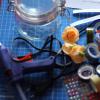
§ 2. Equipment required for the preparation of micro
Desktop.
In the absence of a special table, any table (preferably with drawers) can be adapted with success (preferably with drawers) with a work surface area of \u200b\u200bat least 60 * 120cm.
If the table cover does not have a special coating, then it should be made from any moisture-resistant material. However, the table of the table, intended for direct preparation work, in any case, it is necessary to cover with glass and arrange small (9 * 12 cm) sheets of white or black paper under it. This creates an appropriate background that facilitates work with painted (white sheet) and not colored (black sheet) objects. It is also recommended on both sheets to apply the surface of the slide glass with the designation of the location and size of the covering glass.
In order to make it more convenient to arrange the necessary equipment, you should have a bunk shelf for reagents, solutions and dishes, which is installed either before working (along the back edge of the table), or on the side depending on the location of the table relative to the light source.
Glassware.
Wedding jars with fit plugs A different capacity from 50 to 200 ml is used to prepare histological batteries designed to prepare pieces of tissues to fill with various media. Larger banks are used to fix and storing pieces of tissues in fixing liquids, processing of object glasses, preparation of neutral formalin, etc.
In place of cans with trimming traffic jams, small household banks with tin-screwing lids can be used, of different volumes.
Fuces - Small round glass cups of different diameters and heights with grinded lids.
Biological Cookies - Round, oval or quadrangular (as well as high fuces) are used for wiring histological sections mounted on slide glasses. To impart stability and ensure order in the arrangement, they are placed in special racks made of wood or plastics, several pieces in a row depending on the processing technique.
Petri dishes - Wide, flat glass cups with lids - suitable for various manipulations (painting freely floating and glued on slide slides, use as supports under the fuces, etc.).
Measuring dishes - Cylinders and mennisks of various capacities (from 10 to 250-500 ml) funnels of various sizes.
Chemical cups - Round glass cups without covers with a capacity of 50-100 ml - are widely used when carrying out histochemical reactions, color cuts of glued on glass, etc.
Flask (flat) - with a capacity of 50 ml to 2 liters. Small flasks are used to prepare and storing solutions of various dyes, large - under distilled water and other liquids spent in large quantities.
Pipettes Ordinary (intended for injection of drugs) are used to accumulate dyes and various liquids, graded (with a capacity of 0.1-100 ml) are used to measure small quantities of different liquids. You can use currently used automatic pipettes of different capacity.
Skin glass - Rectangular plates with a size of 76 * 25 mm with a thickness of 1.2-1.4 mm, intended for the placement of sections during the preparation of microcrets. Thicker glasses do not allow a sharp image of the edges of the illuminator's diaphragm in the plane of the drug, as it turns out to be in the thickness of the glass, and this disrupts the focusing of the condenser and sharply reduces the clarity of the image.
POWER GLASS - are thin (0.15-0.17 mm thickness, thicker glasses worsen image quality) plates of various sizes. Serve to cover treated sections located on slide glasses. The size of the covering glasses is chosen depending on the area of \u200b\u200bthe object.
Instruments.
The tools used in the histological laboratory include tweezers, scalpels, hemostatic clamps, Corncang, bacteriological hinges Spatulas, vaccination needles are straight and curved, metal and glass. Glass needles are necessary when silver impregnation, when metal needles cannot be used.
It is also necessary to have an alcohol, hair brush for removing cuts with a microtomic knife, filter paper, needles, threads, dense paper for labeling material, leucoplasty and a glass pencil.
Preparation of object glasses.
Skin glasses used to obtain histological preparations must be prepared to prepare. The exception is ready for use and specially packed imported glass slides.
Skin glasses are washed in warm soapy water or boil 15 minutes in a 2-3% sodium bicarbonate solution, then rinsed with hot water and washed for several hours in running water. Washing glasses wipe with clean cotton cloth and placed in 96% alcohol for several days. Degreased glasses are removed by tweezers from this mixture, wipe with clean cloth and fold into the box.
Skin glasses are also well degreased in a strong hydrochloric acid solution. After a few days, they are washed with running water and dried.
The quality of degreasing can be checked by drunk on the slide water from the pipette: the water spreads the water with a thin layer, and not going to the drop.
For better fixation of cuts on the glass, it is pre-lubricated with a mixture of protein with glycerin. Fresh egg white whipped and filtered through a large-porous filter moistened with distilled water, then stirred with an equal volume of glycerol and several thymol crystals are added. The mixture is stored for several months. The mixture is also used, which consists of 15 ml of blood serum, 5 ml of distilled water and 6 ml of 5% formalin. After filtration, the mixture is ready to apply to the slide. Its use gives the best results than the use of egg protein, as the background is not formed when painting.
To apply a protein on skimming slide glass in one hand, 5-6 glasses are taken in the form of a fan, and to another - a clean glass wand, which is applied by a protein, touching each glass. Then the protein is triturated with a degreased alcohol with a finger on the surface of the glass to its middle, accompanying a slight effort. Some authors are recommended to warm up the glass in a thermostat, but the experience shows that it is unnecessary, since after the transfer of cuts on the glasses they are placed in a thermostat or on a special table for drying, where protein coagulation is at the same time.
A method for fixing the cut to the subject glass without pre-rubbing the last protein with glycerin is developed.
In the bath with warm distilled water, several drops of liquid casein glue are dripped and stirred. Slices are lowered into the resulting turntable liquid, we can make the vacuine needle and catch on a clean low-fat glass.
This method gives the consistently good effect and there is no painted background around the cut, as often happens when used protein.
Thus, special equipment and tools are required for the preparation of microcreparations. The main ones are the substantive and coating glass. Before starting to prepare a micro-product, it is necessary to properly prepare slide glasses. Special requirements are also presented to their storage. But the preparation of one equipment is not enough to prepare microcracy, it is also necessary to prepare materials for research. This is stated in detail in the next paragraph.
§ 3. Preparation of material for the preparation of microdrugs
MicroPreparations are divided into temporary and constant. Preparation of material for temporary preparations includes fixing and painting.
Fixation is the process of rapid conservation of cellular structures in which all physiological biochemical processes stop, and water-soluble substances go into an insoluble state. Consequently, fixation allows you to maintain intracellular structures unchanged for a long time. However, when fixing in cells, artifacts can appear - new structures that are absent in a living cell, for example, a variety of vacuoles. To prevent the appearance of artifacts, it is necessary to use specially selected chemical solutions - clamps, and the fixation itself should be carried out under certain conditions. In particular, it is desirable to use chilled locks (up to 2-3 degrees); For fixing, you need to take individual cells or pieces of fabrics are not thicker than 5mm; The scope of the retainer must exceed the volume of the fixed material of 50-100 times; The retainer should not be used for long-term storage of material; The retainer should not be reused.
Consider the composition and application of the most widespread clamps.
Formaline (formaldehyde, or ant aldehyde). The simplest and most common retainer. It is used in the form of aqueous solutions with a concentration of 4-10%, while 100% the concentration of sales formalin is taken. The sale formalin comprises a mixture of formic acid, which is neutralized by carbon dioxide for 24 hours. Fixation time from 1 hour to 24 hours. For long-term storage, the material is transferred to a fresh 10% formalin. Pure formalin is used if further study of the localization and activity of enzymes is planned. More often, formalin is included in the formulation of more complex clamps.
Alcohol clamps. Contain ethyl or methyl alcohol. The aqueous solutions of alcohols in pure form (70%, 96% or 100%) are relatively rarely used. 100% alcohols are used more often, which is mixed with other substances. It should be borne in mind that methyl alcohol (methanol) and its pairs are poisonous.
Various compositions based on formal, alcohols, organic acids and inorganic substances are used as universal locks.
Acetic alcohol (acetal industry). This is one of the most simple locks. It consists of 3 parts of absolute alcohol (ethyl, and better - methyl) and 1 part of the icy acetic acid. For the preparation of a 100% (absolute) alcohol, the initial alcohols are dehydrated. Anhydrous copper sulfate is used to dehydrate ethyl alcohol (ethanol), and for dehydration of methyl alcohol (methanol), calcium oxide is used. Store them in hermetic dishes. It should be borne in mind that all absolute alcohols are especially poisonous. For the preparation of ice acetic acid, the initial concentrated acid is cooled in the refrigerator; At the same time, acid freezes, earlier than water. The liquid is drained, and the frozen acid is thawing and used to prepare the retainer. The retainer is ready for use after 24 hours. Store the retainer in a dark cold place. Locking time - 2 ... 24 hours. Then the material is transferred to the fresh retainer in which it can be stored up to 1 month in the refrigerator. For longer storage, the material is transferred to 70% alcohol.
Carnuis lock. Composition - Absolute alcohol - 10 cm3 ; chloroform - 3 cm3 ; Acetic acid - 1 cm3 . Lock fixation 1-3 hours.
Fixators containing picric acid, for example, a mixture of beean - 15 ml of a saturated aqueous solution of picric acid: 5 parts of formalin: 1 part of ice acetic acid. This retainer is prepared immediately before use. Fixation time from 1 to 24 hours (sometimes a few days). Fixed material is stored in 70% alcohol.
Fixators containing a suleix (mercury chloride (II) - HGCL2 ). Sulema is used in the form of a saturated aqueous solution in a mixture with an icy acetic acid (25: 1), formalin (8.5: 1) and more complex compositions (the "SUSA" lock, the cenker lock, etc.). Locking time from 1 to 24 hours. Then, the suleix is \u200b\u200bremoved with an alcoholic solution of iodine (6 parts of 70% alcohol: 1 part of the iodine tincture), and the iodine is removed with 70% alcohol. Fixed material is stored in 70% alcohol.
Fixators containing osmium (four-pointed osmium, or osmium acid). Give the best results, used in the manufacture of drugs, both for light and electron microscopy. You can use a 1-2% solution of osphic acid, but more often the compositions are used, for example, a floping retainer - 15 parts of 2% osmisic acid: 1 part of ice acetic acid. Fixation takes place slowly (from 24 hours to several days).
In addition to the listed general applicators, there are also special clamps. For example, the mitochondrial retainer contains 4 parts of a 3% solution of potassium dichromate and 1 part of 40% formalin. The lock for chloroplasts contains 15 parts of a saturated solution of copper sulfate, 1 part of the 40% formalin and 5 parts of water.
In some cases, instead of chemical fixation, a rapid freezing of samples is used, for example, at a liquid nitrogen temperature (-1960 ) or at dry ice temperatures (-780 ). Frozen objects can be dehydrated by sublimation of water in vacuum at temperatures below -400 (This process is called lyophilization).
Staining allows you to identify intracellular structures with increased affinity for certain dyes. Dyes are relatively low molecular weight organic substances with increased affinity for certain chemical components of the cell.
There are many dyes that are used for various purposes. It should be borne in mind that the choice of dye is associated with the character of fixation and various methods of pretreatment of cells.
The names of the dyes can correspond to the resulting color (ruby, carmine, methyl blue, methylene blue, gential purple, methyl green, orange gold). Sometimes the Russian names of the colors are replaced by German, for example: methylblau, gential air. In other cases, the names are displeasted, historically developed, for example: Pyronin, Fuchsin, Safranin, Floreoglucine, Sudan III. Sometimes the name of the dye does not match the resulting color, for example, blue thionine gives purple-red staining. The chemical nomenclature names of dyes are quite rare, for example: dimethylaminobenzaldehyde, 8-amino-1-naphal-5-sulfonic acid.
There are basic (alkaline), acidic and neutral dyes. The main dyes selectively color the basophilic cell structures (that is, the structures with acid properties). Acid dyes selectively color acidophilic, or oxyfly cellular structures (that is, structures with alkaline properties). Neutral dyes are painted and basophilic, and acidophilic structures.
The least toxic dyes are used for the lifetime color of the cells. These dyes are usually used in the form of aqueous solutions, for example: methylene blue (concentration of 1: 1000 to 1: 10000), tripan blue (0.5% solution), neutral red (from 1: 50000 to 1: 200000).
Dyes for fixed cells can be used in pure form (aqueous or alcoholic solutions, a concentration from 0.1% to 1%), for example: eosin, fuchsin. Mixtures of dyes are often used, for example, a mixture of Romanovsky-gimme (contains methylene-azur, methylene purple, methylene blue and eosin), painting according to Mulldari (sequential use of acid fuchsin S, and then mixtures of aniline blue and orange gold G), Azur Eosin , Methylblau-eosin.
However, more often the dye is formed during its preparation. For example, the well-known substance of vegetable origin hematoxylin becomes dye only after its oxidation to hemathein. For staining of nuclei and chromosomes, dyes are widely used in combination with organic acids. Consider the methods of cooking some of the most simple dyes.
Cooking 2% acetophuxin. 1 gram of the main fuchsine is dissolved in 50 ml of 40% acetic acid when heating in a water bath.
Preparation of 1% acetorcein. 50 ml of ice acetic acid is added to 1 ralm and insist about 12 hours. The mixture is then heated to boil and add 50 ml of distilled water. Then heated to a boil and cooled, repeating this procedure 10 times. After a day, the dye filtered. 1 part 1 n are added before staining on 9 parts of the dye. HCl.
Preparation of 4% acetocarmin. The solution is prepared in a heat-resistant flask with a water refrigerator. 4 grams of acetocarmin are mixed with 100 ml of 50% acetic acid, and boil 1 hour. After a day, the dye filtered. Similarly, acetolakmoid is prepared.
Instead of acetic acid, other organic acids are often used, for example, 40% milk acid.
All dyes after cooking are filtered and stored in a dark cool place. The time of dyeing drugs depends on the temperature (usually from 20 minutes to 1 day). When heated or boiling, the coloring time is reduced.
In addition to staining with organic dyes, individual structures can be allocated using their impregnation with silver and other metals.
Thus, when preparing some temporary microcrets, fixation and painting of the material under study is necessary. Thanks to this, it is better to consider the object under the microscope. Specifically, we will tell about the methods of preparation of microcraparations in the fourth paragraph.
§ 4. Methods for the preparation of micro-repair
In the manufacture of temporary microcrets, it is necessary to follow the following sequence of operations:
1. Wash and carefully wipe the substantive and coating glass. In order not to break a very fragile coating glass, it is necessary to put it in the folds of the napkins between the big and index fingers of the right hand and carefully wipe it with the circular movements of the fingers.
2. Apply a drop of liquid (water, glycerol, solution, reagent or dye to the slide glass.
3. Make a cut of the studied organ with the help of the blade. The blade should be very sharp.
4. Select the thinnest cut, transfer it using a vacuum needle or a thin brush to the center of the slide glass of the liquid.
5. Close the bottom with the covering glass so that the air does not hit it. To do this, cover the glass with two fingers abandoned and bring down the lower line to the edge of the liquid drop and smoothly lower it.
6. If there is a lot of fluids, and it follows from under the coating glass, remove it using the filter paper. If the places filled with air remained under the coating glass, then add liquid, placing it with a drop next to the edge of the covering glass, and from the opposite side the filter paper.
In front of the teachers of biology and heads of circles, it is too early or late the task of making a training microcreparation. What a substance capable of fixing the biological object for a long time, and how to make this procedure simple and affordable. Famous balsams (resin-locking) have never been treated with easily accessible substances, especially in the distance from large cities. In addition, they say that these substances are not harmless. And finally, the process of their use is quite difficult.
For the manufacture of the drug you can use PVA glue. It is important that the drug is wet, well moistened, and the glue is fresh and slightly diluted with clean cold boiled water to the desired concentration (the glue represents the emulsion and is easily divorced). After multiple samples and errors, the desired concentration will be made and determine without difficulty.
Then, a drop of water - boiled or distilled on the pure glass. Water should be accurately removed with clean, not leaving hairs with a cloth or filter paper so that the glass is slightly humid. This, as well as the moisture of the sample, contributes to uniform (without bubbles) wetting. On the prepared surface, you need to apply a small drop in advance of the prepared glue of PVA so that air bubbles do not appear. They sometimes do not interfere, but the appearance of the drug spoil. In this drop, a pre-prepared slice or sample is neatly transferred, for example, pre-sacked hot water Daphny. Then with a smooth inclined movement, it is necessary to put the coating glass, also clean and slightly humid. The glue layer between the glasses should be as thin as possible.
If something failed, a sample is valuable and rather large, almost always, unlike the resin, it is possible to wash it with simple water and repeat the procedure. Surplus glue neatly washed off by a thin flowing of water; It is necessary to follow so that it does not register between the glasses. The coating glass must be held. A slightly turbid water remnants can be carefully removed by filter paper or a strip of thin fabric without a pile.
Ready drugs need to be decomposed in a warm dry place. The preparation indicator of the preparation is its transparency. Depending on very many factors, drying to a transparent state continues from one to four weeks. It happens that a too thick layer of glue or glue contaminated by impurities is completely transparent, it does not worsen the image somewhat, but thanks to the small depth of the sharpness of the microscope, even such drugs are available for study.
There is no guarantee that this method can be used for the manufacture of any drugs, since some of them require staining, and dyes can interact with glue.
So-called Lashkin, teacher of biology and ecology of secondary school number 23, from Syzran suggests the following method of cooking microcracy. You can take ordinary gelatin, pour it with water for swelling. Then in the tablespoon to dial some swell gelatin (without water) and heat over fire. When gelatin disperse (it is not necessary for it to boil), drop it on the slide. In this drop, put the sample and close the coating glass, to press it well for the uniform distribution of gelatin. MicroPreparation ready.
Instead of covered glass, you can use cellophane if there are no coating glasses. In addition, Cellofan has one advantage: the micro-processing cannot be crushed, because Cellofan is elastic and does not crack like a coating glass.
With gelatin, it is necessary to work quickly, otherwise it freezes. But if this happens, it is enough to hold the glass over fire - and gelatin will again become liquid. Gelatin is harmless, accessible and very economical.
Preparation of sections of plants
For the manufacture of sections, the object takes large and index fingers of the left hand and with the help of a scalpel or knife align its surface. At the right hand take the blade or razor and make them a smooth fast movement on the object to yourself and to the right (see Appendix Fig. 1, a). Sections should be small and thin. They are removed from the blades with a soft tassel and transferred to the slide glass in a drop of medium (see Appendix Fig. 1, b).
For the manufacture of sections of small objects, the latter are placed in the core of the elder (see Appendix Fig. 2), having previously done a cut or recess in it, and the operation is done above. The blade used to produce sections should be sharp (it should easily cut the hair).
MicroPreparations on Botanica
Epiderma cutoff preparation on the bottom of the Virgin Tradesska leaf in a drop of water.
For the manufacture of the drug, the leaf of the tradescans will ovell around the index finger of the left hand so that the lower side of the purple color is turned outward. With the right hand with the help of a vacant needle to relieve the epiderm with the middle vest in the middle of the sheet and tweezers to remove its piece. At the same time, part of the leaf pulp (mesophyll) is involuntarily captured, but you can usually find a thin section on the periphery consisting of one row of epidermal cells. Torn slice put on the slide in a drop of water with outdoor side up and cover with coating glass.
Eldine Cell Cells Canadian Cells.
In the upper part of the Eldine escape with the help of a tweezers, tear off the sheet and move the water to the glass slide. The sheet should be put on the bottom side of the subject glass.
Preparation of chromoplasts in the cells of mature fruits.
With the help of the prepar needle, take small pieces of the pulp of mature valley fruits, rowan and rosehip. Place each piece of water in a drop of water on the slide and gently divide the cells. Cover with coating glass.
The preparation of starch grains of potatoes.
Cut the potato tuber. With a fresh cut surface, take a small amount of fluid to the scalpel and transfer to a drop of water to the slide glass, cover the cover glass.
Preparation of rocklodes of pear oily cells.
On a slice of fresh or fixed with alcohol of octoplodes of pears, find a group of stony cells, remove them. Place on the slide and give the tip of the scalpel.
Apply to the stony cells of a drop of a 1% solution of florerhlucine in a 50% ethanol, then add a few drops of concentrated hydrochloric acid (when working with concentrated acid, safety regulations should be followed).
Warm shells will acquire a cherry-red or crimson color.
After the appearance of staining, remove the reagent with filter paper, add a drop of glycerol and cover the preparation with coating glass.
Preparation of stomach cells of the plant.
Several sections of the bottom epiderm of sheet are prepared and put them for 2 hours in a 5% glycerin solution. Sections are placed on the slide in the same solution. Consider the drug.
Then replace the glycerin on the water, pulling it out from under the glass with filter paper. Watching what has changed.
After that, the water is replaced with a 20% glycerol solution or a 1M solution of sucrose. Watch changes (see Appendix Fig. 3).
Preparation of microdrugs on zoology
Preparation of the drug of the simplest organisms of the Dainted infusion.
With the help of a glass stick, place a drop of methylcellulose solution on the slide glass.
Pipette drip into this solution with a hay infusion. Cover the drop with coating glass. Consider a microscope[ 6 ] .
Preparation of microtapers in human physiology
Drug smear blood for the study of leukocyte formula.A drop of blood is applied to a dry glass glass (see Appendix Fig. 4, a). Grinding glass is installed at an angle of 45 ° to the subject. Blood when contacting with grinding glass spreads along its edge (see Appendix Fig. 4, b). After that, the grinding glass is advanced to the rapid movement, sliding on the surface of the slide glass. In this case, the blood is smeared with a thin uniform layer along the subject glass (see Appendix Fig. 4, B).
A well-cooked blood smear looks like a clearance yellowish, uniform and transparent. In this case, uniform elements are located in it in one layer.
Preparation of microbiologists
Preparation of drugs of dead cells of microorganisms.
For a detailed study of microorganisms, fixed microdrugs are used. When fixing the microbes are killed and then stained.
The preparation of fixed preparations is made up of the following operations: Preparation of the smear, drying the drug, its fixation, coloring and drying.
1 Stage: Masowing Technology (see application Fig. five).
- Preparation of smear with a dense nutrient medium
- a small drop of the physiological solution is applied to the skim slope glass;
- in the right hand take a bacteriological loop, in the left - a test tube with a culture;
- the loop sterilize, bringing it into the flame of the burner in a vertical position (Glowing of the Sausage) (see Appendix Fig. 5-1);
- remove the tube from the test tube, capturing her with the little finger of the right hand, and burned the edge of the test tube on the alcohol (see Appendix Fig. 5-2.3);
- the loop is brought into the tube, cooled it, touching the walls, after which a small amount of culture is removed from the surface of the medium (see Appendix Fig. 5-4);
- the loop is removed without touching the walls of the tube, burn the edges of the test tube over the alcohol and close the plug (see Appendix Fig. 5-5,6);
- the captured microbial culture is made in a drop of physiological solution, carefully stirred and evenly distributed over the glass in the form of an oval, circle or square with an area of \u200b\u200b1-1,5 cm2 (see Appendix Fig. 5-7);
- at the end of the preparation of the smear, the loop is again sterilized (see Appendix Fig. 5-8).
- In the manufacture of a smear of crops from liquid nutrient media on the slide, 1-3 loops of the material under study are applied and distributed evenly on it; In this case, the use of saline is not required. Next arrive as described above.
Stage 2: Drying of smears.
The cooked stroke is dried in air, over the flame of the burner (but not in the flame), in the stream of warm air or in the thermostat.
It is necessary to know that the heating can disrupt the structure of the microbes, as well as the fact that not to the end of the dried preparation during fixation will be spoiled.
3 Stage: Fixing the smears.
There are physical and chemical methods. When fixing the physical method of glass with a stroke facing upwards, slowly spend 3-4 times through the flame. At the same time, microorganisms die, the smear is attached to the glass and is not washed away. From the under-contaminated smear of the microbes are washed away, the destruction of microorganisms is observed in the replicated, that is, the decompression of separate fragments.
Fixation with a chemical method is achieved by immersion of smears into a fixing liquid, which can be:
Alcohol (15-20 min)
Alcohol-Ether (10-15 min)
Methyl alcohol (5 min)
Chloroform (a few seconds)
Chilled acetone (5 min)
4 Stage: Coloring smears.
Coloring microorganisms has a large diagnostic value. It is a complex physico-chemical process, in the mechanism of which the phenomena of adsorption, capillarity, chemical affinity between dyes and the painted object, the pH of the medium in which they are located are played.
There are simple (approximate) and complex (differential) methods of coloring microorganisms.
Simple coloring.
A simple coloring of bacteria reveals only their morphology (shape, size and mutual arrangement of microbes). Usually only one dye (methylene blue or gential violet) is used. Mix positive and negative coloring methods.
Positive:
- The cooked and fixed smear is placed in a special bridge over the bath.
- Apply any dye for a certain time (the amount of paint should be so to cover the entire surface of the smear, a few drops). At the same time, it is necessary to ensure that the dye does not swell on the smear, if necessary, new portions of the dye are poured on the smear.
- Then carefully and quickly washed with water.
- Dried with filter paper.
- Apply a drop of immersion oil and microscopic.
The smear is considered high-quality if bacteria are arranged isolated from each other and evenly painted.
Negative:
In this case, the method is painted with a background of smear, and microorganisms remain unpainted. It is used to study microorganisms whose shells are poorly perceived by aniline dyes (leptospira, spirochetes).
- A drop of carcasses is applied to the edge of the subject glass and mix her loop with a drop of culture.
- The smear is made by the edge of ground slide glass similarly to the preparation of blood smear (see Appendix Fig. 6). The subject glass is put on the horizontal surface and stick to the left hand; Right hand to the drop is moved at an angle of 450 Grinding glass, along the edge of which a drop is evenly spread; Immediately, tightly pressing the grinding glass in the same position at an angle, promoted it to the left in the subject glass, getting a uniform smear.
- They give dry and microscopy.
Differential (challenging) coloring.
Complex, or differential methods of painting bacteria are based on the peculiarities of the physico-chemical structure of the microbial cell. Allow, applying several solutions of dyes and reagents, determine the morphological properties and structural components of the cell (disputes, capsules, flagelves, etc.).
One of the most important methods is to paint microbes by gram. This is a universal differential diagnostic coloring method proposed by the Danish scientist in 1884. It is used as one of the keys in the determinants of the microbes.
Coloring microbes in gram.
The microbial grappectivity depends on the chemical composition of the bacterial cell and the structure of the cell wall.
- A small piece of filter paper is placed on a fixed stroke and a solution of the genecioviolate is poured for 1-2 minutes.
- Remove the paper, drain the paint and without washing with water, poured a lugol solution for 1 minute (smear stuffing).
- Framework with alcohol for 30 seconds (immersed in a cup of 2-3 times).
- Washed with water.
- Additionally diluted with diluted fuchsin for 1-2 minutes.
- Paint the paint, washed with water, dry with filter paper, are examined with an immersion system.
Gram-positive microorganisms are painted with purple, are not discolored with alcohol and do not perceive the main fuchsin. Gram-negative microorganisms are discolored with alcohol and acquire pink-red color from fuchic staining.
Detection of capsules by negative contrast.
A small number of cells with a solid nutrient medium are placed on the loop on the slide of the diluted fuchsine, mixed with a drop of carcasses, covered with coating glass and viewed under the microscope. On the gray background of the drug, colorless capsules, surrounding cells of microorganisms painted in pink color.
Coloring Endospore.
- A methylene blue is poured on a fixed smear (by Lefelera).
- The dye is adjusted to a boil, holding a glass glass over a flame burner. As the dye evaporates, it is added. Duration of color three-time, 10-15 seconds.
- The slide is cooled and washed thoroughly with water.
- Additionally, within 30 seconds, a 0.5% aqueous solution of saffranin (or fucene cying) is styled.
- The dye is drained, the drug is washed with water, dried and viewed under the microscope.
With proper staining, vegetative cells have red, and the controversy is blue (light pink).
5 Stage: drying.
The water remaining on the glass after washing is carefully removed by filter paper, or sucking it with the edge of the paper, or slightly pressing a piece of paper to the smear. The painted smear must be completely dry, as the water residue forms with cedar oil applied to the smear, turbid emulsion, which interferes with microscopation.
Preparation of drugs of alive cells of microorganisms.
The method of studying microbes in a lively angry state has the following advantages:
- Microbes are studied in intact form.
- The observation of microbes in a living form allows you to identify a number of properties that are not detected from the killed microbes (active mobility).
At the same time, the method has a number of flaws:
- The difficulty of studying unpainted microbes due to their small magnitude.
- The inability to focus on the microbe for a long time, as it quickly moves from the field of view.
In the living condition of the microbes, they are examined in a "crushed drop", "hanging drop" and "imprint", also consider "agar film"; In some cases, it is used by the lifetime (vital) microbial color.
The drug "crushed drop".
A drop of water is applied to the glass glass, a small number of cells of the microorganisms studied are placed in it, stirred and covered with coating glass. Microorganisms grown and on flat and in the liquid nutrient medium are carried away in a drop of water bacteriological loop; Grown in a liquid medium can be transferred as sterile pipette. With a continuous study of the microscopable object of the edge of the cover glass, it is recommended to pour nail polish, which will prevent the rapid drying of the drug (see Appendix Fig. 7).
The drug "hanging a drop".
A drop of a suspension of microorganisms of the loop or a conventional pen is applied to the cover glass, which turn the drop down and placed on a special slide with a deepening (well) in the center. The drop should hang freely, without touching the edges and the bottom of the well. The edges of the wells are pre-lubricated with vaseline. The drop turns out to be sealed in a wet chamber, which makes it possible a multi-day observation of the object. For long-term observations, sterile glass uses, and the suspension of microorganisms is prepared in a liquid nutrient medium. When working with bacteria, it is rarely used (see Appendix Fig. 8).
Preprint preparation.
From an agar medium, on which microorganisms are growing with a solid lawn or in the form of individual colonies, cut a small cube with a scalpel and transfer it to the slide so that the surface with microorganisms is facing up. Then the pure coating glass is applied to the lawn or colony, slightly pressed the loop or tweezers on it and take it off, trying not to move away. The resulting drug is placed imprint down in a drop of water or methylene blue (1:40) on the slide. The imprint can also be obtained on the subject glass, if we touch the surface of the colony or lawn with a glass glass. Used mainly in the study of the spioning of streptomycetes.
The preparation "Agarov film" (or "microculture").
The thin sterilized and heated glass slide is applied with a sterile heated pipette 0.2-0.3 ml of a hot agarized nutrient medium and distributed over the entire surface of the glass. After the wedge of the medium, the loop is removed excess agar, leaving two thin sections of the film, the size of the coating glass. A drop of liquid culture or suspension of cells of microorganism cells is applied to the center of squares of bacterial loop or pipette. The glass is placed in a wet chamber (a cup of Petri with a layer of wet filter paper), which is put into the thermostat. In front of the microcopying on the film with a grew up micro force, a drop of a dye or a drop of water in the event of a film drying and then carefully covered with coating glass.
Lifting painting of microbes.
When working with an crushed drop for better visibility of microbes, the liquid can be slightly tinned by entering a small drop of dye under the covering glass. At the same time, bacteria will be painted and will be clearer visible in the field of view of the microscope.
1 coloring method:
- A saturated aqueous solution of methylene blue is applied to the pure glass.
- Give dry.
- The glass is wiped with a glass until the flare will not accept the light blue shade.
- A drop of the material under study is applied to the slide and covers with coating glass.
- Microbes are gradually painted in a blue color, remaining alive.
2 way painting:
A drop of the material under study is mixed on the glass glass with paint (0.1-0.01%), cover with coating glass and are considered under the microscope.
Thus, we studied which there are methods for the preparation of both temporary and constant microcreparations, from which organisms they can be prepared. It was found out that microsperats are widely used in all biological sciences.
Conclusion
The study of scientific and methodical literature allowed us to draw conclusions that microsperats are widely used in biology lessons and for this you need to be able to manufacture them.
MicroPreparations are divided into temporary and constant. Temporary preparations are not stored, they are prepared during laboratory work, immediately before learning the object. Permanent drugs can be stored for many years. They are mainly manufactured by industrial, but you can cook them yourself.
Micro-processing is presented with certain requirements for the manufacture, storage and use that must be observed.
Before class, the teacher should instruct the students how to properly handle the microcracy.
Special equipment and tools are needed for the preparation of microcreparations on biology: Skin glasses, coating glasses, pipettes, Petri dishes, various flasks, vaccination needles, tweezers, scalpels, alcohol, etc. To the equipment used in the preparation of microcrets, as well as to themselves Micropreparats are presented to storage and preparation requirements, which also need to be observed.
Sometimes, the material for microcracy needs to be fixed and painted, for which special dyes and clamps are used.
MicroPreparations can be prepared in various ways. This is the preparation of sections, smears, prints, and the preparation of microcreparations of living cells of organisms (hanging a drop, crushed drop, imprint, agar film) - All these are temporary micro. The material for them can be various parts of plants, fabrics, microorganisms.
Bibliographic list
- Bavto G.A. Workshop on the anatomy and morphology of plants [Text] / G.A. Bavto, L.M. Erey. - M.: New Edition, 2002. - 464С. : IL.
- Microbiology [Text]: Methodical recommendations for laboratory classes for biologists / Sost students. N. V. Sharypova.- Act., Dopol. - Shadrinsk, 2009. - 47 s., Il.
- Workshop on the anatomy and morphology of plants [Text]: studies. Manual for studies Higher. studies. establishments / V.P. Viktorov, M.A. Gulelenkova, L.N. Dorokhina et al; Ed. L.N. Dorokhina. - M.: Academy, 2001. - 176 p.
- Workshop on microbiology [text]: studies. Manual for studies Higher. studies. establishments / A.I. Netrusov, MA Egorova, L.M. Zakharchuk et al.; Ed. A.I. Neturova. - M.: Academy, 2005. - 608 p.
- Workshop on plant physiology [text]: studies. Manual for studies Higher. Ped. studies. institutions / I.V. Plotnikova, E.A. Zhurukhina, O.B. Mikhalevskaya et al.; Ed. VB Ivanova. - M.: Academy, 2001. - 144 p.
- Sukhanova L.V. Notebook for laboratory work (7-8 class) [Text] / L.V. Sukhanova // September 1: Ed. House on September 1. - 2004. - № 25-26. - P. 36-46.
- Barinov OG [Electronic resource]. - Access mode:http://bio.1september.ru/articlef.php?id\u003d200104304.. - Title from the screen.
- Wikipedia [Electronic resource]. - Access mode:http://ru.wikipedia.org/wiki/Mikropreparat - Title from the screen.
- Methodical recommendations [Electronic resource]. - Access mode:http://freecode.pspo.perm.ru/080/Work\u003e Meeting recommendedness. Htm. - Title from the screen.
- http://labx.narod.ru/documents/micropreparaty.html. - Title from the screen.
- Microscopic technique in biology [electronic resource]. - Access mode:http://labx.narod.ru/documents/microscope_slides.html. - Title from the screen.
- Microscopic technique in biology [electronic resource]. - Access mode:http://labx.narod.ru/documents/prigotovlenie_micropreparatov.html - Title from the screen.
- Website of the biology teacher [Electronic resource]. - Access mode:http://tana.ucoz.ru/load/436-1-0-354 - Title from the screen.
- Corner of Russia. The nature of Kuzbass [Electronic resource]. - Access mode:http://prirodakem.narod.ru/biblioteka/my_st/doci/kl_ob.htm. - Title from the screen.
- Firm "New Lyceum" [Electronic resource]. - Access mode:http://novylicey.narod.ru/microprep_3_5.htm - title from the screen.
- Electronic Library Gaga (Gorno-Altai State University) [Electronic resource]. - Access mode:http://elib.gasu.ru/eposobia/papina/malprak1/r_1_2.html - Title from the screen.
application
Fig. 1. Hand position in the manufacture of cut (A) and removal of cuts from a razor (b)
Fig. 2. Bookmark an object into the core of the elder.

Fig. 3. Preparation of the microcracy.
Covering the object with coating glass.

Fig. four. Blood blasting scheme

Fig. 5. Preparation of the drug-smear

Fig. 6. Preparation of smear.

Fig. 7. "Restable Drop": top view and side

Fig. 8. "Hanging Drop": top view and side
For the preparation of temporary microcrats, it is necessary to have a set of substantive and coating glasses, preparation needles, razors, scalpels, glass wands for water, tweezers, filter paper, some reagents.
The substantive and coating glass is washed with water and wipe the dryness with a soft cloth. The thin slice of the vegetable object is placed in a drop of water on the slide and cover with coating glass. If the liquid on the preparation protrudes beyond the edges of the cover glass, then the excess is removed by the filter paper strips. If the water does not cover the entire area under the coating glass, the pipette is applied near the edge of the coverage glass, which itself is drawn under the glass.
If it is necessary to introduce any painting reagent water from under the covering glass, sucking with filter paper, and the drip of the reagent is applied from the opposite side to the edge of the coating glass.
The following substances can be paint reagents:
1) iodine, dissolved in potassium iodide (for starching starch grains in cells);
2) chlorine-zinc-iodine (for staining cellulose cell membranes);
3) Floroglyucine and hydrochloric acid (for staining of the weathered shells);
4) fuchsin (for cytoplasm color);
5) hematoxylin (for dyeing nuclei);
6) glycerin (for the enlightenment of the drug).
Cell - the main structural and functional unit of the body of the plant. At unicellular plants, the cell functions as a whole organism, in multicellular organisms there is differentiation of cells. Therefore, the sizes, the form and structure of cells in such organisms are very diverse. Adult live vegetable cell consists of a protoplast surrounded by a cellular shell and containing non-residential inclusions (spare substances and final metabolism products).
Protoplast- Live contents of the cell - consists of organoids, or organelle surrounded by hyaloplasm. Orgella can be divided into three groups: two-paved - core, plastids, mitochondria; Single-grams - endoplasmic reticulum (endoplasmic network - EPS), the device (complex) of the Golgi, vacuol, lysosomes, plasma; Unmampled - ribosomes, microtubule, microfilaments. Galoplasmas. It is a continuous colloidal cell phase having a certain viscosity. It surrounds all organelles and ensures their interaction. Hyaloplasm with organelles, minus kernel and plastids, called cytoplasma.
Core- Mandatory part of the eukaryotic cell. This is the place of storage and reproduction of hereditary information. The kernel also serves as the metabolic control center and almost all the processes occurring in the cell. Outside the core is covered with a double membrane - nuclear sheath, permeated by pores, on the edges of which the outer membrane goes into the inner. The internal contents of the kernel - karioplasm with chromatin and nuclei immersed in it, and ribosomes.
Mitochondria - present in all living eukaryotic cells. Their inner membrane forms growing in the cavity of mitochondria in the form of plates or tubes, called cristes. The space between the crystams is filled with a homogeneous matrix. The matrix meet ribosomes and their own DNA. The main function of mitochondria is to ensure the energy needs of the cell by breathing.
Platids- Organelles found only in the plant cell. They are represented by chloroplasts (green), chromoplasts (yellow, orange, red-orange) and leukoplasts (colorless). Chloroplasts have a two-world shell. The inner membrane is going to the cavity of the chloroplast with a few growing outgrowths. Between the growth is the stroma. Grows and stromes form in the cavity of chloroplast complex system of membrane surfaces, separating special flat bags, called tylacoids or lamella. Tillacoids form stacks - graars. In the membranes of Tylakoids, the main pigment of green plants is concentrated - chlorophyll and auxiliary pigments - carotenoids.
Endoplasmic reticulum - three-dimensional system of vacuoles and tubules having a shape of flat bags or tanks. Rough EPS is the site of protein synthesis and covered with numerous ribosomes. Smooth EPS is deprived of Ribosomes and serves as a place of formation of lipids.
Vacuole - cavities in protoplastic eukaryotic cells. Vacuoles are derivatives of EPS, limited membrane - tonoplast and filled with watery content - cellular juice. In young plant cells, vacuoles are the semicite of the tubules and bubbles (provakoli), as they increase the cells increase and merge into one greater vacuol. It occupies 70-90% of the cell volume, and the protoplast is located in the form of a thin post-headed layer. Mostly an increase in the size of the cell
Comes due to the growth of vacuole. As a result, tour pressure occurs and the elasticity of cells and tissues is maintained.
Cellular juice It is an aqueous solution of mineral salts and various organic compounds: carbohydrates (mono-, di- and polysaccharides), proteins, organic acids and their salts (the most common lemon, apple, amber, oxalic acids and their derivatives), alkaloids (nitrogen-containing compounds Many of which are vegetable poisons, some are used by a person - caffeine, Atropine, Cinene, Morphine, Codeine), Tanins (phenolic derivatives), glycosides (derivatives of sukharov). Among the latter, the most interesting group of flavonoids (these are pigments of two main colors: flavones - yellow and anthocyans - red-purple). The most often flavonoids are contained in the cells of the perianth of flowers, which they give a variety of color. Interestingly, anthocyans are able to change color depending on the reaction of the cell juice: with weakly acid - red, and with neutral or main - blue-purple. Changing the color of the anthocyanov can be observed when opening the flowers forget-me-not, meduse or sprinkling roughening. The buds of these plants are pink whites, and the blurred flowers are blue or purple. In addition to the anthocian petals may be contained in other parts of the plant - leaves, stems, roots, giving them a characteristic color.
Machine Golgi. consists of separate docyomes and Golgi bubbles. Dokiosomes are a stacks of flat, non-contacting disk-hair tanks, limited by membranes. Golgi bubbles are stated from the edges of the dontiomic plates or the ends of the tubes and are directed towards the plasmamama or vacuole. Golgi bubbles transport the resulting polysaccharides.
Laboratory work number 1Subject: "Cooking and a description of microfrets of cells of various organisms."
Purpose of work: Secure the ability to prepare microscaps and view them under a microscope, find the features of the structure of cells of various organisms, own the topic terminology.
Equipment:skin scales of bulbs, human mouth epithelial cells, honeystand culture, glass with water, microscope, tea spoon, cover and slide, blue ink, iodine, micro-milking animal cells, notebook, handle, simple pencil, line,
Progress:
Work 1.
1. Consider in the figure the sequence of preparation of the peel scales of flashes.
2. Prepare glass glass, thoroughly rub it gauze.
3. Pipette apply 1-2 drops of water on the slide.
4. With the help of a vacuic needle, carefully remove a small piece of transparent skin from the inner surface of the scales of the bow. Put a piece of peel in a drop of water and straighten the needle tip.
5. Cover the skin with covered glass, as shown in the figure.
6. Consider the prepared drug at a small increase. Mark which parts of the cell you see.
7. Color the drug with a solution of iodine. To do this, apply an iodine solution on the slide glass. Filter paper on the other hand pull out the extra solution.
8. Consider the painted drug. What changes occurred?
9. Consider the drug with a large increase. Find chloroplasts on it in the sheet cells, the dark strip, the surrounding cell, the shell; Under it, a golden substance is a cytoplasm (it can occupy the entire cell or be near the walls). In the cytoplasm, the core is clearly visible. Find a vacuole with cellular juice (it differs from cytoplasm in color).
10. Draw 2-3 cells of the robe of the bow. Denote the shell, cytoplasm, core, vacuol with cellular juice.
In the cytoplasm of the plant cell there are numerous small tales - plasts. With a great magnification, they are clearly visible. In the cells of different organs, the number of plastids is different.
In plastics plants can be of different colors: green, yellow or orange and colorless. In the cells of the skin scales, for example, plastics are colorless.
Work 2.
1. Prepare the bacteria of the hay stick.
2. Consider drugs under the microscope.

3. Consider ready-made microsperats of cells of a multicellular animal organism.
4. Suspension seen with the image of the object in the picture.
Work 3.
Consider ready-made microspects of multicellular animal cells.
Match seen with the image of the object in the picture.

3. Indicate the cells of the cell depicted in Fig. four
Laboratory work number 2
Subject: "Observation of the phenomenon of plasmolysis and deplasmolysis"
Purpose:ensure that there is a phenomenon of plasmolysis and deplasmolysis in living plants cells and the speed of physiological processes.
Equipment: Microscopes, subject and coating glasses, glass sticks, water glasses, filter paper, salt salt, onion.
Progress
Remove the lower skin of the scales of the bow (4mm 2);
Prepare a micro-process, consider and sketch 4-5 cells seen;
On one side of the cover glass, apply a few drops of the solution of the table salt, and on the other hand, pull the water with a strip of filter paper;
Consider a micro-process within a few seconds. Pay attention to changes that occurred with cell membranes and the time for which these changes occurred. Draw a changed object.
Apply a few drops of distilled water at the edge of the cover glass and pull it out on the other side by filter paper, flushing the plasmolysis solution.
For a few minutes, consider the microscope under the microscope. Note the changes in the position of the cell membranes and the time for which these changes occurred.
Match seen with the image of the object in Figure 1.
Draw a studied object.
Make a conclusion in accordance with the aim of work, noting the speed of plasmolysis and deplasmolysis. Explain the difference in the speed of these two processes.
1. Where did the water move (in cells or from them) when placing a tissue into a salt solution?
2. Why can you explain the direction of the water movement?
3. Where did the water move when placing the fabric into the water? What is this explained?
4. What do you think it would happen in cells if they were left in solid solution for a long time?
5. Can I use a salt solution to destroy weeds?
6. Give the definition of the Terminams - Plasmoliz, Defosmoliz, Osmosis, Turgor.
7. Explain why apples are less juicy in jam?

Figure 1. Plasmolysis and deplasmolysis
Laboratory work number 3
Subject: "Comparison of the structure of plants and animal cells, mushrooms, bacteria."
Purpose:learn the features of the structure of cells of various organisms, compare them among themselves; Own the topic terminology.
Equipment:microscopes, subject and coating glasses, glasses with water, glass sticks, elephant plant leaf, yeast, honeystand culture, micro-milking animal micro-cells.
Work 1.
1. Prepare the drug of the eldine leaf cells. To do this, separate the sheet from the stem, put it in a drop of water on the slide and cover the cover glass.
2. Consider the preparation under the microscope. Find chloroplasts in cells.
3. Draw the structure of the cell of the eldine sheet. Make inscriptions to your drawing. 4.Reat Figure 1. Take a conclusion about the form, cell sizes different organs of plants

Fig. 1. Color, shape and sizes of cells of different organs of plants
Work 2.
1. Cut the tea spoon a little mucus from the inside of the cheek. 2. Place the mucus on the slide and tun the blue ink diluted in water. Cover the preparation with coating glass. 3. Consider the drug under the microscope.
Work 3.
Consider the finished micro-cell of the cells of a multicellular animal organism.
Compare these cells among them.
Comparison results Enter Table 1
Answer the questions:
What is the similarity and difference of cells?
What are the reasons for the similarities and differences in cells of different organisms?
Practical work
Subject : "Drawing up the simplest crossing schemes."
Purpose: learn to write out the types of weights formed by organisms with specified genotypes; briefly record the condition of genetic tasks; solve situational problems of genetics; Use genetic terminology skills.
Equipment: Tutorial, Notebook, Terms of Tasks, Handle.
Progress:
Exercise 1
Write all types of weights formed by organisms having the following genotypes: AABB, AA, MMPP, PPKK, AABBCC, AABBCCPP, AABBCC.
By writing the gametes, it is necessary to remember that the body is homozygous one (AA) or several (AABBCC) genes, all genets are the same for these genes, as they carry the same allele.
In the case of heterozygousness, one gene (AA), the body forms two types of hemes, carrying its different alleles. DiGaterozygous organism (AAVB) forms four types of weights. In general, the body forms the more types of hamets than at a greater number of genes he is heterosigoten. The total number of types of weights is 2 to the degree n, where N is the number of genes in the heterozygous state. By writing down gametes, it is necessary to be guided by the law of "cleanliness" of Games, in accordance with which each gamet carries one of each pair of allele genes.
Task 2.
Learn to briefly record the condition of the genetic situational problem and its solution.
With a brief record, the conditions of the genetic problem, the dominant feature is designated (a), and a recessive - a lower case (a) letter with the designation of the corresponding feature. The genotype of the body having a dominant sign, without additional instructions on its homo- or heterozygency in the condition of the problem, is denoted by a?, Where the question reflects the need to establish the genotype during the solution of the problem. The organism's genotype with recessive signs is always homozygoten on a recessive allele - AA. Symptoms adhered to the floor are denoted in case of x - linked inheritance as xª or ha
An example of a brief record Conditions and solutions
A task. In humans, the role of karelo color eye dominates over a blue color option. Blue-eyed woman marries a heterozygous carrage male. What is the eye color of the eye?
Summary Conditions Summary Solution
A - Karya eye color parents - p aa x aa
A - Blue eye color Gameta - G A, and
Parents: AA X AA offspring - F aa aa
Offspring? Karya color blue color
Task 3.
Briefly record the condition of the genetic situational problem and its solution.
Task: Accounting Myopia dominates normal vision. A child with normal vision has been born from myntic parents. What is the genotype of parents? What other children can be from this marriage?
Practical work
Subject : "Solution of genetic tasks."
Purpose: learn to solve genetic tasks; explain the effect of external factors on the manifestation of a trait; Use genetic terminology skills.
Equipment: Tutorial, Notebook, Terms of Tasks, Handle.
Progress:
1. Recall the basic laws of inheritance of signs.
2. Collective analysis of tasks on monohybrid and digabid crossing.
3. Independent solution of problems on mono-librid and digibrid crossing, describing in detail the course of the solution and formulate a complete answer.
4. Collective discussion of solving tasks between students and teacher.
5. Make output.
Tasks for mono-librid crossing
Task No. 1. In cattle, the gene that causes the black wool color dominates the genome determining the red color. What offspring can we expect from the crossing of a homozygous black bull and a red cow?
We will analyze the solution to this task. Initially, we introduce notation. In genetics for genes, alphabetic symbols were adopted: dominant genes denote by capital letters, recessive - line. The black color gene is dominated, so it is denoted by A. Men Red Coloring Wool Recessive - a. Consequently, the genotype of the black homozygous bull will be AA. What is the genotype of a red cow? It has a recessive sign that can manifest a phenotypically in a homozygous state (organism). Thus, its genotype AA. If in the genotype of the cow was at least one dominant gene A, then she would not have red wool. Now that genotypes of parental individuals are determined, it is necessary to compile a scheme of theoretical crossing
Black Bull forms one type of weights according to the genuine - all genital cells will contain only gene A. For the convenience of counting, we write only the types of weights, and not all sex cells of this animal. The homozygous cow also has one type of Games - a. When merging such games, one, the only possible genotype - AA, i.e. All offspring will be uniformly and will bear a sign of a parent who has a dominant phenotype - black bull ..
Thus, you can write down the following answer: when crossing the homozygous black bull and the red cow in the offspring, you should expect only black heterozygous calves
The following tasks should be solved on their own, describing in detail the course of the solution and formulating a complete answer.
Task # 2. What offspring can be expected from crossing the cow and bull, heterozygous wool painting?
Task # 3. In guinea pigs, rigging wool is determined by the dominant genome, and smooth - recessive.
1. The crossing of two swirls between themselves gave 39 individuals with a vortex wool and 11 smooth-haired animals. How many individuals having a dominant phenotype should be homozygous on this basis?
2. A guide pig with a swirl wool when crossing with a person with smooth wool, given in the offspring of 28 vortexes and 26 smooth-haired descendants. Determine the genotypes of parents and descendants.
Tasks for di- and polygibrid crossing
Task number 7. Write the gamets of organisms with the following genotypes: AAVA; aabb; Aai; AAVA; AAVA; Aabb; AAV; AAVVSS; Aajss; AAVSS; AAVSS
We will analyze one of the examples. When solving such tasks, it is necessary to be guided by the law of purity of Games: Gameta genetically cleaned, since only one gene from each allelic pair falls into it. Take, for example, a person with AAVBSS genotype. From the first pair of genes - pairs A - in each sex cell falls in the process of meiosis or gene A, or gene a. In the same Games from the pair of genes in, located in another chromosome, the gene in or b is entered. The third steam is also in each sex cell supplying a dominant gene with or his recessive allele - with. Thus, Gameta may contain either all dominant genes - ABC, or recessive - ABC, as well as their combinations: ABC, ABC, ABE, ABC, ABC, and BS.
In order not to be mistaken in the amount of grants formed by the body with the studied genotype, it is possible to use the formula n \u003d 2n, where n is the number of types of Games, and n is the number of heterozygous pairs of genes. In the correctness of this formula, it is easy to verify examples: heterozygotaaa has one heterozygous pair; Consequently, n \u003d 21 \u003d 2. It forms two grades: a and a. DiGeterosigotaAvoAvo contains two heterozygous pairs: n \u003d 22 \u003d 4, four types of Games are formed: AB, AB, AB, AB. The trigenosigotaahavs in accordance with this should form 8 varieties of genital cells n \u003d 23 \u003d 8), they are already discharged above.
Task number 8. In cattle, the rose gene is dominated over the height genome, and the black coat gene is over the red color genome. Both pairs of genes are in different pairs of chromosomes.
1. What are the calves, if we crossed the heterozygous on both pairs
Signs of bull and cow?
2. What offspring should be expected from crossing the black comole bull, heterozygous on both pairs of signs, with a red horned cow?
Additional tasks for laboratory work
Task # 1. At the Belliferma, the range of 225 mart was obtained. Of these, 167 animals have brown fur and 58 mink bluish-gray color. Determine the genotypes of the source forms, if it is known that the brown color gene dominates the genome determining the bluish-gray wool color.
Task # 2. A person has a brown eye of the eye dominates the genome due to blue eyes. Blue-eyed man, one of whose parents had a brown eyes, married a carbohylase woman, who had a father to be a brown eyes, and the mother was blue. What offspring can you expect from this marriage?
Task # 3. Albinism is inherited in humans as a recessive sign. In the family, where one of the spouses Albino and the other has pigmented hair, there are two children. One child albino, the other - with painted hair. What is the probability of the birth of the next child-albino?
Task number 4. In dogs, the black color of the wool dominates the coffee, and short wool - over long. Both pairs of genes are in different chromosomes.
1. What percentage of black short-haired puppies can be expected from crossing two individuals, heterozygous on both grounds?
2. The hunter bought a black dog with a short wool and wants to be sure that it does not carry the genes of long coffee wool. What partner in the phenotype and genotype should be chosen for crossing to check the genotype of the purchased dog?
Task number 5. Human, the human eye gene dominates the genome determining the development of blue eye painting, and the gene, which causes the ability to better own the right hand, prevails over the genome determining the development of left-handedness. Both pairs of genes are located in different chromosomes. What could be children if their parents are heterozygous?
Task number 6. A person has a recessive gene and detects innocent deafness. Hereditary deaf-and-dumb man married a woman having a normal rumor. Is it possible to determine the genotype of the mother of the child?
Task number 7. From the yellow pea seed, a plant was obtained, which gave 215 seeds, of which 165 yellow and 50 green. What are the genotypes of all forms?
Task number 8. Father and mother felt the bitter taste of phenylthioma. Two of four children do not feel the taste of this drug. Taking that the differences in sensitivity to the phenylthiomo-independent monogenne, determine the dominant or recessive insensitivity to the phenylthiomoopevine.