The structure of human erythrocytes. Erythrocyte: structure, shape and function. Features of the structure of erythrocytes Erythrocytarian life cycle
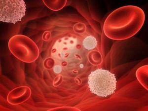
The erythrocyte is called capable of transporting oxygen to the tissues due to hemoglobin, and carbon dioxide to the lungs. This is a simple cell structure that is of great importance for the vital activity of mammals and other animals. Erythrocyte is the most numerous organism: about a quarter of all body cells are red blood tales.
General patterns of erythrocyte existence
Erythrocyte is a cell that occurred from a red sprout of blood formation. For a day, such cells produced about 2.4 million, they fall into the bloodstream and begin to perform their functions. In the course of experiments, it was determined that an erythrocyte adult, the structure of which is significantly simplified compared to other cells of the body, will live 100-120 days.
All vertebrates (with rare exceptions) from the respiratory organs to tissues oxygen is transferred by hemoglobin of red blood cells. There are exceptions: all representatives of the "white-level" fish exist without hemoglobin, although they can synthesize it. Since, at a temperature of their habitat, oxygen is well soluble in water and blood plasma, then more massive carriers, which are erythrocytes, are not required by these fish.

Erythrocytes chordovyov
In such a cell, like erythrocyte, the structure is different depending on the class of chord. For example, fish, birds and amphibious morphology of these cells look like. They differ only in size. The form of erythrocytes, the volume, size and absence of some organelle is distinguished by mammalian cells from others that the other chordines have. There is also its own pattern: mammalian erythrocytes do not contain extra organelles and they are much smaller, although they have a large surface of contact.

Considering the structure and man, general features can be revealed immediately. Both cells contain hemoglobin and participate in oxygen transport. But human cells are smaller, they are oval and have two concave surfaces. Erythrocytes of frogs (as well as birds, fish and amphibians, except Salamandra) spherical, they have core and cellular organelles that can be activated if necessary.
In human erythrocytes, as in the red blood cells of higher mammals, no cores and organelles. The size of the erythrocytes of the goat is 3-4 microns, a person is 6.2-8.2 microns. Amphiums have a cell size of 70 microns. Obviously, the size here is an important factor. Human erythrocyte albeit less, but has a large surface due to two concaves.
The small size of the cells and their large amount allowed to repeatedly increase the ability of blood to bind oxygen, which now depends only on external conditions. And such features of the structure of human erythrocytes are very important, because they allow you to comfortably feel in a certain area of \u200b\u200bhabitat. This is a measure of adaptation to life on land, which began to develop more of amphibians and fish (unfortunately, not all fish in the process of evolution got the opportunity to settle land), and reached the peak of development from higher mammals.
The structure of blood taurus depends on the functions that are assigned to them. It is described with three angles:
- Features of the external structure.
- Component composition of red blood cell.
- Inner morphology.
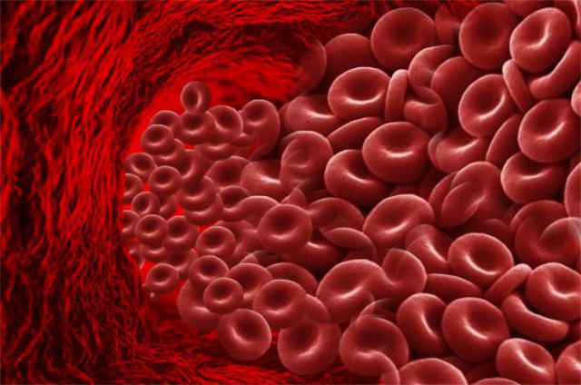
Externally, in the profile, the erythrocyte looks like a biconed disk, and in the face - as a round cell. The diameter is normal 6.2-8.2 microns.
More often in serum cells are present with small differences in size. With a lack of iron, there is a sieve decrease, and anisocytosis is recognized in the blood smear (many cells with different sizes and diameter). With a deficit folic acid Or vitamin B 12 red blood cell increases to Megaloblast. Its size is approximately 10-12 microns. The volume of the normal cell (normocite) is 76-110 cubic meters. μm.
The structure of erythrocytes in the blood is not the only feature of these cells. Much more important than their number. Small dimensions allowed to increase their number and, therefore, the area of \u200b\u200bthe contact surface. Oxygen is actively captured by human erythrocytes than frogs. And the most easily in the tissues is given from human erythrocytes.
The number is really important. In particular, an adult in a cubic millimeter contains 4.5-5.5 million cells. The goat has about 13 million erythrocytes in millilitress, and in reptiles - only 0.5-1.6 million, fish 0.09-0.13 million in millilitress. In a newborn child, the number of erythrocytes is about 6 million in millilitress, and the elderly is less than 4 million per milliliter.
Erythrocyte functions
Red blood tales - erythrocytes, quantity, structure, functions and features of the development of which are described in this publication, are very important to humans. They implement some very important features:
- transport oxygen to fabrics;
- carry carbon dioxide from tissue to easy;
- bind toxic substances (glycated hemoglobin);
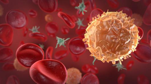
- participate in immune reactions (immune reactions (immune to viruses and due to active forms of oxygen are able to destructively influence blood infections);
- capable of carrying some drugs;
- participate in the sale of hemostasis.
Continue consideration of such a cell as an erythrocyte, the structure is maximally optimized to implement the above functions. It is the maximum light and movable, has a large contact surface for gas diffusion and flowing chemical reactions with hemoglobin, and is also quickly divided and replenishes losses in peripheral blood. This is a narrow specialized cell, to replace the functions of which is not yet possible.

Erythrocyte membrane
In such a cell, like erythrocyte, the structure is very simple, which does not apply to its membrane. It is 3-layer. The mass fraction of the membrane is 10% of cell. In its composition 90% of proteins and only 10% of lipids. This makes the erythrocytes with special cells of the body, since almost all other membranes lipids are dominated over proteins.
The bulk form of erythrocytes due to the flow of the cytoplasmic membrane may vary. Outside the membrane itself is located a layer of surface proteins having a large amount of carbohydrate residues. It is glycopeptides under which there is a bilayl lipid addressed by hydrophobic ends into and outward red blood cells. Under the membrane, on the inner surface, a layer of proteins that do not have carbohydrate residues are located again.
Receptor Erythrocyt complexes
The membrane function is to ensure the deformability of the erythrocyte, which is necessary with the capillary passage. At the same time, the structure of human erythrocytes provides additional possibilities - cellular interaction and electrolyte current. Squirrels with carbohydrate residues are the receptor molecules, thanks to which the erythrocytes do not "hunt" CD8 leukocytes and macrophages of the immune system.
Erythrocytes exist thanks to receptors and are not destroyed by their own immunity. And when, as a result of multiple pushing on capillaries or due to mechanical damage, red blood cells lose some receptors, the macrophages of the spleen "remove" them from blood flow and destroy.
The internal structure of the Erythrocyt
What is the erythrocyte? The structure is not less interest, rather than the function. This cell is similar to a hemoglobin bag, a limited membrane, on which receptors are expressed: differentiation clusters and a variety of blood groups (along the landscaner, in the rhesus, by Duffy and the other). But inside the cell is special and very different from other cells of the body.
Differences are as follows: Erythrocytes in women and men do not contain kernels, they have no ribosomes and an endoplasmic network. All of these organelles were removed after the hemoglobin filling. Then the organelles were unnecessary, because for pushing the capillars, a cell with minimal sizes needed. Therefore, inside it contains only hemoglobin and some auxiliary proteins. Their role is not yet clarified. But due to the lack of an endoplasmic network, ribosomes and the kernel, it has become light and compact, and most importantly, it can be easily deformed with the fluid membrane. And these are the most important features of the structure of red blood cells.
Erythrocytarian life cycle
The main features of the erythrocytes are in their short life. They cannot share and synthesize the protein due to the nucleus remote from the cell, and therefore the structural damage to their cells accumulate. As a result, erythrocyte is characteristic of aging. However, hemoglobin, which is captured by the spleen macrophages during the death of red blood cell, will always be sent to the formation of new oxygen carriers.
The erythrocyte life cycle begins in the bone marrow. This organ is present in the plate substance: in the sternum, in the wings of the ileum bones, in the bones of the base of the skull, as well as in the cavity of the femoral bone. Here, from the stem cell of blood under the action of cytokines, the predecessor of myeloposhes with code (CDA-Gamm) is formed. After dividing, she will give the hematopoix to the hematopois, denoted by the code (Boy E). The erythropoese predecessor is formed from it, which is indicated by the code (CFU).
The same cell is called the colony-forming cell of the red blood sprout. It is sensitive to erythropoetina - a substance of hormonal nature, highlighting the kidneys. Increasing the number of erythropoietin (according to the principle of positive feedback in functional systems) accelerates the processes of division and production of red blood cells.
Education Erythrocyte
The sequence of cell bone transformations of Koe E is: an erythroblast is formed from it, and from it - priormocyte, giving rise to the basophilic Normoblast. As the protein accumulates, it becomes a polychromatophilic normosoblast, and then oxyfly normosoblast. After removing the kernel, it becomes reticulocyte. The latter gets into blood and differentiates (matures) to normal erythrocyte.
Erythrocyte destruction
Approximately 100-125 days the cell circulates in the blood, constantly tolerates oxygen and removes metabolic products from tissues. It transports carbon dioxide tied with hemoglobin and sends it back into the lungs, along the way, filling its protein molecules with oxygen. And as damage gains, phosphatidylserine molecules and receptor molecules. Because of this, the erythrocyte hits the "under the sight" of the macrophage and is destroyed by them. And the gem obtained from all overwhelmed hemoglobin is again sent to the synthesis of new erythrocytes.
And then they will spread it (oxygen) by the body of the animal.
Encyclopedic YouTube.
-
1 / 5
Erythrocytes are highly specialized cells whose function is the transfer of oxygen from the lungs to the body tissues and carbon dioxide transport (CO 2) in the opposite direction. Vertebrates, except mammals, red blood cells have a kernel, there is no mammalian erythrocytes.
The most specialized erythrocytes of mammals, devoid of the core and organelle and having a two-way disk, which causes the high attitude of the area to the volume, which facilitates gas exchange. The features of the cytoskeleton and the cell membrane allow the erythrocytes to undergo significant deformations and restore the form (human erythrocytes with a diameter of 8 microns pass through capillaries with a diameter of 2 -3 μm).
The oxygen transport is provided by hemoglobin (HB), which accounts for ≈98% of the mass of proteins of the erythrocyte cytoplasm (in the absence of other structural components). Hemoglobin is a tetramer in which each protein chain carries gem - the protoporphyrin IX complex with a 2-blade iron ion, oxygen is reversibly coordinated with the hemoglobin FE 2+ ion, forming HBO 2 oxemoglobin:
HB + O 2 HBO 2
The feature of the oxygen binding is hemoglobin is its alto-cellular regulation - the stability of oxymemoglobin falls in the presence of 2,3-diphosphoglycerolic acid - the intermediate product of glycolysis and, to a lesser extent, carbon dioxide, which contributes to the release of oxygen in the tissues in need.
Carbon dioxide transport by erythrocytes occurs with participation carboangendresses 1.contained in their cytoplasm. This enzyme catalyzes the reversible formation of bicarbonate from water and carbon dioxide diffusing in red blood cells:
H 2 O + CO 2 ⇌ (\\ DisplayStyle \\ Rightleftharpoons) H + + HCO 3 -
As a result, hydrogen ions accumulate in the cytoplasm, however, the decrease in this is slightly due to the high buffer tank of hemoglobin. Due to the accumulation in the cytoplasm of bicarbonate ions there is a concentration gradient, however, bicarbonate ions can leave the cell only under the condition of preserving the equilibrium charge distribution between the inner and external medium, separated by the cytoplasmic membrane, that is, the bicarbonate ion erythrocyte must be accompanied by either the way out of the cation or an anion output. The erythrocyte membrane is practically impenetrable for cations, but contains chloride ion channels, as a result, the yield of bicarbonate from the erythrocyte is accompanied by the input of chloride anion (chloride shift).
The formation of red blood cells
The colony-forming unit of erythrocytes (CO-E) gives the erythrobustos beginning, which is already given morphologically distinguishable cells-descendants of norroblasts (consistently passing stages):
- Erythroblast. Distinctive features of it are as follows: diameter 20-25 μm, large (more than 2/3 of the entire cell) core with 1-4 clearly decorated nucleoli, brightly basophilic cytoplasm with a purple tint. Around the kernel there is an enlightenment of the cytoplasm (so-called "perinuclear enlightenment"), and the cytoplasm can be formed on the periphery (so-called "ears"). The last 2 features although they are characteristic of the ethyrtoscience, but they are not observed.
- Proroamcite. Distinctive features: a diameter of 10-20 μm, the kernel is deprived of the nucleoli, chromatin grubets. The cytoplasm begins to brighten, perinuclear enlightenment increases in size.
- Basophilic normoblast. Distinctive features: diameter 10-18 microns, devoid of nucleol core. Chromatin begins to segmented, which leads to uneven perception of dyes, the formation of oxide and bascohromatin zones (T.N. "Wheel-like kernel").
- Polychromatophilic normoblast. Distinctive features: diameter 9-12 μm, picnotic (destructive) changes begin in the core, but the wheels are preserved. Cytoplasm acquires oxilicity due to high concentration Hemoglobin.
- Oxyfly Normoblast. Distinctive features: diameter 7-10 μm, the kernel is subject to picnosis and shifted to the periphery of the cell. The cytoplasm is clearly pink, near the nucleus in it are detected by chromatin fragments (tales).
- Reticulocyte. Distinctive features: Diameter 9-11 μm, with a supravital color has a yellow-green cytoplasm and a blue-violet reticulum. When painting on Romanovsky-Gymzem, no distinctive features are not detected compared to mature erythrocyte. In the study of the fullness, speed and adequacy of erythropois, a special analysis of the number of reticulocytes is carried out.
- Normocyte. Mature red blood cell, with a diameter of 7-8 microns, not having a nucleus (in the center - enlightenment), cytoplasm - pink-red.
Hemoglobin begins to accumulate already at the stage of Costa, however its concentration becomes quite high to change the color of the cell only at the level of polychromatophilic norms. Also, the nucleus is also occurring (and later the destruction) of the nucleus - with something, but it is displaced only in the later stages. In this process, hemoglobin is played in this process in this process (its main type - Hb-a), which is in high concentration toxic for the cell itself.
Structure and composition
Most groups of vertebrates red blood cells have core and other organides.
Mammals, mature erythrocytes are deprived of nuclei, internal membranes and most organides. The nuclei is thrown out of precursor cells during erythropois. Typically, mammalian erythrocytes have a form of a two-way disk and contain mainly breathing pigment hemoglobin. In some animals (for example, camels), red blood cells have an oval shape.
The contents of the red blood cell is represented mainly by the respiratory pigment hemoglobin caused by the red blood color. However, on early stages The amount of hemoglobin is small in them, and at the stage of the erythroblasts, the color of the cell blue; Later, the cage becomes gray and, only completely mature, acquires red color.
An important role in the erythrocyte is performed by cellular (plasma) membrane, transmitting gases (oxygen, carbon dioxide), ions (,) and water. The membrane permeates transmembrane proteins - glycoforins, which, due to the large number of residues of N-acetylneiramine (sialovaya) acid, are responsible for approximately 60% of the negative charge on the surface of the erythrocytes.
On the surface of the lipoprotein membrane there are specific antigens of glycoprotein nature - agglutinogen - factors of blood groups (currently more than 15 blood groups systems are studied: AB0, Rh Factor, Duffy Antigen (eng.)russian, Kell Antigen, Kidd Antigen (eng.)russian), resulting in agglutination of red blood cells under the action of specific agglutinins.
The effectiveness of the functioning of hemoglobin depends on the size of the surface of the contact of the erythrocyte with the medium. The total surface of all blood erythrocytes in the body is the greater, the smaller their size. At the lower vertebrate erythrocytes are large (for example, in the tailed amphiferous amphium - 70 μm in diameter), the erythrocytes of the highest vertebrate smallers (for example, the goat is 4 microns in diameter). In humans, the erythrocyte diameter is 6.2-8.2 microns, the thickness is 2 microns, the volume is 76-110 μm³.
- in men - 3,9-5,5 ⋅10 12 per liter (3.9-5.5 million in 1 mm³),
- in women - 3.9-4,7 ⋅10 12 per liter (3.9-4.7 million in 1 mm³),
- in newborns - up to 6,0 ⋅10 12 per liter (up to 6 million in 1 mm³),
- the elderly - 4,0 €10 12 per liter (less than 4 million in 1 mm³).
Blood transfusion
The average life expectancy of the human erythrocyte - 125 days (about 2.5 million erythrocytes is formed every second and their number is collapsed), dogs - 107 days, at home rabbits and cats - 68.
Pathology
With different blood diseases, it is possible to change the color of red blood cells, their size, quantity, as well as forms; They can take, for example, a sickle, oval, spherical or target form.
Changing the form of erythrocytes is called poikilocytosis. Spherocytosis (spherical form of erythrocytes) is observed in some forms of hereditary
The first school lessons about the device human organism Meet the main "blood inhabitants: red cells - red blood cells (ER, RBC), which determine the color due to, contained, and white (leukocytes) in them, which are not visible to the eye, because they do not affect the eye.
Human erythrocytes, unlike animals, do not have a nucleus, but before losing it, they must pass the way from the erythrobusta cell, where the hemoglobin synthesis begins, reach the last nuclear stage - accumulating hemoglobin, and turn into a mature nuclear-free cell, the main The component of which is a red blood pigment.

What only people did not do with red blood cells, studying their properties: and around the globe tried to wrap them (it turned out 4 times), and to lay them (52 \u200b\u200bthousand kilometers) in the coin columns (52 thousand kilometers), and the area of \u200b\u200bthe erythrocytes is compared with the surface area of \u200b\u200bthe human body (red blood cells have exceeded all expectations Their area was higher than 1.5 thousand times).
These unique cells ...
Another important feature of the erythrocytes lies in their two-way form, but if they were spherical, the total area of \u200b\u200btheir surface would be less than 20% of the present. However, the abilities of the erythrocytes are not only in the magnitude of their total area. Thanks to a biconged disk form:
- Erythrocytes are capable of carrying more oxygen and carbon dioxide;
- To show plasticity and freely pass through narrow holes and curved capillary vessels, that is, for young full-fledged cells, there are practically no obstacles in the bloodstream. The ability to penetrate the most remote corners of the body is lost with the age of erythrocytes, as well as with their pathological conditions, when their form and size changes. For example, spherocytes, sickle, weights and pears (cachylocytosis), do not have such high plasticity, cannot be brought into narrow capillaries macrocytes, and moreover, megalocytes (anisocytosis), therefore, the tasks of their changed cells are not so flawless.

The chemical composition of ER is presented to a greater degree of water (60%) and a dry residue (40%) in which 90 - 95% occupies a red blood pigment -, And the remaining 5 - 10% is distributed between lipids (cholesterol, lecithin, kefalin), proteins, carbohydrates, salts (potassium, sodium, copper, iron, zinc) and, of course, enzymes (carboangeerase, cholinesterase, glycolithic, etc.).
Cellular structures that we used to celebrate in other cells (core, chromosome, vacuoles), ER is missing as unnecessary. Erythrocytes live up to 3 - 3.5 months, then constituted with the help of erythropoietic factors, which are allocated during the destruction of the cell, serve the team that they are time to replace with new ones - young and healthy.
The beginning of its erythrocyte takes from predecessors, which, in turn, originate from the stem cell. Red blood tales are reproduced if everything is fine in the body, in the bone of the plane bones (skull, spine, chest, ribs, pelvic bones). In cases where for any reason bone marrow It cannot produce them (tumor defeat), the red blood cells "recall" other organs (liver, fork iron, spleen) were engaged in the intrauterine development (liver, fork iron, spleen) and force the organism to start erythropoes in the forgotten places.
How many of them should be normal?
The total number of erythrocytes contained in the body as a whole, and the concentration of red cells, cruising on the bloodstream - the concepts of different. The total number includes cells that have not yet left the bone marrow, went to the depot in case of unforeseen circumstances or went into swimming to fulfill their immediate duties. The combination of all three populations of red blood cells is called - erytron. Erytron contains from 25 x 10 12 / l (tera / liter) to 30 x 10 12 / l of red blood cells.

The rate of erythrocytes in the blood of adults is different in gender, and in children, depending on age. In this way:
- The norm in women ranges from 3.8 - 4.5 x 10 12 / l, respectively, hemoglobin they also have less;
- That for a woman is normal indicator, the men are called an easy degree anemia, since the lower and upper boundary of the erythrocyte norms are noticeably above: 4.4 x 5.0 x 10 12 / l (the same applies to hemoglobin);
- In children, before the year, the concentration of red blood cells is constantly changing, so for each month (in newborns - every day) there is its own norm. And if suddenly, in the analysis of blood, erythrocytes in a child of two weeks of one week are increased to 6.6 x 10 12 / l, then it cannot be regarded as pathology, simply in newborns such a norm (4.0 - 6.6 x 10 12 / l).
- Some oscillations are observed and after the year of life, but normal values They do not particularly differ from those in adults. In adolescents 12-13 years old, hemoglobin content in red blood cells and the level of the erythrocyte themselves corresponds to the norm of adults.
Increased content of red blood cells in the blood is called erythrocytosiswhich is absolute (true) and redistribution. Rediatric erythrocytosis of pathology is not and occurs when erythrocytes in the blood are increased under certain circumstances:
- Stay in the mountainous location;
- Active physical work and sports;
- Psycho-emotional arousal;
- Dehydration (loss of fluid in diarrhea, vomiting, etc.).
The high indicators of the content of erythrocytes in the blood are a sign of pathology and true erythrocytosis, if they have become the result of the reinforced formation of red blood cells caused by an unlimited proliferation (reproduction) of the predecessor cells and its differentiation into mature erythrocyte forms ().
Reducing the concentration of red blood cells called erythroenia. It is observed in the blood loss, inhibition of erythropoese, decay of erythrocytes () under the action of adverse factors. Low red blood cells in the blood and reduced content HB in red blood cells is a sign.
What does abbreviation mean?
Modern hematological analyzers, in addition to hemoglobin (HGB), reduced or elevated content of blood erythrocytes (RBC), (HCT) and other familiar analyzes, can count other indicators that are denoted by Latin abbreviation and are not at all clear to the reader:

In addition to all the listed advantages of red blood cells, I would like to note one more thing:
Erythrocytes are considered a mirror reflecting the state of many organs. A peculiar indicator capable of "feeling" problems or allowing to monitor the course of the pathological process is.
Big Ship - Great Swimming
Why are red blood cells are so important to diagnose many pathological conditions? Their special role follows and is formed by virtue of unique opportunities, and so that the reader can imagine the true significance of the erythrocytes, let's try to list their duties in the body.

Truly, functional problems of red blood cells are wide and diverse:
- They carry out the transportation of oxygen to the tissues (with the participation of hemoglobin).
- Carbon dioxide (with participation, in addition to hemoglobin, carbanese and ion exchanger enzyme CL- / HCO 3).
- Perform a protective function, since it is capable of adsorbing harmful substances and transfer the antibody (immunoglobulins) on its surface (immunoglobulins), the components of the complementary system, formed immune complexes (AT-AG), as well as to synthesize the antibacterial substance called eryitrine.
- Participate in the exchange and regulation of water-salt equilibrium.
- Provide tissue nutrition (erythrocytes adsorb and transfer amino acids).
- Participate in maintaining informational bonds in the body by transferring macromolecules that these bonds provide (creator function).
- It contains thromboplastin, which comes out of the cell during the destruction of red blood cells, which is a signal for the coagulation system to begin hypercoagulation and education. In addition to thromboplastin, red blood cells carry heparin that prevents thrombosis. Thus, the active participation of erythrocytes in the process of blood coagulation is obvious.
- Red blood cells are capable of suppressing high immunoreactivity (perform the role of suppressors), which can be used in the treatment of various tumor and autoimmune diseases.
- Participate in the regulation of the production of new cells (erythropoes) by exemption from destroyed old erythrocytes of erythropoietic factors.
Red blood calves are destroyed mainly in the liver and spleen with the formation of decay products (, iron). By the way, if we consider each cage separately, it will not be so red, rather, yellowish - red. Accumulating into huge millionth masses, they, thanks to hemoglobin, in them being, are becoming as we used to see them - a rich-red color.
Video: Lesson on red blood cells and blood functions
Erythrocytes also known called red blood tales, - human blood cells. Erythrocytes are highly specialized cells whose function is the transfer of oxygen from the lungs to the body tissues and carbon dioxide transport (CO 2) in the opposite direction. Vertebrates, except mammals, red blood cells have a kernel, there is no mammalian erythrocytes.
The most specialized erythrocytes of mammals, devoid of the core and organelle and having a two-way disk, which causes the high attitude of the area to the volume, which facilitates gas exchange. Features of the cytoskeleton and cell membrane allow the erythrocytes to undergo significant deformations and restore the form (human erythrocytes with a diameter of 8 μm pass through the capillaries with a diameter of 2-3 microns).
The oxygen transport is provided by hemoglobin (HB), which accounts for ≈98% of the mass of proteins of the erythrocyte cytoplasm (in the absence of other structural components). Hemoglobin is a tetramer, in which each protein chain carries a gem - Protoporphyrin IX complex with an ion bivalent glaze, oxygen is reversibly cordinized with the hemoglobin Fe 2+ ion, forming oxymemoglobin HBO 2:
The feature of the oxygen binding is hemoglobin is its alto-cellular regulation - the stability of oxymemoglobin falls in the presence of 2,3-diphosphoglycerolic acid - the intermediate product of glycolysis and, to a lesser extent, carbon dioxide, which contributes to the release of oxygen in the tissues in need. The contents of the red blood cell is represented mainly by the respiratory pigment hemoglobin caused by the red blood color. However, in the early stages, the number of hemoglobin in them is not enough, and at the stage of the erythroblasts, the color of the cell is blue; Later, the cage becomes gray and, only completely mature, acquires red color.
An important role in the erythrocyte is performed by cellular (plasma) membrane, transmitting gases (oxygen, carbon dioxide), ions (Na, k) and water. The transmembrane proteins are permeated with glycoforins, which, due to the large number of sialic acid residues, are responsible for approximately 60%. Negative charge on the surface of the red blood cells.
On the surface of the lipoprotein membrane there are specific antigens of glycoprotein - agglutinogens - the factors of blood group systems (more than 15 blood group systems are studied: AB0, RUS factor, Antigen Daffi (English) Russian, Kell Antigen, Antigen Kidd (eng.) Russian), resulting in agglutination of red blood cells under the action of specific agglutinins.
The effectiveness of the functioning of hemoglobin depends on the size of the surface of the contact of the erythrocyte with the medium. The total surface of all blood erythrocytes in the body is the greater, the smaller their size. In humans, the diameter of the erythrocyte is 7.2-7.5 μm, the thickness is 2 microns, the volume - 76-110 μm³ of the erythrocyte membrane is a plastic molecular mosaic consisting of proteins, lipoproteins and glycoproteins and, possibly, purely lipid plots. Its thickness is about 10 nm, it is about a million times more permeable for anions than for cations. The transfer of substances through the membrane is committed depending on their chemical properties different ways: Hydrodynamically (by diffusion), when substances, as in solution, pass through the membrane pores filled with water, or, if substances are soluble in fats, by penetrating through lipid sections. Some substances can enter into easily reversible connections with molecules built into the membrane - carriers, and in the future they or passively, or as a result of the so-called active transport pass through the membrane.
45.Education of red blood cells. Factors involved in the formation of erythrocytes and hemoglobin, regulation of erythropois. ESO, key factors that determine the value of ESO.
the main incentive of the development of erythrocytes of Jav-Xia Hypoxia. Hypoxia is a decrease in oxygen soda in tissues. O2 deficiency promotes ORG-JU erythropoetins in the kidney epithelium. Erythropoetins enter blood, then in KKM, where the dip-ku and the development of stem cells in red blood cells are stimulated. The regulation of erythropoese in-Xia vitamin B12 and folic acid. These vitamins are necessary for the maturation and development of the coder of the cell. Vitamin B12 is binding to the stomach with a protein carrier and the transcribe transcribe and turns into 12 pk. There it is subjected to hydrolysis, and WIT. B12 with the internal phlegant factor posts in the ileum. In this department, the presences of Ca2 + is associated with the membrane of enterocyte. Bunds blood, and transported to targets. Vitamin B12 is in the synthesis of DNA in the erythroblasts. Vitamin B6 - Coenzyme, Uch-Ii in the Obr-Imy in Erythroblast. Vitamin C contributes to the metabolism of folic acid in the erythroblast. SE - a non-presumption for the presence of a disease, because The level of blood plasma proteins increases and the erythrocyte sedimentation rate increases. Normally from 5 to 10 mm / hour.
1The minibor provides information about the main results of the study of erythrocyte proteins. The structure and functions of the complexes of protein 4.1.r and the protein 3 of the bands are discussed, the results of riskers - conveyors, including the role of Aquaporin 1 in carbon dioxide transport. The ideas about the mechanism of the GárDOS effect in red blood cells are discussed. Information about the interactoma of proteins of cytosol of erythrocytes is given. The issues of development of oxidative stress in red blood cells are discussed, including the role of Peroxidoxin protein 2. The participation of hemoglobin in the aging mechanisms of red blood cells is shown.
erythrocytes
hemoglobin
oxidative stress
1. Minddukshev I.V., Krivoshlik V.V., Dobrylko I.A. et al. // Biological membranes. - 2012. - T.27, No. 1. - P. 23-28.
2. Barvitenko N.N., ADRAGNA N.C., WEBER R.E. ErythroCyte Signal Transduction Pathways, Their Oxygenation Dependence and Functional Significance // Cell Physiol Biochem - 2005. - No. 15. -p. 1-18.
3. Baines A.J. Evolution of Spectrin Function in Cytoskeletal and Membrane Networks // Biochem Soc Trans. - 2009. - Vol. 37 (Pt 4). - P. 796-803.
4. Blank Me, Ehmke H. Aquaporin-1 and HCO3 (-) - Cl- Transporter-Mediated Transport of CO2 Across the Human ErythroCyte Membrane // Physiol. - 2003. - Vol. 550 (Pt 2). - P. 419-429.
5. Brazhe N.A., ABDALI S, Brazhe A.R., Luneva O.G. et al. // Biophys J. - 2009. - Vol. 97 (12). - P. 3206-3214.
6. Bruce L.j., Beckmann R., Ribeiro M.L., Peters L.L. et al. // Blood. - 2003. - Vol. 101, No. 10. - P. 4180-4188.
7. Burak çimen M.Y. Free Radical Metabolism in Human ErythroCytes // Clinica Chimica Acta. - 2008. - Vol. 390, No. 1-2. -P. 1-11.
8. Blodgett DM, Graybill C, Carruthers A. Analysis of Glucose Transporter Topology and Structural Dynamics // J Biol Chem. - 2008. - № 283: 36416-36424.
9. Campanella M.E., Chu H., Low P.S. Assembly and regulation of a Glycolytic Enzyme Complex on the Human ErythroCyte Membrane // PNAS. - 2005. -VOL. 102, No. 7. - P. 2402-2407.
10. Davies J.A. Degradation of Oxidized Proteins by The 20s Proteasome // Biochimie. - 2001. - Vol. 83. - P. 301-310.
11. D'Alessandro A, Rightti PG, Zolla L. The Red Blood Cell Proteome and Interactome: An Update // Proteome Res. - 2010. - Vol. 9 (1). - P. 144-163.
12. Endeward, V., Musa-Aziz, R., Cooper, G. J., Chen, L. et al. // The FASEB Journal. - 2006. - Vol. 20, No. 12. - P. 1974-1981.
13. Gauthier E, Guo X, Mohandas N, An X. Phosphorylation-Dependent PERTURBATIONS OF THE 4.1R-Associated Multiprotein Complex of the ErythroCyte Membrane // Biochemistry. - 2011. -VOL. 50 (21). - P. 4561-4567.
14. GOODMAN S.R. Kurdia A., Ammann L., Kakhniashvili D., Daescu O. // Exp Biol Med. - 2007. - Vol. 232, №11. - P. 1391-1408.
15. Ian A. Lewis, M. Estela Campanella, John L. Markley and Philip S. Low Role of Band 3 in Regulating Metabolic Flux of Red Blood Cells // PNAS. - 2009. -VOL. 106, No. 44. - P. 18515-18520.
16. Lang P.A., Kaiser S., Myssina S., Wieder T., Lang F., Huber S.M. // Am J Physiol Cell Physiol. - 2003. - Vol. 285 (6). -P. 1553-1560.
17. Lang F., Lang K.S., Wieder T., Myssina S. et al. // Pflugers Arch. - 2003. - Vol. 447 (2). - P. 121-125.
18. Li H.T., Feng L., Jiang W.D., Liu Y. et al. // AQUAT TOXICOLOL. - 2013. -VOL. 126. - P. 169-179.
19. Low F.m., Hampton M.B., Peskin A.V., WinterBourn C.C. PEROXIREDOXIN 2 FUNCTIONS AS A NONCATALYTIC SCAVENGER OF LOW-LEVEL HYDROGEN PEROXIDE IN THE ERYTHROCYTE // Blood. - 2007. -VOL. 109 (6). - P. 2611-2617.
20. Low F.m., Hampton M.B., WinterBourn C.C. PEROXIREDOXIN 2 AND PEROXIDE METABOLISM IN THE ERYTHROCYTE // ANTIOXID REDOX SIGNAL. - 2008. - Vol. 10 (9). - P. 1621-1630.
21. Mairbäurl H., Weber R.E. Oxygen Transport by Hemoglobin // Physiol. - 2012. - Vol. 2. - P. 1463-1489.
22. Maher A.D., Kuchel P.W. The Gárdos Channel: A Review of the Ca2 +ActiveVated K + Channel in Human ErythroCytes // INT J Biochem Cell Biol. - 2003. - Vol. 35 (12). - P. 1182-1197.
23. Manno S., Takakuwa Y., Mohandas N. MODULATION OF ERYTHROCYTE MEMBRANE MECHANICAL FUNCTION BY PROTEIN 4.1 Phosphorylation. // J Biol Chem. - 2005. - Vol. 280. - P. 7581-7582.
24. MANTA B., HUGO M, ORTIZ C., Ferrer-Sueta G., Trujillo M., Denicola A. // Arch Biochem Biophys. - 2009. - Vol. 484 (2). - P. 146-154.
25. Matarrese P., Straface E., Pietraforte D., Gambardella L., et al. // FASEB J. - 2005. - Vol. 19, No. 3. - P. 416-418.
26. METERE A, IORIO E, PIETRAFORTE D, PODO F, MINETTI M. Arch Biochem Biophys. - 2009. - Vol. 484 (2). - P. 173-82.
27. Neelam S., KakhniaShvili D.G., Wilkens S., Levene S., Goodman S.R. // Exp Biol Med - 2011. - Vol. 236, No. 5. - P. 580-591.
28. Nunomura W., Takakuwa Y., Parra M., Conboy J. Mohandas N. // J. Biol. Chem. - 2000. - Vol. 275. - P. 24540-24546.
29. Nunomura W., Takakuwa Y. Regulation of Protein 4.1R Interactions with Membrane Proteins by Ca2 + and Calmodulin // Front BioSci. - 2006. - Vol. 11. - P. 1522-1539.
30. Puchulu-Campanella E, Chu H, Anstee D.J et al. // j.biol Chem. - 2013. - Vol. 288 (2). - P. 848-858.
31. Takakuwa Y. PROTEIN 4.1, A MULTIFUNCTIONAL PROTEIN OF THE ERYTHROCYTE MEMBRANE SKELETON: STRUCTURE AND FUNCTIONS IN ERYTHROCYTES AND NONERYTHROID CELLS. // INT J Hematol. - 2000. - Vol. 72 (3). - P. 298-309.
32. Rinehart J., Gulcicek E.E., Joiner C.H., Lifton R.P., Gallagher P.G. // Curr Opin Hematol. -2010. - Vol. 17 (3). - P. 191-197.
33. Rocha S., Costa E., Coimbra S., Nascimento H. et al. // Blood Cells MOL DIS. - 2009. - Vol. 43 (1). - P. 68-73.
34. Salomao M., Zhang X., Yang Y., Lee S. et al. // PROC NATL ACAD SCI. - 2008. - Vol. 10, No. 105 (23). - P. 8026-8031.
Proteomic achievements have significantly expanded our ideas about individual proteins, the structure and functions of macromolecular protein complexes in red blood cells. Macromolecular associates were found on the erythrocyte membrane, which are called the protein complex 4.1.r and the complex of the 3 strip protein. A model of the organization of the macromolecular complex of cytoskeletal and transmembrane proteins with the participation of protein 4.1 R is proposed. The horizontal protein 4.1 R. interacts with actin, spectrine and protein P55, and the latter determines the nodal connections between the membrane and the cytoskeleton components. The vertical of the protein 4.1 R interacts with the cytoplasmic domain of the transmembrane protein of Glycoforin C, protein 3 of the strip and CD44, which creates a kind of bridge between the protein network and the membrane bislock. The main function of the protein complex 4.1 R is the determination of mechanical properties and deformability of erythrocyte membranes. It was suggested that the violations of this complex are determined not only the instability of erythrocyte membranes, but also remodeling the surface of the red cells. . Studies on identifying factors regulating multiple protein - protein interactions in a protein complex 4.1 R. One of such factors is phosphorylation of the protein 4.1 r with the participation of protein kinase C. As a result, the ability of the protein 4.1 r is reduced to form a complex with spectrine and actin, dissociation from Glycoforina with which leads to a change in the mechanical properties of the erythrocyte membranes. It was suggested that the elasticity of the erythrocyte membrane to a greater extent depends on the dynamic restructuring of the spectrine dimer / spectrine tetramer on the effect of the voltage shift in the bloodstream.
Protein 3 bands forms the base (Cor) for the macromolecular complex of integral and peripheral proteins of the erythrocyte membrane. It was originally assumed that this complex functions as an integrated structural unit (metabolone) for exchange CO2 / O2 in red blood cells. Later studios showed that the tetramer of protein 3 bands is associated with Ankirin, which, in turn, interacts with the spectrin. Transmembrane glycoproteins GPA, RH, RHAG are associated with a protein of 3 bands, while CD47 and LW interact with RH / RHAG. Two cytoplasmic protein domains 3 bands have soluble protein binding sites. Moreover, a large N -Conal terminal domain has binding sites for both deoxyhemoglobin and for a number of glycolysis enzymes (glyceraldehyde-3-phosphatehydrogenase and aldolaza). Presumably, the interaction of glycolysis enzymes with a domain of a protein 3 bands passes with the participation of docking proteins. C-terminal plot links carboangendresses II. The binding of carboniferresis II leads to two events: the absorption of carbon dioxide and the release of oxygen from hemoglobin. Under conditions of high oxygenation, the binding of glycolithic enzymes with protein 3 bands inhibits glycoliz when heinsing the pentosophosphate path. Under conditions of low oxygenation, the interaction of deoxyhemoglobin with a protein of 3 bands leads to an increase in glycolysis and a decrease in the pentosophosphate path. Expanded representations of role 2, 3-diffline phosphoglycerat. This metabolite interacts with the complex of spectrometry-actin protein 4.1, contributes to the interaction with the spectrine-anthlen-protein complex 4.1.
New data on membrane proteins - conveyors were obtained. Along with known conveyors, such as Na +, K + -atf-Aza and Ca2 + -atf-Aza, the presence of Na + / K + / 2Cl-conveyor and glucose conveyor is shown. Regarding the last opinion diverge. According to one understanding, glucose conveyor is represented by GLUT1 1, according to others - GLUT1, GLUT3, GLUT4. There is information about participating in the transfer of glucose Glycoporin a. There was also an existence of other conveyors, in particular, the hydrogen-lactate of the cat carrier. The data confirming the presence of a protein - the XK conveyor participating in the transfer of amino acids and oligopeptides.
The Erythrocyte membranes found the presence of Aquaporin 1. Blank Me and Ehmke H. showed that not only HCO3 (-) - CL-conveyor, but also an Aquaporin 1 red blood cells takes part in the transport of carbon dioxide through the erythrocyte membrane. Endeward V. The data that demonstrates that through Aquavoporin 1 is transferred to 60% of carbon dioxide, which allows you to consider Aquavoporin as the main way of receipt of CO2 in red blood cell.
For erythrocytes, a phenomenon of the output of potassium ions (CA (2 +) - Dependent K (+) Efflux) was found. Responsible for this effect (Gárdos Effect) is a specific channel membrane protein (Gárdos Channel), the activator of which are calcium ions. One of the features of Ca (2 +) - Dependent K (+) channels is their participation in the regulation of apoptosis of red blood cells. The study of the function of non-selective cation channels in the regulation of the volume of the cell is launched. According to the representations of Lang F et altogether. . In human erythrocytes, non-selective cationic channels are opened with osmotic cell wrinkling. Also among channel activation stimulants are the oxidative stress and hypoenergetic state. Cationic channels are permeable for calcium and their discovery leads to an increase in calcium level in cytosol. Calcium ions entering through the cationic channel, stimulate the activation of the grinding, which leads to the destruction of phosphatidylserine asymmetry in the erythrocyte membranes and stimulates Ca (2 +) - the dependent yield K (+), which leads to the loss of potassium ions and cell wrinkling. The impaired asymmetry of phosphatidylserine is confirmed by the binding of annexin, which is a sign of apoptotic cells. The exposition of phosphatidylserine on the outside of the erythrocyte membrane stimulates phagocytes to absorb apoptotic erythrocytes.
Rinehart J et al. The view was expressed that KCL Kotransport and the activation of Gardos channels plays a large role in the regulation water salt balance in red blood cells.
The cytosol of erythrocytes contain a large number of proteins. According to data, 751 protein is identified using proteoma technologies. This made it possible to determine the degree of interaction and mutual influence of these proteins (interact). Draws attention to the presence of certain clusters, one of which the authors called Rod Box (Repair or Destroy). This box contains proteins that, using ATP energy, participate in the refolding of damaged proteins. The composition of this box includes shaperons and proteins of proteasome subunits, proteins of thermal shock. The study shows the presence of the 20s proteos (independent of ATP and UBIBITINE) in mature red blood cells. The authors conclude a lawsager question about the reasons for the preservation of these proteos in mature red blood cells. It was suggested that 20S proteasomas are more resistant to oxidative stress. Another question is the existence of a dusty-dependent pratolytic protein degradation in red blood cells.
Presence in the membranes of polyunsaturated fatty acids, Wednesday rich in oxygen and containing iron makes erythrocytes subject to oxidative stress. The source of AFC in the erythrocytes is the autocidant of hemoglobin, as a result, superoxidanions are formed (O2.-). At the same time, the hemoglobin turns the methemoglobin. In addition to the superoxide, hydrogen peroxide and other active oxygen forms (reactions of the Gaure Wece and Fenton) are formed. Active forms of oxygen induce activation of lipid peroxidation, oxidative damage to red blood cell proteins, i.e. Promote the development of oxidative stress.
The formation of MDA contributes to the formation of cross-strokes between phospholipids and membrane proteins. The result is a violation of the function of the membrane, cell deformability and the restriction of erythrocyte life. The most sensitive to the formation of MDA proteins - conveyors of ions and protein 3 bands, as well as glyceraldehyde-3 - phosphatehydhydehyd-3 - phosphofukinase. It is assumed that the critical link for the survival of the erythrocyte is the oxidizing damage of Ca2 + ATP-PS. An increase in the formation of hydrogen peroxide contributes to the increase in methemoglobin, gem and spectrine complexes - hemoglobin. When the interaction of state-inxidanions with nitrogen oxide is formed peroxynitrite. Peroxinitrite causes multiple intricate changes, including damage to the cytoskeleton, membrane proteins, induces the formation of methemoglobin and contributes to the activation of various proteases. In addition, under the action of peroxynitrite, phosphatidylserine is exposed on the outer layer of the erythrocyte membrane. Peroxinitrite induces phosphorylation of tyrosine protein 3 bands and at the same time inhibits the activity of the membrane-bound protein, phosphothirin phosphatase. The result of these parallel effects of peroxyntrite is the activation of glycolysis. In addition to peroxynitrite, the phenomenon induction of apoptosis of red blood cells was shown for hydroxyl radicals.
From oxidative stress, erythrocytes protect membrane-bound proteinases, AHO enzymes and other proteins. Currently, much attention is paid to the study of the peroxideoxin 2 protein (PRX2), as one of the most important antioxidant proteins of red blood cells. PRX2 is thiol-dependent peroxidase. In combination with catalase and glutathioneer peroxide, PRx2 constitute an effective system for utilization of hydrogen peroxide formed in low concentrations during the autocusation of hemoglobin. The reduced form of peroxidexin is supported by Tyoracedoxinuruction, but the activity of the latter is quite low. PRx2 has a high sensitivity to oxidation of hydrogen peroxide. The model of the PRX2 catalytic cycle is proposed, consisting of three stages. It is interesting to note that this cycle requires 2 conformational conditions: Full Folding with the formation of an active center and a local defolding form required to restore PRX2. In addition to the function of the noncatalithic hydrogen peroxide function, peroxide-cellin regulates the transport of ions, binding to the erythrocyte membrane and activating the GárDOS channels, but the mechanism of this process is not yet clear. An increase in hydrogen intracellular peroxide leads to an increase in the proportion of the membrane-bound hemoglobin and activation of lipid peroxidation. The binding of PRx2 with the membrane also increased by increasing the concentration of hydrogen peroxide. The value of this phenomenon is not clear. Nevertheless, according to the authors, although the growth of the membrane-bound hemoglobin and the membrane-bound PRX2 are two independent processes, but both of these events are markers of the oxidative stress of erythrocytes.
New data appeared on the localization of hemoglobin inside the red blood cell. According to Brazhe Na and et al. There are 2 hemoglobin populations in red blood cells: submembrane and cytosol. In this case, the conformation of the submembrane hemoglobin molecules differs from the such cytosolic fraction. Requires further studies of this phenomenon. Expanded ideas about alto-solid oxygen binding regulators with hemoglobin. According to Mairbäurl and Weber, regulation is due to changes in such alto-solid effectors as protons (H +), carbon dioxide (CO2), organic phosphates and chlorides (CL-).
Of great interest is the discussion of the question of the role of hemoglobin in the aging of red blood cells. It is shown that aging erythrocytes accumulate redox-detected hemoglobin, reproduced lipids, high molecular weight aggregates of proteins lose silic acids. These processes lead to a decrease in phospholipid symmetry, the formation of cross-hemoglobin crossbones, protein aggregation of 3 bands, an increase in the glycated finite products. It is assumed that the interaction of hemoglobin, especially under hypoxia with protein 3, erythrocyte membrane bands is critical to change the erythrocyte membrane, which in turn is a trigger mechanism for removing hemocirculation cells. These rearrangements of the membrane include the exposure of antigenic sites, an increase in the calcium closure into erythrocytes, dried potassium leakage from erythrocytes, which leads to wrinkling cells and loss of deforility. An unresolved problem is the probable oxidative damage to the specific proteins of membranes during redox reactions, which take place when binding hemoglobin with a membrane. Further proteomal research can identify specific proteins involved in the aging mechanisms of red blood cells.
There are actual data on the development of apoptosis of red blood cells. The review provides enough detailed description Signal paths include apoptosis of red cells. According to, the first path is associated with the activation of cyclooxygenase, the formation of prostaglandin E2 and the formation of cationic channels. The second way is associated with cascade activation of spingomelinase. In addition, the process of apoptosis of erythrocytes can be induced by peroxynitrite, hydroxyl radicals, as well as methemoglobin. The results of the study showing the relationship between the change in the deformation properties of the erythrocyte membranes and the launch of the apoptosis program.
Thus, data are accumulated, expanding the ideas about metabolic processes in red blood cells. In the future, these results can be used in interpreting and predicting changes in the structure and functions of erythrocytes under various pathological conditions.
Bibliographic reference
Muravlev L.E., Molotov-Luchansky V.B., Klyuev D.A., Ponamareva O.A., Kalina A.S., Kolebaeva G.T. Erythrocyte proteins. Mini-review // Uspekhi modern natural science. - 2013. - № 4. - p. 28-31;
URL: http://natural-sciences.ru/ru/article/view?id\u003d31639 (date of handling: 12/13/2019). We bring to your attention the magazines publishing in the publishing house "Academy of Natural Science"


