Phases of mitosis and their features. mitosis - phases of mitosis. Changes that occur in prophase
Mitosis- the main method of division of eukaryotic cells, in which the doubling first occurs, and then the hereditary material is evenly distributed between daughter cells.
Mitosis is a continuous process with four phases: prophase, metaphase, anaphase and telophase. Before mitosis, the cell prepares for division, or interphase. The period of cell preparation for mitosis and mitosis itself together constitute mitotic cycle. Below is a brief description of phases of the cycle.
Interphase consists of three periods: presynthetic, or postmitotic, - G 1, synthetic - S, postsynthetic, or premitotic, - G 2.
Presynthetic period (2n 2c, Where n- number of chromosomes, With- number of DNA molecules) - cell growth, activation of biological synthesis processes, preparation for the next period.
Synthetic period (2n 4c) - DNA replication.
Postsynthetic period (2n 4c) - preparation of the cell for mitosis, synthesis and accumulation of proteins and energy for the upcoming division, increase in the number of organelles, doubling of centrioles.
Prophase (2n 4c) - dismantling of nuclear membranes, divergence of centrioles to different poles of the cell, formation of spindle filaments, “disappearance” of nucleoli, condensation of biromatid chromosomes.
Metaphase (2n 4c) - alignment of maximally condensed bichromatid chromosomes in the equatorial plane of the cell (metaphase plate), attachment of spindle threads at one end to the centrioles, the other to the centromeres of the chromosomes.
Anaphase (4n 4c) - division of two-chromatid chromosomes into chromatids and the divergence of these sister chromatids to opposite poles of the cell (in this case, the chromatids become independent single-chromatid chromosomes).
Telophase (2n 2c in each daughter cell) - decondensation of chromosomes, formation of nuclear membranes around each group of chromosomes, disintegration of spindle threads, appearance of a nucleolus, division of the cytoplasm (cytotomy). Cytotomy in animal cells occurs due to the cleavage furrow, in plant cells - due to the cell plate.
1 - prophase; 2 - metaphase; 3 - anaphase; 4 - telophase.
Biological significance mitosis The daughter cells formed as a result of this method of division are genetically identical to the mother. Mitosis ensures the constancy of the chromosome set over a number of cell generations. It underlies processes such as growth, regeneration, asexual reproduction, etc.
is a special method of dividing eukaryotic cells, as a result of which the cells transition from a diploid state to a haploid state. Meiosis consists of two successive divisions preceded by a single DNA replication.
First meiotic division (meiosis 1) is called reduction, since it is during this division that the number of chromosomes is halved: from one diploid cell (2 n 4c) two haploid (1 n 2c).
Interphase 1(at the beginning - 2 n 2c, at the end - 2 n 4c) - synthesis and accumulation of substances and energy necessary for both divisions, increase in cell size and number of organelles, doubling of centrioles, DNA replication, which ends in prophase 1.
Prophase 1 (2n 4c) - dismantling of nuclear membranes, divergence of centrioles to different poles of the cell, formation of spindle filaments, “disappearance” of nucleoli, condensation of bichromatid chromosomes, conjugation of homologous chromosomes and crossing over. Conjugation- the process of bringing together and intertwining homologous chromosomes. A pair of conjugating homologous chromosomes is called bivalent. Crossing over is the process of exchange of homologous regions between homologous chromosomes.
Prophase 1 is divided into stages: leptotene(completion of DNA replication), zygotene(conjugation of homologous chromosomes, formation of bivalents), pachytene(crossing over, recombination of genes), diplotene(detection of chiasmata, 1 block of oogenesis in humans), diakinesis(terminalization of chiasmata).

1 - leptotene; 2 - zygotene; 3 - pachytene; 4 - diplotene; 5 - diakinesis; 6 — metaphase 1; 7 - anaphase 1; 8 — telophase 1;
9 — prophase 2; 10 — metaphase 2; 11 - anaphase 2; 12 - telophase 2.
Metaphase 1 (2n 4c) - alignment of bivalents in the equatorial plane of the cell, attachment of spindle filaments at one end to the centrioles, the other to the centromeres of the chromosomes.
Anaphase 1 (2n 4c) - random independent divergence of two-chromatid chromosomes to opposite poles of the cell (from each pair of homologous chromosomes, one chromosome goes to one pole, the other to the other), recombination of chromosomes.
Telophase 1 (1n 2c in each cell) - the formation of nuclear membranes around groups of dichromatid chromosomes, division of the cytoplasm. In many plants, the cell goes from anaphase 1 immediately to prophase 2.
Second meiotic division (meiosis 2) called equational.
Interphase 2, or interkinesis (1n 2c), is a short break between the first and second meiotic divisions during which DNA replication does not occur. Characteristic of animal cells.
Prophase 2 (1n 2c) - dismantling of nuclear membranes, divergence of centrioles to different poles of the cell, formation of spindle filaments.
Metaphase 2 (1n 2c) - alignment of bichromatid chromosomes in the equatorial plane of the cell (metaphase plate), attachment of spindle filaments at one end to the centrioles, the other to the centromeres of the chromosomes; 2 block of oogenesis in humans.
Anaphase 2 (2n 2With) - division of two-chromatid chromosomes into chromatids and the divergence of these sister chromatids to opposite poles of the cell (in this case, the chromatids become independent single-chromatid chromosomes), recombination of chromosomes.
Telophase 2 (1n 1c in each cell) - decondensation of chromosomes, formation of nuclear membranes around each group of chromosomes, disintegration of the filaments of the spindle, appearance of the nucleolus, division of the cytoplasm (cytotomy) with the resulting formation of four haploid cells.
Biological significance of meiosis. Meiosis is the central event of gametogenesis in animals and sporogenesis in plants. Being the basis of combinative variability, meiosis provides genetic diversity of gametes.
Amitosis
Amitosis- direct division of the interphase nucleus by constriction without the formation of chromosomes, outside the mitotic cycle. Described for aging, pathologically altered and doomed cells. After amitosis, the cell is not able to return to the normal mitotic cycle.
Cell cycle
Cell cycle- the life of a cell from the moment of its appearance until division or death. An essential component of the cell cycle is the mitotic cycle, which includes the period of preparation for division and mitosis itself. In addition, in the life cycle there are periods of rest, during which the cell performs its inherent functions and selects future fate: death or return to the mitotic cycle.
Go to lectures No. 12"Photosynthesis. Chemosynthesis"
Go to lectures No. 14"Reproduction of Organisms"
Among all the interesting and quite complex topics in biology, it is worth highlighting two processes of cell division in the body - meiosis and mitosis. At first it may seem that these processes are the same, since in both cases cell division occurs, but in fact there is a big difference between them. First of all, you need to understand mitosis. What is this process, what is interphase of mitosis and what role do they play in human body? This will be discussed in more detail in this article.
Difficult biological process, which is accompanied by cell division and the distribution of chromosomes between these cells - all this can be said about mitosis. Thanks to it, chromosomes containing DNA are evenly distributed between the daughter cells of the body.
There are 4 main phases in the process of mitosis. They are all interconnected, since the phases smoothly transition from one to another. The prevalence of mitosis in nature is due to the fact that it is it that is involved in the process of division of all cells, including muscle, nerve, and so on.
Briefly about interphase
Before entering the state of mitosis, a cell that divides goes into interphase, that is, it grows. The duration of interphase can occupy more than 90% of the total time of cell activity in normal mode.
Interphase is divided into 3 main periods:
- phase G1;
- S-phase;
- phase G2.
They all take place in a certain sequence. Let's look at each of these phases separately.
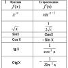
Interphase - main components (formula)
Phase G1
This period is characterized by the preparation of the cell for division. It increases in volume for the further phase of DNA synthesis.
S-phase
This is the next stage in the interphase process, during which the body's cells divide. As a rule, the synthesis of most cells occurs over a short period of time. After division, the cells do not increase in size, but the last phase begins.
Phase G2
The final stage of interphase, during which cells continue to synthesize proteins while increasing in size. During this period, there are still nucleoli in the cell. Also, in the last part of the interphase, duplication of chromosomes occurs, and the surface of the nucleus at this time is covered with a special shell that has a protective function.
On a note! At the end of the third phase, mitosis occurs. It also includes several stages, after which cell division occurs (this process in medicine is called cytokinesis).
Stages of mitosis
As noted earlier, mitosis is divided into 4 stages, but sometimes there can be more. Below are the main ones.
Table. Description of the main phases of mitosis.
| Phase name, photo | Description |
|---|---|
| During prophase, spiralization of chromosomes occurs, as a result of which they take on a twisted shape (it is more compact). All synthetic processes in the body’s cell stop, so ribosomes are no longer produced. |
| Many experts do not distinguish prometaphase as a separate phase of mitosis. Often all the processes that occur in it are referred to as prophase. During this period, the cytoplasm envelops the chromosomes, which move freely throughout the cell until a certain point. |
| The next phase of mitosis, which is accompanied by the distribution of condensed chromosomes on the equatorial plane. During this period, microtubules are renewed on an ongoing basis. During metaphase, the chromosomes are arranged so that their kinetochores are in a different direction, that is, directed towards opposite poles. |
| This phase of mitosis is accompanied by the separation of the chromatids of each chromosome from each other. The growth of microtubules stops, they now begin to disassemble. Anaphase does not last long, but during this period of time the cells manage to disperse closer to different poles in approximately equal numbers. |
| This is the last stage during which chromosome decondensation begins. Eukaryotic cells complete their division, and a special shell is formed around each set of human chromosomes. When the contractile ring contracts, the cytoplasm separates (in medicine this process is called cytotomy). |


Important! The duration of the complete mitosis process, as a rule, is no more than 1.5-2 hours. The duration may vary depending on the type of cell being divided. Also, the duration of the process is influenced by external factors, such as light conditions, temperature, and so on.
What biological role does mitosis play?
Now let's try to understand the features of mitosis and its importance in the biological cycle. First of all, it ensures many vital processes of the body, including embryonic development.
Mitosis is also responsible for tissue repair and internal organs body after various types damage, resulting in regeneration. In the process of functioning, cells gradually die, but with the help of mitosis, the structural integrity of tissues is constantly maintained.
Mitosis ensures the preservation of a certain number of chromosomes (it corresponds to the number of chromosomes in the mother cell). read on our website.
Video - Features and types of mitosis
Lecture No. 10
Number of hours: 2
MITOSIS
1. Cell life cycle
2. Mitosis. Stages of mitosis, their duration and characteristics
3. Amitosis. Endoreproduction
1. Cell life cycle
The cells of a multicellular organism are extremely diverse in the functions they perform. Cells have different life spans depending on their specialization. So, after embryogenesis is completed, nerve cells stop dividing and function throughout the life of the organism. Cells of other tissues (bone marrow, epidermis, epithelium) small intestine) in the process of performing their function, they quickly die and are replaced by new ones as a result of cell division.Cell division underlies the development, growth and reproduction of organisms. Cell division also ensures self-renewal of tissues throughout the life of the body and restoration of their integrity after damage. There are two ways to divide somatic cells: amitosis And mitosis. Indirect cell division (mitosis) is predominantly common. Reproduction by mitosis is called asexual reproduction, vegetative reproduction or cloning.
Cell life cycle (cell cycle) is the existence of a cell from division until the next division or death. The duration of the cell cycle in multiplying cells is 10-50 hours and depends on the type of cells, their age, the hormonal balance of the body, temperature and other factors. The details of the cell cycle vary among different organisms. In single-celled organisms, the life cycle coincides with the life of the individual. In continuously reproducing tissue cells, the cell cycle coincides with the mitotic cycle.
Mitotic cycle - a set of sequential and interconnected processes during the period of cell preparation for division and the period of division (Figure 1). In accordance with the above definition, the mitotic cycle is divided into interphase And mitosis (Greek “mitos” - thread).
Interphase- the period between two cell divisions - is divided into phases G 1, S and G 2 (their duration is indicated below, typical for plant and animal cells.). In terms of duration, interphase makes up the majority of the cell's mitotic cycle. Most variable over time G 1 and G 2 periods.
G 1 (from English.grow- grow, increase). The duration of the phase is 4–8 hours. This phase begins immediately after the formation of the cell. During this phase, RNA and proteins are intensively synthesized in the cell, and the activity of enzymes involved in DNA synthesis increases. If the cell does not further divide, it enters the phase G 0 – period of rest. Taking into account the resting period, the cell cycle can last weeks or even months (liver cells).
S (from English)synthesis- synthesis).The duration of the phase is 6–9 hours. The cell mass continues to increase, and chromosomal DNA is doubled. The two helices of the old DNA molecule separate, and each becomes a template for the synthesis of new DNA strands. As a result, each of the two daughter molecules necessarily includes one old helix and one new one. However, the chromosomes remain single in structure, although doubled in mass, since two copies of each chromosome (chromatids) are still connected to each other along their entire length. After completing the phase S During the mitotic cycle, the cell does not immediately begin to divide.
G2.In this phase, the cell completes the process of preparation for mitosis: ATP accumulates, achromatin spindle proteins are synthesized, and centrioles double. The cell's mass continues to increase until it is approximately twice its original mass, and then mitosis occurs.
Rice. Mitotic cycle: M- mitosis, P - prophase, Mf - metaphase, A - anaphase, T- telophase, G 1 - presynthetic period, S - synthetic period, G 2 - postsynthetic
2. Mitosis. Stages of mitosis, their duration and characteristics. Mitosis is conditional divided into four phases: prophase, metaphase, anaphase and telophase.
Prophase.The two centrioles begin to diverge towards opposite poles of the nucleus. The nuclear membrane is destroyed; at the same time, special proteins combine to form microtubules in the form of threads. Centrioles, now located at opposite poles of the cell, have an organizing effect on microtubules, which as a result line up radially, forming a structure resembling appearance aster flower (“star”). Other filaments of microtubules extend from one centriole to another, forming a spindle. At this time, the chromosomes spiral and, as a result, thicken. They are clearly visible in a light microscope, especially after staining. Reading genetic information from DNA molecules becomes impossible: RNA synthesis stops and the nucleolus disappears. In prophase, the chromosomes split, but the chromatids still remain attached in pairs at the centromere. Centromeres also have an organizing effect on the spindle filaments, which now stretch from centriole to centromere and from it to another centriole.
Metaphase.In metaphase, chromosome spiralization reaches its maximum, and shortened chromosomes rush to the equator of the cell, located at an equal distance from the poles. Formed equatorial, or metaphase, plate. At this stage of mitosis, the structure of the chromosomes is clearly visible, they are easy to count and study their individual characteristics. Each chromosome has a region of primary constriction - the centromere, to which the spindle thread and arms are attached during mitosis. At the metaphase stage, the chromosome consists of two chromatids, connected to each other only at the centromere.

Rice. 1. Mitosis of a plant cell. A - interphase;
B, C, D, D-
prophase; E, F-metaphase; 3, I - anaphase; K, L, M-telophase
IN anaphase the viscosity of the cytoplasm decreases, the centromeres are separated, and from this moment the chromatids become independent chromosomes. The spindle threads attached to the centromeres pull the chromosomes to the poles of the cell, while the chromosome arms passively follow the centromere. Thus, in anaphase, the chromatids of the chromosomes doubled in interphase precisely diverge to the poles of the cell. At this moment, the cell contains two diploid sets of chromosomes (4n4c).
Table 1. Mitotic cycle and mitosis
| Phases | Process occurring in the cell |
|
| Interphase | Presynthetic period (G1) | Protein synthesis. RNA is synthesized on despiralized DNA molecules |
| Synthetic period (S) | DNA synthesis is the self-duplication of a DNA molecule. Construction of the second chromatid into which the newly formed DNA molecule passes: bichromatid chromosomes are obtained |
|
| Postsynthetic period (G2) | Protein synthesis, energy storage, preparation for division |
|
| Phases mitosis | Prophase | Bichromatid chromosomes spiral, nucleoli dissolve, centrioles separate, nuclear envelope dissolves, spindle filaments are formed |
| Metaphase | The spindle strands are attached to the centromeres of the chromosomes; bichromatid chromosomes are concentrated at the equator of the cell |
|
| Anaphase | Centromeres divide, single-chromatid chromosomes are stretched by spindle filaments to the cell poles |
|
| Telophase | Single-chromatid chromosomes despiral, a nucleolus is formed, the nuclear membrane is restored, a partition between cells begins to form at the equator, and the spindle filaments dissolve |
|
IN telophase chromosomes unwind and despiral. The nuclear envelope is formed from the membrane structures of the cytoplasm. At this time, the nucleolus is restored. This completes nuclear division (karyokinesis), then division of the cell body (or cytokinesis) occurs. When animal cells divide, a groove appears on their surface in the equatorial plane, gradually deepening and dividing the cell into two halves - daughter cells, each of which has a nucleus. In plants, division occurs through the formation of a so-called cell plate that separates the cytoplasm: it arises in the equatorial region of the spindle, and then grows in all directions, reaching the cell wall (i.e., grows from the inside out). The cell plate is formed from material supplied by the endoplasmic reticulum. Then each of the daughter cells forms on its side cell membrane and finally, cellulose cell walls are formed on both sides of the plate. Features of the course of mitosis in animals and plants are given in Table 2.
Table 2. Features of mitosis in plants and animals
| plant cell | animal cell |
| There are no centrioles No stars are formed A cell plate is formed During cytokinesis, no furrow is formed Mitoses are predominantly occur in meristems | Centrioles are present Stars are formed No cell plate is formed During cytokinesis, a furrow is formed Mitoses occur in various tissues of the body |
Thus, from one cell two daughter cells are formed, in which the hereditary information exactly copies the information contained in the mother cell. Starting from the first mitotic division of a fertilized egg (zygote), all daughter cells resulting from mitosis contain the same set of chromosomes and the same genes. Therefore, mitosis is a method of cell division that involves the precise distribution of genetic material between daughter cells. As a result of mitosis, both daughter cells receive a diploid set of chromosomes.
The entire process of mitosis takes in most cases from 1 to 2 hours. Mitosis frequency in different fabrics and at different types different. For example, in red bone marrow In humans, where 10 million red blood cells are formed every second, 10 million mitoses should occur every second. And in nervous tissue mitoses are extremely rare: for example, in the central nervous system cells generally stop dividing in the first months after birth; and in the red bone marrow, in the epithelial lining of the digestive tract and in the epithelium of the renal tubules, they divide until the end of life.
Regulation of mitosis, the question of the trigger mechanism of mitosis.
The factors that induce a cell to undergo mitosis are not precisely known. But it is believed that the factor of the ratio of the volumes of the nucleus and cytoplasm (nuclear-plasma ratio) plays a major role. According to some data, dying cells produce substances that can stimulate cell division. The protein factors responsible for the transition to the M phase were initially identified based on cell fusion experiments. The fusion of a cell at any stage of the cell cycle with a cell in the M phase leads to the entry of the nucleus of the first cell into the M phase. This means that in a cell in the M phase there is a cytoplasmic factor capable of activating the M phase. Later, this factor was secondarily discovered in experiments on the transfer of cytoplasm between frog oocytes at different stages of development, and was called the “maturation promoting factor” MPF (maturation promoting factor). Further study of MPF showed that this protein complex determines all M-phase events. The figure shows that nuclear membrane breakdown, chromosome condensation, spindle assembly, and cytokinesis are regulated by MPF.
Mitosis is inhibited by high temperature, high doses of ionizing radiation, and the action of plant poisons. One such poison is called colchicine. With its help, you can stop mitosis at the stage of the metaphase plate, which allows you to count the number of chromosomes and give each of them an individual characteristic, i.e., carry out karyotyping.
4. Amitosis. Endoreproduction
Amitosis (from the Greek a - negative particle and mitosis) -direct division of the interphase nucleus by ligation without transformation of chromosomes. During amitosis, uniform divergence of chromatids to the poles does not occur. And this division does not ensure the formation of genetically equivalent nuclei and cells. Compared to mitosis, amitosis is a shorter and more economical process. Amitotic division can occur in several ways. The most common type of amitosis is the lacing of the nucleus into two parts. This process begins with the division of the nucleolus. The constriction deepens and the core splits in two. After this, the separation of the cytoplasm begins, but this does not always happen. If amitosis is limited only to nuclear division, then this leads to the formation of bi- and multinucleated cells. During amitosis, budding and fragmentation of nuclei can also occur.
A cell that has undergone amitosis is subsequently unable to enter the normal mitotic cycle.
Amitosis occurs in cells of various tissues of plants and animals. In plants, amitotic division occurs quite often in the endosperm, in specialized root cells and in storage tissue cells. Amitosis is also observed in highly specialized cells with weakened viability or degenerating, during various pathological processes, such as malignant growth, inflammation, etc.
The main process in preparing a cell for mitosis is DNA replication and chromosome duplication. But DNA synthesis and mitosis are not directly related, because final DNA synthesis is not the direct cause of the cell entering mitosis. Therefore, in some cases, cells do not divide after chromosome doubling; the nucleus and all cells increase in volume and become polyploid. This phenomenon - reduplication of chromosomes, without division, developed in the process of evolution as a method to ensure the growth of organs without increasing the number of cells. All cases where chromosome reduplication or DNA replication occurs, but mitosis does not occur, are called endoreproductions. The cells become polyploid. As a constant process, endoreproduction is observed in liver and epithelial cells urinary tract mammals. When endomitosis chromosomes become visible after reduplication, but the nuclear membrane is not destroyed.
If dividing cells are cooled for a while ortreat them with any substance that destroys microtubulesspindles (for example, colchicine), then cell division will stopXia. In this case, the spindle will disappear, and the chromosomes without divergence tothe poles will continue the cycle of their transformations: they will beginto swell, to be covered with a nuclear membrane. This arises due tounions of all undiverged sets of chromosomes largenew kernels. They will naturally contain a 4n number at the beginningchromatid and, accordingly, 4c the amount of DNA. A-priory,it is no longer a diploid, but a tetraploid cell. Such polyplo idnyecells can go out of stage gi go to the S-period and, if remove colchicine, divide again mitotically, givingdescendants with 4 n number of chromosomes. As a result, you can getpolyploid cell lines of different ploidy values.This technique is often used to produce polyploid plants.
As it turned out, in many organs and tissues of normal diploidy organisms of animals and plants contain cellswith large nuclei, the amount of DNA in which is many times greater2 p. When such cells divide, it is clear that the number of chromosomesthey also have a multiple increase in comparison with ordinary diploside cells. These cells are the result of somaticskoy polyploidy. This phenomenon is often called endoreproduct tion- - the appearance of cells with increased DNA content.The appearance of such cells occurs as a result of the lackin general or incompleteness of individual stages of mitosis. ExistingThere are several points in the process of mitosis, the blockade of whichwill lead to its stop and to the appearance of polyploid cells.A block may occur during the transition from the C 2 period to the properbut mitosis, arrest can occur in prophase and metaphase, inIn the latter case, there is often a violation of the integrity of thefission retena. Finally, cytotomy abnormalities may alsostop fission, which will lead to the appearance of binuclear and polyploid cells.
With a natural blockade of mitosis at its very beginning, with transition G 2 - prophase, cells begin the next cyclereplication, which will lead to a progressive increase inamount of DNA in the nucleus. In this case, no morphologicallogical features of such kernels, in addition to their large sizes.When the nuclei are enlarged, mitoti chromosomes are not detected in them chetical type. Often this type of endoreproduction without mitotic condensationsation of chromosomes occurs in invertebrate animals, revealing It is also found in vertebrates and plants.In invertebrates, as a result of a block of mitosis, the degree of polyploidy can reach enormous values. So, in giantneurons of the mollusk tritonia, the nuclei of which reach the size up to 1 mm (!), contains more than 2-10 5 haploid sets of DNA.Another example of a giant polyploid cell isresulting from DNA reduplication without the entry of cellscurrent into mitosis, can serve as a silk gland cellsilkworm. Its core has a bizarre branchingshape and can contain huge amounts of DNA. GiganticAscaris esophageal gland cells can contain up to 100,000c DNA.
A special case of endoreproduction is an increaseploidy reduction by polythenia. When poured in S -period during replication of DIC new toblack chromosomes continue to remain despiralizedcondition, but are located near each other, do not diverge anddo not undergo mitotic condensation. In suchIn a truly interphase form, the chromosomes again enter the next replication cycle, double again and do not diverge. Bygradually as a result of replication and nondisjunction of chromosomalthreads, a multifilamentous, polytene chromoso structure is formedwe have an interphase nucleus. The last circumstance is necessary underdraw a line, since such giant polytene chromosomes are neitherwhen they do not participate in mitosis, moreover, this is truly interphasenal chromosomes involved in the synthesis of DNA and RNA.They differ sharply from mitotic chromosomes in size.frames: several times thicker than mitotic chromosomes due tothat they consist of a bundle of multiple unseparated chromatid - 1000 times the volume of Drosophila polytene chromosomes “more mitotic. They are 70-250 times longer than mitotic due to the fact that in the interphase state the chromosomes are less condensed densified (coiled) than mitotic chromosomes.In addition, in Diptera their total number in cells is equal to haploid due to the fact that during polytenization there is a volume formation, conjugation of homologous chromosomes. So, in DrosophilaThere are 8 chromosomes in a diploid somatic cell, and in a giant cellsalivary gland cell - 4. There are giant polyploid nuclei with polytene chromosomes in some larvae of dipteran insects in the cellsalivary glands, intestines, malpighian vessels, fat bodies, etc. Polytene chromosomes in the macronucleus infuso are described ria stilonychia. This type of endoreproduction has been best studied in insects.It has been calculated that in Drosophila, in the cells of the salivary glandsup to 6-8 cycles of reduplication may occur, leading tototal cell ploidy equal to 1024. In some chironomids(their larva is called a bloodworm) ploidy in these cells is up toreaches 8000-32000. In cells, polytene chromosomes beginbe visible after reaching polyteny at 64-128 p, before thatsuch nuclei do not differ in anything except size from surrounding onesdiploid nuclei.
Polytene chromosomes also differ in their structure: they structurally heterogeneous along the length, consist of disks, betweenkovy areas and poufs. Location drawingdisks is strictly characteristic of each chromosome and differseven in closely related animal species. The disks are areas of condensed chromium matina. Discs may vary in thickness. Their total number in polytene chromosomes of chironomids reaches 1.5-2.5 thousand.Drosophila has about 5 thousand discs.The discs are separated by interdiscal spaces, which, like discs, consist of chromatin fibrils, only looser packed. On the polytene chromosomes of dipterans, swellings are often visible,poufs. It turned out that poufs appear in places of some diskov due to their decondensation and loosening. Reveals in poufsRNA is present and synthesized there.The pattern of arrangement and alternation of discs on polytene chromosomes is constant and does not depend on either the organ or age animal. This is a good illustration of the sameness the quality of genetic information in every cell of the body.Puffs are temporary formations on chromosomes, and during the development of the organism there is a certain sequence in their appearance and disappearance on the genetically different parts of the chromosome. This afterbirthThe effectiveness varies for different fabrics. It has now been proven thatthe formation of puffs on polytene chromosomes is an expressiongene activity: RNA necessary for for carrying out protein synthesis at different stages of insect development. Under natural conditions, Diptera are especially active inin relation to RNA synthesis, the two largest puffs, the so-calledwashed rings by Balbiani, who described them 100 years ago.
In other cases of endoreproduction, polyploid cells aredisappear as a result of violations of the division apparatus - spindle:In this case, mitotic condensation of chromosomes occurs. This the phenomenon is called endomitosis, because chromo condensationmosome and their changes occur inside the nucleus, without disappearingnuclear shell.For the first time, the phenomenon of endomitosis was well studied in cells:various tissues of the water bug - Guerria. At the beginning of endomiAs a result, chromosomes condense, causing them to become homogeneousclearly visible inside the nucleus, then the chromatids separate, stretch out. These stages, according to the state of the chromosomes, can correspond to promote prophase and metaphase of normal mitosis. Then the chromosomesin such nuclei disappear, and the nucleus takes the form of an ordinary interphase core, but its size increases in accordance with the increasedetermination of ploidy. After the next DNA reduplication, this cycle of endomitosis is repeated. As a result, there may bepolyploid (32 p) and even giant nuclei.A similar type of endomitosis has been described during the development of macronucleiowls in some ciliates and in a number of plants.
Endoreproduction result: polyploidy and increase in cell size.
The value of endoreproduction: cell activity is not interrupted. So, for example, inthe destruction of nerve cells would lead to their temporary shutdownfunctions; endoreproduction allows without interruption in functionin order to increase cell mass and thereby increase the volumeThis is the amount of work performed by one cell.
increasing cell productivity.
Time course of mitosis and cytokinesis typical of a mammalian cell. The exact numbers vary for different cells. Cytokinesis begins in anaphase and ends, as a rule,
by the end of telophase
The phase of the cell cycle corresponding to cell division is called M phase. The M-phase is conventionally divided into six stages, gradually and continuously transforming into one another. The first five - prophase, prometaphase, metaphase, anaphase and telophase - constitute mitosis, and the process of separation of the cell cytoplasm, or cytokinesis, which begins in anaphase, proceeds until the completion of the mitotic cycle and, as a rule, is considered part of telophase.
The duration of individual stages is different and varies depending on the type of tissue, the physiological state of the body, external factors. The longest stages are associated with the processes of intracellular synthesis: prophase and telophase. The most rapid phases of mitosis, during which the movement of chromosomes occurs: metaphase and anaphase. The actual process of chromosome divergence to the poles usually does not exceed 10 minutes.
Prophase

The main events of prophase include the condensation of chromosomes inside the nucleus and the formation of a division spindle in the cytoplasm of the cell. The disintegration of the nucleolus in prophase is a characteristic, but not obligatory, feature for all cells.
Conventionally, the beginning of prophase is taken to be the moment of the appearance of microscopically visible chromosomes due to the condensation of intranuclear chromatin. Chromosome compaction occurs due to multi-level DNA helixing. These changes are accompanied by an increase in the activity of phosphorylases that modify histones directly involved in DNA composition. As a consequence, the transcriptional activity of chromatin sharply decreases, nucleolar genes are inactivated, and most of the nucleolar proteins dissociate. Condensing sister chromatids in early prophase remain paired along their entire length with the help of cohesin proteins, but by the beginning of prometaphase, the connection between chromatids is maintained only in the centromere region. By late prophase, mature kinetochores are formed on each centromere of sister chromatids, necessary for chromosomes to attach to the microtubules of the spindle in prometaphase.
Along with the processes of intranuclear condensation of chromosomes, the mitotic spindle begins to form in the cytoplasm, one of the main structures of the cell division apparatus, responsible for the distribution of chromosomes between daughter cells. Polar bodies, microtubules and chromosome kinetochores take part in the formation of the division spindle in all eukaryotic cells.
The onset of mitotic spindle formation in prophase is associated with dramatic changes in the dynamic properties of microtubules. The half-life of the average microtubule decreases approximately 20-fold from 5 minutes to 15 seconds. However, their growth rate increases approximately 2 times compared to the same interphase microtubules. Polymerizing plus ends are “dynamically unstable” and change abruptly from uniform growth to rapid shortening, in which the entire microtubule often depolymerizes. It is noteworthy that for the proper functioning of the mitotic spindle, a certain balance between the processes of assembly and depolymerization of microtubules is necessary, since neither stabilized nor depolymerized spindle microtubules are able to move chromosomes.
Along with the observed changes in the dynamic properties of the microtubules that make up the spindle filaments, division poles are formed in prophase. Centrosomes replicated in the S phase diverge in opposite directions due to the interaction of pole microtubules growing towards each other. With their minus ends, the microtubules are immersed in the amorphous substance of the centrosomes, and polymerization processes occur from the plus ends facing the equatorial plane of the cell. In this case, the probable mechanism of pole separation is explained as follows: dynein-like proteins orient the polymerizing plus ends of polar microtubules in a parallel direction, and kinesin-like proteins, in turn, push them towards the division poles.
In parallel with the condensation of chromosomes and the formation of the mitotic spindle, during prophase, fragmentation of the endoplasmic reticulum occurs, which breaks up into small vacuoles, which then diverge to the periphery of the cell. At the same time, ribosomes lose connections with the ER membranes. The cisternae of the Golgi apparatus also change their perinuclear localization, breaking up into individual dictyosomes distributed in the cytoplasm in no particular order.
Prometaphase

Prometaphase
The end of prophase and the onset of prometaphase are usually marked by the disintegration of the nuclear membrane. A number of lamina proteins are phosphorylated, as a result of which the nuclear envelope fragments into small vacuoles and the pore complexes disappear. After the destruction of the nuclear membrane, the chromosomes are located in the nuclear region without any particular order. However, soon they all begin to move.
In prometaphase, intense but random movement of chromosomes is observed. Initially, individual chromosomes rapidly drift to the nearest pole of the mitotic spindle at a speed reaching 25 μm/min. Near the division poles, the probability of interaction of newly synthesized spindle microtubule plus ends with chromosome kinetochores increases. As a result of this interaction, kinetochore microtubules are stabilized from spontaneous depolymerization, and their growth partly ensures the removal of the chromosome connected to them in the direction from the pole to the equatorial plane of the spindle. On the other side, the chromosome is overtaken by strands of microtubules coming from the opposite pole of the mitotic spindle. By interacting with kinetochores, they also participate in chromosome movement. As a result, sister chromatids become associated with opposite poles of the spindle. The force developed by microtubules from different poles not only stabilizes the interaction of these microtubules with kinetochores, but also ultimately brings each chromosome into the plane of the metaphase plate.
In mammalian cells, prometaphase usually occurs within 10-20 minutes. In grasshopper neuroblasts, this stage takes only 4 minutes, and in the endosperm of Haemanthus and in newt fibroblasts it takes about 30 minutes.
Metaphase

Metaphase
At the end of prometaphase, the chromosomes are located in the equatorial plane of the spindle at approximately equal distances from both poles of division, forming a metaphase plate. The morphology of the metaphase plate in animal cells, as a rule, is distinguished by the ordered arrangement of chromosomes: the centromeric regions face the center of the spindle, and the arms face the periphery of the cell. In plant cells, chromosomes often lie in the equatorial plane of the spindle without strict order.
Metaphase occupies a significant part of the period of mitosis, and is characterized by a relatively stable state. All this time, the chromosomes are held in the equatorial plane of the spindle due to the balanced tension forces of kinetochore microtubules, performing oscillatory movements with insignificant amplitude in the plane of the metaphase plate.
In metaphase, as well as during other phases of mitosis, active renewal of spindle microtubules continues through intensive assembly and depolymerization of tubulin molecules. Despite some stabilization of the bundles of kinetochore microtubules, there is a constant reassembly of interpolar microtubules, the number of which reaches a maximum in metaphase.
By the end of metaphase, a clear separation of sister chromatids is observed, the connection between which is maintained only in the centromeric regions. The chromatid arms are parallel to each other, and the gap separating them becomes clearly visible.
Anaphase

Anaphase is the shortest stage of mitosis, which begins with the sudden separation and subsequent separation of sister chromatids towards opposite poles of the cell. Chromatids diverge at a uniform speed reaching 0.5-2 µm/min, and they often take a V-shape. Their movement is driven by significant forces, estimated at 10 dynes per chromosome, which is 10,000 times the force required to simply move a chromosome through the cytoplasm at the observed speed.
Typically, chromosome segregation in anaphase consists of two relatively independent processes called anaphase A and anaphase B.
Anaphase A is characterized by the separation of sister chromatids to opposite poles of cell division. The same forces that previously held the chromosomes in the plane of the metaphase plate are responsible for their movement. The process of chromatid separation is accompanied by a reduction in the length of depolymerizing kinetochore microtubules. Moreover, their decay is observed mainly in the region of kinetochores, from the plus ends. Probably, depolymerization of microtubules at kinetochores or in the region of division poles is a necessary condition for the movement of sister chromatids, since their movement stops with the addition of taxol or heavy water, which have a stabilizing effect on microtubules. The mechanism underlying chromosome segregation in anaphase A remains unknown.
During anaphase B, the poles of cell division themselves diverge, and, unlike anaphase A, this process occurs due to the assembly of polar microtubules from the plus ends. The polymerizing antiparallel filaments of the spindle, when interacting, partly create a force pushing the poles apart. The magnitude of the relative movement of the poles in this case, as well as the degree of overlap of polar microtubules in the equatorial zone of the cell, varies greatly among individuals of different species. In addition to pushing forces, the division poles are affected by pulling forces from astral microtubules, which are created as a result of interaction with dynein-like proteins on the plasma membrane of the cell.
The sequence, duration, and relative contribution of each of the two processes that make up anaphase can be extremely different. Thus, in mammalian cells, anaphase B begins immediately after the start of chromatid divergence to opposite poles and continues until the mitotic spindle lengthens by 1.5-2 times compared to the metaphase one. In some other cells, anaphase B begins only after the chromatids reach the division poles. In some protozoa, during anaphase B, the spindle lengthens 15 times compared to the metaphase one. Anaphase B is absent in plant cells.
Telophase

Telophase
Telophase is considered the final stage of mitosis; its beginning is taken to be the moment the separated sister chromatids stop at the opposite poles of cell division. In early telophase, decondensation of chromosomes and, consequently, an increase in their volume are observed. Near the grouped individual chromosomes, the fusion of membrane vesicles begins, which begins the reconstruction of the nuclear envelope. The material for constructing the membranes of newly formed daughter nuclei are fragments of the initially disintegrated nuclear membrane of the mother cell, as well as elements of the endoplasmic reticulum. In this case, individual vesicles bind to the surface of the chromosomes and fuse together. The outer and inner nuclear membranes are gradually restored, the nuclear lamina and nuclear pores are restored. During the process of nuclear membrane restoration, discrete membrane vesicles probably connect to the surface of chromosomes without recognizing specific nucleotide sequences, since experiments have shown that nuclear membrane restoration occurs around DNA molecules borrowed from any organism, even a bacterial virus. Inside the newly formed cell nuclei, chromatin becomes dispersed, RNA synthesis resumes, and nucleoli become visible.
In parallel with the processes of formation of the nuclei of daughter cells in telophase, the disassembly of spindle microtubules begins and ends. Depolymerization proceeds in the direction from the division poles to the equatorial plane of the cell, from minus ends to plus ends. In this case, microtubules persist longest in the middle part of the spindle, which form the residual Fleming body.
The end of telophase predominantly coincides with the separation of the mother cell body by cytokinesis. In this case, two or more daughter cells are formed. The processes leading to the separation of the cytoplasm begin in the middle of anaphase and can continue after the completion of telophase. Mitosis is not always accompanied by division of the cytoplasm, therefore cytokinesis is not classified as a separate phase of mitotic division and is usually considered as part of telophase.
There are two main types of cytokinesis: division by transverse cell constriction and division by formation of a cell plate. The plane of cell division is determined by the position of the mitotic spindle and runs at right angles to the long axis of the spindle.
When a cell divides by a transverse constriction, the site of cytoplasmic division is preliminarily laid down during anaphase, when a contractile ring of actin and myosin filaments appears in the plane of the metaphase plate under the cell membrane. Subsequently, due to the activity of the contractile ring, a cleavage furrow is formed, which gradually deepens until the cell is completely divided. At the end of cytokinesis, the contractile ring completely disintegrates, and the plasma membrane contracts around a residual Fleming body, consisting of an accumulation of remnants of two groups of polar microtubules, closely packed together with dense matrix material.
Division by formation of the cell plate begins with the movement of small membrane-bound vesicles towards the equatorial plane of the cell. Here they merge to form a disc-shaped, membrane-surrounded structure called the early cell plate. The small vesicles originate primarily from the Golgi apparatus and move toward the equatorial plane along the residual pole microtubules of the spindle, forming a cylindrical structure called the phragmoplast. As the cell plate expands, the microtubules of the early phragmoplast simultaneously move to the periphery of the cell, where, due to new membrane vesicles, the growth of the cell plate continues until its final fusion with the membrane of the mother cell. After the final separation of the daughter cells, cellulose microfibrils are deposited in the cell plate, completing the formation of a rigid cell wall.
| Prevost, Jean-Louis |
Mitosis (indirect division) is the division of somatic cells (cells of the body). The biological significance of mitosis is the reproduction of somatic cells, the production of copy cells (with the same set of chromosomes, with exactly the same hereditary information). All somatic cells in the body are derived from a single parent cell (zygote) through mitosis.
1) Prophase
- chromatin spirals (twists, condenses) into chromosomes
- nucleoli disappear
- the nuclear envelope disintegrates
- Centrioles diverge to the cell poles, a spindle is formed
2) Metaphase- chromosomes line up along the equator of the cell, a metaphase plate is formed
3) Anaphase- daughter chromosomes separate from each other (chromatids become chromosomes) and move towards the poles
4) Telophase
- chromosomes despiral (unwind, decondense) to the state of chromatin
- the nucleus and nucleoli appear
- spindle filaments are destroyed
- cytokinesis occurs - the division of the cytoplasm of the mother cell into two daughter cells
The duration of mitosis is 1-2 hours.
Cell cycle
This is the period of a cell’s life from the moment of its formation through the division of the mother cell until its own division or death.
The cell cycle consists of two periods:
- interphase(the state when the cell does NOT divide);
- division (mitosis or).
Interphase consists of several phases:
- presynthetic: the cell grows, active synthesis of RNA and proteins occurs in it, and the number of organelles increases; in addition, preparation for DNA doubling occurs (accumulation of nucleotides)
- synthetic: doubling (replication, reduplication) of DNA occurs
- postsynthetic: the cell prepares for division, synthesizes substances necessary for division, for example, spindle proteins.
MORE INFORMATION: ,
PART 2 ASSIGNMENTS:
Tests and assignments
Choose one, the most correct option. The process of reproduction of cells of organisms of different kingdoms of living nature is called
1) meiosis
2) mitosis
3) fertilization
4) crushing
Answer
1. All of the following features, except two, can be used to describe the processes of interphase of the cell cycle. Identify two characteristics that “drop out” from the general list and write down the numbers under which they are indicated in the table.
1) cell growth
2) divergence of homologous chromosomes
3) arrangement of chromosomes along the equator of the cell
4) DNA replication
5) synthesis of organic substances
Answer
2. All of the following features, except two, can be used to describe the processes occurring in interphase. Identify two characteristics that “drop out” from the general list and write down the numbers under which they are indicated in the table.
1) DNA replication
2) formation of the nuclear membrane
3) chromosome spiralization
4) ATP synthesis
5) synthesis of all types of RNA
Answer
3. The processes listed below, except two, are used to characterize interphase of the cell cycle. Identify two processes that “fall out” from the general list and write down the numbers under which they are indicated.
1) formation of the spindle
2) ATP synthesis
3) replication
4) cell growth
5) crossing over
Answer
Choose one, the most correct option. At what stage of life do chromosomes spiral into cells?
1) interphase
2) prophase
3) anaphase
4) metaphase
Answer
Choose three options. Which cell structures undergo the greatest changes during mitosis?
1) core
2) cytoplasm
3) ribosomes
4) lysosomes
5) cell center
6) chromosomes
Answer
1. Establish the sequence of processes occurring in a cell with chromosomes in interphase and subsequent mitosis
1) arrangement of chromosomes in the equatorial plane
2) DNA replication and formation of two-chromatid chromosomes
3) chromosome spiralization
4) divergence of sister chromosomes to the cell poles
Answer
2. Establish the sequence of processes occurring during interphase and mitosis. Write down the corresponding sequence of numbers.
1) spiralization of chromosomes, disappearance of the nuclear envelope
2) divergence of sister chromosomes to the cell poles
3) formation of two daughter cells
4) doubling of DNA molecules
5) placement of chromosomes in the plane of the cell equator
Answer
3. Establish the sequence of processes occurring in interphase and mitosis. Write down the corresponding sequence of numbers.
1) dissolution of the nuclear membrane
2) DNA replication
3) destruction of the fission spindle
4) divergence of single-chromatid chromosomes to the cell poles
5) formation of a metaphase plate
Answer
4. Establish the correct sequence of processes occurring during mitosis. Write down the numbers under which they are indicated.
1) decay of the nuclear shell
2) thickening and shortening of chromosomes
3) alignment of chromosomes in the central part of the cell
4) the beginning of the movement of chromosomes towards the center
5) divergence of chromatids to the cell poles
6) formation of new nuclear membranes
Answer
5. Establish the sequence of processes occurring during mitosis. Write down the corresponding sequence of numbers.
1) chromosome spiralization
2) chromatid divergence
3) formation of a fission spindle
4) despiralization of chromosomes
5) division of the cytoplasm
6) location of chromosomes at the equator of the cell
Answer
6. Establish the sequence of processes occurring during mitosis. Write down the corresponding sequence of numbers.
1) spindle threads are attached to each chromosome
2) the nuclear envelope is formed
3) doubling of centrioles occurs
4) protein synthesis, increase in the number of mitochondria
5) the centrioles of the cell center diverge to the cell poles
6) chromatids become independent chromosomes
Answer
FORMING 7:
4) disappearance of spindle threads
Choose one, the most correct option. When a cell divides, a spindle is formed in
1) prophase
2) telophase
3) metaphase
4) anaphase
Answer
Choose one, the most correct option. Mitosis does NOT occur in prophase
1) dissolution of the nuclear membrane
2) formation of the spindle
3) chromosome doubling
4) dissolution of nucleoli
Answer
Choose one, the most correct option. At what stage of life do chromatid cells become chromosomes?
1) interphase
2) prophase
3) metaphase
4) anaphase
Answer
Choose one, the most correct option. Unspiralization of chromosomes during cell division occurs in
1) prophase
2) metaphase
3) anaphase
4) telophase
Answer
Choose one, the most correct option. In what phase of mitosis are pairs of chromatids attached by their centromeres to the filaments of the spindle?
1) anaphase
2) telophase
3) prophase
4) metaphase
Answer
Establish a correspondence between the processes and phases of mitosis: 1) anaphase, 2) telophase. Write numbers 1 and 2 in the correct order.
A) the nuclear envelope is formed
B) sister chromosomes diverge to the poles of the cell
C) the spindle finally disappears
D) chromosomes despiral
D) chromosome centromeres separate
Answer
Establish a correspondence between the characteristics and phases of mitosis: 1) metaphase, 2) telophase. Write numbers 1 and 2 in the order corresponding to the letters.
A) Chromosomes consist of two chromatids.
B) Chromosomes despiral.
C) The spindle strands are attached to the centromere of the chromosomes.
D) The nuclear envelope is formed.
D) Chromosomes line up in the equatorial plane of the cell.
E) The division spindle disappears.
Answer
Establish a correspondence between the characteristics and phases of cell division: 1) anaphase, 2) metaphase, 3) telophase. Write numbers 1-3 in the order corresponding to the letters.
A) despiralization of chromosomes
B) number of chromosomes and DNA 4n4c
B) arrangement of chromosomes along the equator of the cell
D) divergence of chromosomes to the poles of the cell
D) connection of centromeres with spindle filaments
E) formation of the nuclear membrane
Answer
All but two of the characteristics listed below are used to describe the phase of mitosis shown in the figure. Identify two characteristics that “drop out” from the general list and write down the numbers under which they are indicated.
1) the nucleolus disappears
2) a fission spindle is formed
3) DNA molecules double
4) chromosomes are actively involved in protein biosynthesis
5) chromosomes spiral
Answer
Choose one, the most correct option. What is accompanied by the spiralization of chromosomes at the beginning of mitosis?
1) acquisition of a dichromatide structure
2) active participation of chromosomes in protein biosynthesis
3) doubling the DNA molecule
4) increased transcription
Answer
Establish a correspondence between the processes and periods of interphase: 1) postsynthetic, 2) presynthetic, 3) synthetic. Write down the numbers 1, 2, 3 in the order corresponding to the letters.
A) cell growth
B) ATP synthesis for the fission process
B) ATP synthesis for the replication of DNA molecules
D) synthesis of proteins to build microtubules
D) DNA replication
Answer
1. All of the following features, except two, can be used to describe the process of mitosis. Identify two characteristics that “drop out” from the general list and write down the numbers under which they are indicated.
1) underlies asexual reproduction
2) indirect division
3) provides regeneration
4) reduction division
5) genetic diversity increases
Answer
2. All of the above characteristics, except two, can be used to describe the processes of mitosis. Identify two characteristics that “drop out” from the general list and write down the numbers under which they are indicated.
1) formation of bivalents
2) conjugation and crossing over
3) constancy of the number of chromosomes in cells
4) formation of two cells
5) preservation of chromosome structure
Answer

All of the signs listed below, except two, are used to describe the process shown in the figure. Identify two characteristics that “drop out” from the general list and write down the numbers under which they are indicated.
1) daughter cells have the same set of chromosomes as parent cells
2) uneven distribution of genetic material between daughter cells
3) provides growth
4) formation of two daughter cells
5) direct division
Answer
All but two of the processes listed below occur during indirect cell division. Identify two processes that “fall out” from the general list and write down the numbers under which they are indicated.
1) two diploid cells are formed
2) four haploid cells are formed
3) somatic cell division occurs
4) conjugation and crossing over of chromosomes occurs
5) cell division is preceded by one interphase
Answer
1. Establish correspondence between stages life cycle cells and processes. Occurring during them: 1) interphase, 2) mitosis. Write numbers 1 and 2 in the order corresponding to the letters.
A) the spindle is formed
B) the cell grows, active synthesis of RNA and proteins occurs in it
B) cytokinesis occurs
D) the number of DNA molecules doubles
D) chromosome spiralization occurs
Answer
2. Establish a correspondence between the processes and stages of the cell life cycle: 1) interphase, 2) mitosis. Write numbers 1 and 2 in the order corresponding to the letters.
A) chromosome spiralization
B) intensive metabolism
B) doubling of centrioles
D) divergence of sister chromatids to the cell poles
D) DNA reduplication
E) increase in the number of cell organelles
Answer
What processes occur in a cell during interphase?
1) protein synthesis in the cytoplasm
2) chromosome spiralization
3) synthesis of mRNA in the nucleus
4) reduplication of DNA molecules
5) dissolution of the nuclear membrane
6) divergence of the centrioles of the cell center to the cell poles
Answer

Determine the phase and type of division shown in the figure. Write two numbers in the order specified in the task, without separators (spaces, commas, etc.).
1) anaphase
2) metaphase
3) prophase
4) telophase
5) mitosis
6) meiosis I
7) meiosis II
Answer

All but two of the characteristics listed below are used to describe the stage of the cell life cycle shown in the figure. Identify two characteristics that “drop out” from the general list and write down the numbers under which they are indicated.
1) the spindle disappears
2) chromosomes form an equatorial plate
3) a nuclear membrane is formed around the chromosomes at each pole
4) separation of the cytoplasm occurs
5) chromosomes spiral and become clearly visible
Answer

Establish a correspondence between the processes and stages of cell division. Write numbers 1 and 2 in the order corresponding to the letters.
A) destruction of the nuclear membrane
B) chromosome spiralization
B) divergence of chromatids to the poles of the cell
D) formation of single chromatid chromosomes
D) divergence of centrioles to the cell poles
Answer

Look at the drawing. Indicate (A) the type of division, (B) the phase of division, (C) the amount of genetic material in the cell. For each letter, select the corresponding term from the list provided.
1) mitosis
2) meiosis II
3) metaphase
4) anaphase
5) telophase
6) 2n4c
7) 4n4c
8) n2c
Answer

All but two of the features listed below are used to describe the cellular structure shown in the figure. Identify two characteristics that “drop out” from the general list and write down the numbers under which they are indicated.
1) type of cell division - mitosis
2) phase of cell division - anaphase
3) chromosomes, consisting of two chromatids, are attached by their centromeres to the filaments of the spindle
4) chromosomes are located in the equatorial plane
5) crossing over occurs
Answer
© D.V. Pozdnyakov, 2009-2019