Chromosomes carry out. The structure and functions of chromosomes. Prokaryotes and chromosomes
eukaryotic chromosomes
Centromere
Primary constriction
X. p., in which the centromere is localized and which divides the chromosome into shoulders.
Secondary constrictions
A morphological feature that allows you to identify individual chromosomes in a set. They differ from the primary constriction in the absence of a noticeable angle between the segments of the chromosome. Secondary constrictions are short and long and are localized at different points along the length of the chromosome. In humans, these are 13, 14, 15, 21 and 22 chromosomes.
Types of chromosome structure
There are four types of chromosome structure:
- telocentric(rod-shaped chromosomes with a centromere located at the proximal end);
- acrocentric(rod-shaped chromosomes with a very short, almost imperceptible second arm);
- submetacentric(with shoulders of unequal length, resembling the letter L in shape);
- metacentric(V-shaped chromosomes with arms of equal length).
The chromosome type is constant for each homologous chromosome and may be constant in all members of the same species or genus.
Satellites (satellites)
Satellite- this is a rounded or elongated body, separated from the main part of the chromosome by a thin chromatin thread, equal in diameter or slightly smaller than the chromosome. Chromosomes that have a companion are commonly referred to as SAT chromosomes. The shape, size of the satellite and the thread connecting it are constant for each chromosome.
nucleolus zone
Zones of the nucleolus ( nucleolus organizers) are special areas associated with the appearance of some secondary constrictions.
Chromonema
A chromoneme is a helical structure that can be seen in decompacted chromosomes through an electron microscope. It was first observed by Baranetsky in 1880 in the chromosomes of Tradescantia anther cells, the term was introduced by Veydovsky. Chromonema may consist of two, four or more threads, depending on the object under study. These threads form spirals of two types:
- paranemic(elements of the spiral are easy to separate);
- plectonemic(the threads are tightly intertwined).
Chromosomal rearrangements
Violation of the structure of chromosomes occurs as a result of spontaneous or provoked changes (for example, after irradiation).
- Gene (point) mutations (changes at the molecular level);
- Aberrations (microscopic changes visible with a light microscope):
giant chromosomes
Such chromosomes, which are characterized by huge sizes, can be observed in some cells at certain stages of the cell cycle. For example, they are found in the cells of some tissues of dipteran insect larvae (polytene chromosomes) and in the oocytes of various vertebrates and invertebrates (lampbrush chromosomes). It was on preparations of giant chromosomes that it was possible to reveal signs of gene activity.
Polytene chromosomes
The Balbiani were first discovered in th, but their cytogenetic role was identified by Kostov, Paynter, Geitz, and Bauer. Contained in the cells of the salivary glands, intestines, trachea, fat body and malpighian vessels of Diptera larvae.
Lampbrush chromosomes
Bacterial chromosomes
There is evidence of the presence of proteins associated with nucleoid DNA in bacteria, but no histones have been found in them.
Literature
- E. de Robertis, V. Novinsky, F. Saez Biology of the cell. - M.: Mir, 1973. - S. 40-49.
see also
Wikimedia Foundation. 2010 .
See what "Chromosomes" are in other dictionaries:
- (from chromo ... and soma), organelles of the cell nucleus, which are carriers of genes and determine inheritances, properties of cells and organisms. They are capable of self-reproduction, have a structural and functional individuality and keep it in a row ... ... Biological encyclopedic dictionary
- [Dictionary of foreign words of the Russian language
- (from chromo... and Greek soma body) structural elements of the cell nucleus containing DNA, which contains the hereditary information of the organism. Genes are arranged in a linear order on chromosomes. Self-duplication and regular distribution of chromosomes along ... ... Big Encyclopedic Dictionary
CHROMOSOMES, structures that carry genetic information about the body, which is contained only in the nuclei of EUKARYOTIC cells. Chromosomes are thread-like, they consist of DNA and have a specific set of GENES. Each type of organism has a characteristic ... ... Scientific and technical encyclopedic dictionary
Chromosomes- Structural elements of the cell nucleus containing DNA, which contains the hereditary information of the organism. Genes are arranged in a linear order on chromosomes. Each human cell contains 46 chromosomes, divided into 23 pairs, of which 22 ... ... Great Psychological Encyclopedia
Chromosomes- * templesomes * chromosomes are self-reproducing elements of the cell nucleus that retain their structural and functional identity and stain with basic dyes. They are the main material carriers of hereditary information: genes ... ... Genetics. encyclopedic Dictionary
CHROMOSOMES, ohm, units chromosome, s, female (specialist.). A permanent component of the nucleus of animal and plant cells, carriers of hereditary genetic information. | adj. chromosomal, oh, oh. H. cell set. Chromosomal theory of heredity. ... ... Explanatory dictionary of Ozhegov
The chromosome set of a person carries not only hereditary characteristics, as it is written in any textbook, but also karmic debts, which can manifest themselves as hereditary diseases if a person, by the time they are presented for payment, has not managed to change his erroneous perception of reality, thereby paying off the next debt. In addition, a person could distort the chromosomes not only with the mistakes of his worldview, but also with an unhealthy diet, lifestyle, being or working in harmful places, etc. All these factors additionally distort the human chromosomes, which is easy to verify if periodically chromosomes, for example, on computer diagnostics Oberon. From the same diagnostics, it can be seen that with healing, the state of the human chromosome set improves. Moreover, the restoration of chromosomes and only partial occurs much later than the restoration of the health of an organ or system of a person if the healing of a person was carried out without working out the root causes. This means that the human chromosomes are the first to take on the “strike of fate”, which then manifests itself at the cellular level, and then in the form of a disease.
So, the accumulated "wealth" of errors is fixed in a person at the level of his chromosomes. Distortions in chromosomes close or distort the superpowers of a person and create illusion of fear, because distort energy and information, cause an illusory perception of oneself, people and the world around.
Large distortions in human chromosomes are the root cause of pride, which arises due to the illusory perception of oneself, starting from 12% distortion. Large distortions of the chromosome set are usually inherent in sorcerers and a diverse audience practicing magic (because they have little energy), NLP, Reiki, hypnosis, dianetics, cosmic energy, "channels". Such professionals themselves constantly have to use it, because. otherwise, the burden of accumulated karma due to the use of harmful methods of pushing problems into the future can be crushed, the same can be said about unintelligent patients who agree to use such methods.
The average value of distortions of the chromosome set in humans is 8%.
Each pair of chromosomes is responsible for its own area of health and life. I will give the data for the 5th, 8th, 17th and 22nd, since they contain the main distortions (85% of 100%) for those who will be present at the April 19 session.
The 5th pair of chromosomes is responsible for childbearing, the relationship of the sexes, the transfer of tribal energies, including karmic rewards for negative tribal karma (ORK).
The 8th pair is responsible for immunity, cleansing from toxins and toxins, the lymphatic system, the defecation and excretion system (including sweat glands), the genitourinary system, the kidneys, the liver, the spleen, the small and large intestines.
The 17th pair is responsible for the production of hormones in the body, including endorphins, the thyroid gland, the pituitary gland, and the entire endocrine system.
The 22nd pair is responsible for the musculoskeletal system and movement control (vestibular apparatus, middle ear and incoordination), lactic acid production (fatigue), physical endurance of the body.
Here are some examples:
- Athletes with distortions in the 22nd pair of chromosomes will never be able to achieve significant sports achievements. More precisely, the magnitude of sporting achievements is inversely proportional to distortions in the 22nd pair of chromosomes.
- A dancer will never become outstanding if she has distortions in the 5th and 22nd pairs of chromosomes.
Distortions in chromosomes are one of the main causes of altered cells.
Chromosome is the organized structure of DNA and protein found in cells. This is one piece of DNA coiled into a spiral, containing many genes, regulatory elements and other nucleotide sequences. Chromosomes also contain DNA-bound proteins that serve to package DNA and control its functions. Chromosomal DNA codes for all or most of an organism's genetic information; some species also contain plasmids or other extrachromosomal genetic elements.
Or Down's disease, also known as trisomy 21 is an inherited disorder caused by the presence of part or all of 3 copies of 21 chromosomes. Usually, it is associated with delayed physical development, facial features, or mild to moderate intellectual...
Chromosomes vary widely between different organisms. The DNA molecule can be round or linear, and it can contain from 100,000 to more than 375,000,000 nucleotides in a long chain. Typically, eukaryotic cells (cells with nuclei) have large linear chromosomes, while prokaryotic cells (cells without defined nuclei) have smaller round chromosomes, although there are many exceptions to this rule. In addition, cells may contain chromosomes of several types; for example, mitochondria in most eukaryotes and chloroplasts in plants have their own little chromosomes.
In eukaryotes, nuclear chromosomes are packed with proteins into a dense structure called chromatin. This allows very long DNA molecules to fit into the cell nucleus. The structure of chromosomes and chromatin varies throughout the cell cycle. Chromosomes are an essential building block for cell division and must reproduce, divide and pass successfully to their daughter cells to ensure genetic diversity and the survival of their offspring. Chromosomes can be either duplicated or non-duplicated. Non-duplicated chromosomes are single linear strands in which the duplicated chromosomes contain two identical copies (called chromatids) united by a centromere.
Compaction of duplicated chromosomes during mitosis and meiosis results in the classic four-arm structure. Chromosomal recombination plays a vital role in genetic diversity. If these structures are mishandled through processes known as chromosomal instability and translocation, the cell may undergo a mitotic catastrophe and die, or it may unexpectedly escape apoptosis, leading to cancer progression.
In practice, "chromosome" is a rather vague term. For prokaryotes and viruses where there is no chromatin, the term genophore is more appropriate. In prokaryotes, DNA is usually organized in a loop that coils tightly around itself, sometimes accompanied by one or smaller round DNA molecules called plasmids. These small, round genomes are also found in mitochondria and chloroplasts, reflecting their bacterial origin. The simplest genophores are found in viruses: these are DNA or RNA molecules - short linear or round genophores, which are often devoid of structural proteins.
Word " chromosome” is formed by the Greek words “χρῶμα” ( chroma, color) and "σῶμα" ( soma, body) due to the property of chromosomes to undergo very strong staining with certain dyes.
History of the study of chromosomes
In a series of experiments begun in the mid-1880s, Theodore Boveri definitely demonstrated that chromosomes are the vectors of heredity. His two principles were subsequence chromosomes and individuality chromosomes. The second principle was very original. Wilhelm Roux suggested that each chromosome carries a different genetic load. Boveri was able to test and confirm this hypothesis. With the help of a rediscovery made in the early work of Gregor Mendel, in the early 1900s, Boveri was able to note the connection between the rules of inheritance and the behavior of chromosomes. Boveri influenced two generations of American cytologists: among them Edmund Beecher Wilson, Walter Sutton, and Theophilus Painter (Wilson and Painter actually worked with him).
In his famous book The cell in development and heredity Wilson linked together the independent work of Boveri and Sutton (circa 1902), calling the chromosome theory of heredity the "Sutton-Boveri Theory" (the names are sometimes interchanged). Ernst Mair notes that the theory has been hotly contested by some famous geneticists such as William Bateson, Wilhelm Johansen, Richard Goldschmidt and T.H. Morgan, they all had a rather dogmatic mindset. In the end, full evidence was obtained from chromosome maps in Morgan's own laboratory.
Prokaryotes and chromosomes
Prokaryotes - bacteria and archaea - usually have one round chromosome, but there are many variations.
In most cases, the chromosome size of bacteria can vary from 160,000 base pairs in an endosymbiotic bacterium Candidatus Carsonella ruddii up to 12,200,000 base pairs in a soil-dwelling bacterium Sorangium cellulosum. Spirochetes of the genus Borrelia are a notable exception to this classification, along with bacteria such as Borrelia burgdorferi(cause of Lyme disease) containing one linear chromosome.
Structure in Sequences
Prokaryotic chromosomes have less sequence-based structure than eukaryotes. Bacteria usually have a single point (origin of duplication) from where duplication begins, while some archaea contain multiple points of origin of duplication. Genes in prokaryotes are often organized into operons and usually do not contain introns, unlike eukaryotes.
DNA packaging
Prokaryotes do not have nuclei. Instead, their DNA is organized into a structure called a nucleoid. A nucleoid is a separate structure that occupies a specific area of a bacterial cell. However, this structure is dynamic, maintained and transformed by the actions of histone-like proteins that bind to the bacterial chromosome. In archaea, DNA in chromosomes is even more organized, with DNA packaged in structures similar to eukaryotic nucleosomes.
Bacterial chromosomes tend to bind to the bacterial plasma membrane. In molecular biology applications, this allows its isolation from the plasmid DNA by centrifugation of the lysed bacterium and sedimentation of the membranes (and attached DNA).
Prokaryotic chromosomes and plasmids are, like eukaryotic DNA, generally supercoiled. The DNA must first be isolated in a weakened state in order to access transcription, regulation, and duplication.
in eukaryotes
Eukaryotes (cells with nuclei found in plants, yeast, and animals) have large, linear chromosomes contained in the cell nucleus. Each chromosome has one centromere, with one or two arms protruding from the centromere, although in most circumstances these arms are not visible as such. In addition, most eukaryotes have a single round mitochondrial genome, and some eukaryotes may have additional small round or linear cytoplasmic chromosomes.
In the nuclear chromosomes of eukaryotes, uncompacted DNA exists in a semi-ordered structure where it is wrapped around histones (structural proteins) to form a composite material called chromatin.
Chromatin
Chromatin is a complex of DNA and protein found in the nucleus of a eukaryote that packages chromosomes. The structure of chromatin varies greatly between different stages of the cell cycle, as required by DNA.
Interfacial chromatin
During interphase (the period of the cell cycle when the cell does not divide), two types of chromatin can be distinguished:
- Euchromatin, which consists of active DNA, that is, expressed as a protein.
- Heterochromatin, which consists mostly of inactive DNA. It appears to serve structural purposes during chromosome stages. Heterochromatin can be further divided into two types:
- Constitutive heterochromatin, never expressed. It is located around the centromere and usually contains repeat sequences.
- Facultative heterochromatin, sometimes expressed.
Metaphase chromatin and division
During the early stages of mitosis or meiosis (cell division), the strands of chromatin become increasingly dense. They cease to function as accessible genetic material (transcription stops) and become a compact transportable form. This compact shape makes the individual chromosomes visible, and they form a classic four-arm structure, with a pair of sister chromatids attached to each other at the centromere. The shorter arms are called " p shoulders" (from the French word " petite"- small), and longer shoulders are called " q shoulders" (letter " q' follows the letter ' p» in the Latin alphabet; q-g "grande" - large). This is the only natural context in which individual chromosomes are visible with an optical microscope.
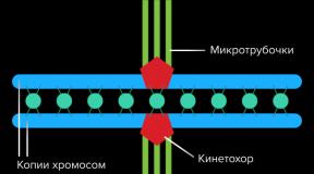
During mitosis, microtubules grow from centrosomes located at opposite ends of the cell and also attach to the centromere in specialized structures called kinetochores, one of which is present on each sister chromatid. A special DNA base sequence in the region of kinetochores, together with special proteins, ensures long-term attachment to this region. The microtubules then pull the chromatids towards the centrosomes so that each daughter cell inherits one set of chromatids. When the cells divide, the chromatids unwind and the DNA can be transcribed again. Despite their appearance, chromosomes are structurally highly compacted, which allows these giant DNA structures to fit into cell nuclei.
human chromosomes
Chromosomes in humans can be divided into two types: autosomes and sex chromosomes. Certain genetic traits are associated with a person's sex and are transmitted through the sex chromosomes. Autosomes contain the rest of the genetic information that is inherited. All act in the same way during cell division. Human cells contain 23 pairs of chromosomes (22 pairs of autosomes and one pair of sex chromosomes), giving a total of 46 per cell. In addition to these, there are many hundreds of copies of the mitochondrial genome in human cells. Sequencing the human genome provided a lot of information about each chromosome. Below is a table that compiles statistics for chromosomes based on the Sanger Institute's human genome information in the VEGA (Vertebrate Genome Comments) database. The number of genes is a rough estimate, as it is partly based on gene prediction. The total length of chromosomes is also a rough estimate based on the estimated size of regions of inconsistent heterochromatins.
|
Chromosomes |
Genes |
Total number of complementary base pairs of nucleic acids |
Ordered complementary base pairs of nucleic acids |
|
X (sex chromosome) | |||
|
Y (sex chromosome) | |||
|
Total |
3079843747 |
2857698560 |
Number of chromosomes in different organisms
eukaryotes
These tables give the total number of chromosomes (including sex chromosomes) in the cell nuclei. For example, diploid human cells contain 22 different kinds of autosomes, each present in two copies, and two sex chromosomes. This gives 46 chromosomes in total. Other organisms have more than two copies of their chromosomes, such as hexaploid bread wheat contains six copies of seven different chromosomes, for a total of 42 chromosomes.
|
The number of chromosomes in some plants |
|
||||
|
plant species |
|
||||
|
Arabidopsis thaliana(diploid) |
|
||||
|
|
|||||
|
garden snail |
|
||||
|
Tibetan fox |
|
||||
|
domestic pig |
|
||||
|
laboratory rat |
|
||||
|
Syrian hamster |
|
||||
|
|
|||||
|
domestic sheep |
|
||||
|
|
|||||
|
|
|||||
|
Kingfisher |
|
||||
|
Silkworm |
|
||||
|
|
|
|
|||
|
Number of chromosomes in other organisms |
|||||
|
Kinds |
Large chromosomes |
Intermediate chromosomes |
microchromosomes |
||
|
Trypanosoma brucei | |||||
|
domestic pigeon ( Columba livia domestics) | |||||
|
2 sex chromosomes | |||||
|
|
|
|
|
|
|
Normal members of individual eukaryotic species have the same number of nuclear chromosomes (see table). Other eukaryotic chromosomes, that is, mitochondrial and plasmid-like small chromosomes, vary greatly in number, and there can be a thousand copies per cell.
Species with asexual reproduction have one set of chromosomes, the same as in the cells of the organism. However, asexual species can be haploid and diploid.
Sexually reproducing species have somatic cells (body cells) that are diploid, having two sets of chromosomes, one from the mother and one from the father. Gametes, the reproductive cells, are haploid [n]: they have one set of chromosomes. Gametes are obtained by meiosis of a diploid germline cell. During meiosis, the corresponding chromosomes of the father and mother can exchange small parts of each other (crossover), and thereby form new chromosomes that are not inherited only from one or the other parent. When male and female gametes combine (fertilization), a new diploid organism is formed.
Some animal and plant species are polyploid: they have more than two sets of homologous chromosomes. Agriculturally important plants, such as tobacco or wheat, are often polyploid compared to ancestral species. Wheat has a haploid number of seven chromosomes found in some cultivated plants as well as in wild ancestors. The more common pasta and bread wheats are polyploid, having 28 (tetraploid) and 42 (hexaploid) chromosomes, compared to 14 (diploid) chromosomes in wild wheat.
prokaryotes
Prokaryotic species as a whole have one copy of each major chromosome, but most cells can easily survive with multiple copies. For example, Buchnera, an aphid symbiont, has many copies of its chromosome, ranging from 10 to 400 copies per cell. However, in some large bacteria such as Epulopiscium fishelsoni, up to 100,000 copies of a chromosome may be present. The number of copies of plasmids and plasmid-like small chromosomes, as in eukaryotes, varies considerably. The number of plasmids in a cell is almost entirely determined by the rate of plasmid division - rapid division generates a high number of copies.
Karyotype
Generally karyotype is a characteristic chromosomal complement of eukaryotic species. The preparation and study of karyotypes is part of cytogenetics.
Although DNA duplication and transcription are highly standardized in eukaryotes, the same cannot be said for their karyotypes, which are usually highly variable. The types of chromosome number and their detailed organization may vary. In some cases, there can be significant variation between species. Often there is:
- fluctuation between the two sexes;
- fluctuation between the germ line and the soma (between the gametes and the rest of the organism);
- fluctuation between members of a population due to balanced genetic polymorphism;
- geographical fluctuation between races;
- mosaic or other abnormalities
Also, fluctuations in the karyotype can occur during development from a fertilized egg.
The technique for determining the karyotype is commonly referred to as karyotyping. Cells can be blocked partially through division (in metaphase) under artificial conditions (in a reaction tube) with colchicine. These cells are then stained, photographed and arranged into a karyogram, with a set of ordered chromosomes, autosomes in length order and sex chromosomes (here X/Y) at the end.
As with many sexually reproducing species, humans have special gonosomes (sex chromosomes, as opposed to autosomes). It is XX for women and XY for men.
Historical note
It took many years to study the human karyotype before the most basic question was answered: How many chromosomes are there in a normal diploid human cell? In 1912, Hans von Winivarter reported 47 chromosomes in spermatogonia and 48 in oogonia, including the XX/XO sex determination mechanism. Painter in 1922 was unsure about a person's diploid number - 46 or 48, initially leaning towards 46. He later revised his opinion from 46 to 48, and correctly insisted that a person has the XX/XY system.
To finally solve the problem, new techniques were needed:
- Use of cells in culture;
- Prepare cells in a hypotonic solution where they swell and spread chromosomes;
- Delay of mitosis in metaphase with a solution of colchicine;
- Crushing the drug on the subject holder, stimulating the chromosomes in a single plane;
- Slicing the micrograph and arranging the results in an irrefutable karyogram.
It was not until 1954 that the human diploid number of 46 was confirmed. Given the techniques of Winivarter and Painter, their results were quite remarkable. The chimpanzee (the closest living relative of modern humans) has 48 chromosomes.
Delusions
Chromosomal abnormalities are disruptions in the normal chromosomal content of a cell and are a major cause of genetic conditions in humans such as Down syndrome, although most abnormalities have little or no effect. Some chromosomal disorders do not cause disease in carriers, such as translocations or chromosomal inversions, although they may lead to an increased chance of having a child with a chromosome disorder. An abnormal number of chromosomes or sets of chromosomes, called aneuploidy, can be lethal or give rise to genetic disorders. Families that may carry a chromosomal rearrangement are offered genetic counseling.
The gain or loss of DNA from chromosomes can lead to a variety of genetic disorders. Examples among people:
- Cat cry syndrome, caused by the division of part of the short arm of chromosome 5. The condition is so named because affected children make high-pitched, cat-like cries. People affected by this syndrome have wide-set eyes, a small head and jaw, moderate to severe mental health problems, and short stature.
- Down syndrome, the most common trisomy, is usually caused by an extra copy of chromosome 21 (trisomy 21). Characteristic features include decreased muscle tone, a stocky build, asymmetrical cheekbones, slanted eyes, and mild to moderate developmental disabilities.
- Edwards syndrome, or trisomy 18, is the second most common trisomy. Symptoms include slowness of movement, developmental disorders, and numerous congenital anomalies that cause serious health problems. 90% of patients die in infancy. They are characterized by clenched fists and overlapping fingers.
- Isodicentric chromosome 15, also called idic(15), partial tetrasomy of the long arm of chromosome 15 or reverse duplication of chromosome 15 (inv dup 15).
- Jacobsen's syndrome occurs very rarely. It is also called a terminal deletion disorder of the long arm of chromosome 11. Those affected have normal intelligence or mild developmental disabilities, with poor speech skills. Most have a bleeding disorder called the Paris-Trousseau syndrome.
- Klinefelter syndrome (XXY). Men with Klinefelter's syndrome are usually sterile, usually taller, their arms and legs are longer than their peers. Boys with the syndrome are usually shy and quiet, and are more likely to have slow speech and dyslexia. Without testosterone treatment, some may develop gynecomastia during adolescence.
- Patau syndrome, also called D-syndrome or trisomy 13 chromosomes. The symptoms are somewhat similar to trisomy 18, without the characteristic folded hand.
- Small extra marker chromosome. This means the presence of an extra abnormal chromosome. The properties depend on the origin of the additional genetic material. Cat's eye syndrome and isodicentric chromosome 15 (or idic15) syndrome are caused by an extra marker chromosome, like Pallister-Killian syndrome.
- Triple X syndrome (XXX). XXX girls tend to be taller, thinner and more likely to have dyslexia.
- Turner syndrome (X instead of XX or XY). With Turner syndrome, female sexual characteristics are present, but underdeveloped. Women with Turner syndrome have a short torso, low forehead, abnormal development of the eyes and bones, and a concave chest.
- Syndrome XYY. XYY boys are usually taller than their siblings. Like XXY boys and XXX girls, they are more likely to have learning difficulties.
- Wolf Hirschhorn Syndrome, which is caused by partial destruction of the short arm of chromosome 4. It is characterized by severe growth retardation and serious mental health problems.
Chromosomes are threadlike molecules that carry hereditary information for everything from height to eye color. They are made from a protein and a single molecule of DNA, which contains the genetic instructions of an organism, passed down from its parents. In humans, animals, and plants, most chromosomes are located in pairs within the cell nucleus. Humans have 22 of these chromosome pairs, called autosomes.
Humans have 22 pairs of chromosomes and two sex chromosomes. Women have two X chromosomes; Men have an X chromosome and a Y chromosome.
How gender is determined
Humans have an extra pair of sex chromosomes, for a total of 46 chromosomes. The sex chromosomes are called X and Y, and their combination determines a person's sex. As a rule, women have two X chromosomes, and men have XY chromosomes. This XY sex determination system is found in most mammals, as well as some reptiles and plants.
The presence of XX or XY chromosomes is determined when the sperm fertilizes the egg. Unlike other cells in the body, the cells in the egg and sperm, called gametes or sex cells, have only one chromosome. Gametes are produced by meiotic cell division, which results in the split cells having half the number of chromosomes as parent or progenitors. In the case of humans, this means that the parent cells have two chromosomes and they have one gamete.
All gametes in the mother's eggs have X chromosomes. The father's sperm contains about half the X and half the Y chromosomes. Sperm is a variable factor in determining the sex of a child. If the sperm carries an X chromosome, it will combine with the egg's X chromosome to form a female zygote. If the sperm carries a Y chromosome, it will result in the birth of a boy.
During fertilization, the gametes from the sperm combine with the gametes from the egg to form a zygote. The zygote contains two sets of 23 chromosomes for the required 46. Most women are 46XX and most men are 46XY, according to the World Health Organization.
However, there are some options. Recent studies have shown that a person can have many different combinations of sex chromosomes and genes, especially those who self-identify as LGBT. For example, a certain X chromosome called Xq28 and a gene on chromosome 8 appear to be found in higher prevalence in gay men, according to a 2014 study in the journal Psychological Medicine.
A few out of a thousand babies are born with one sex chromosome (45X or 45Y), this is called monosomy. Others are born with three or more sex chromosomes (47XXX, 47XYY or 47XXY, etc.), this is called polysomy. “In addition, some men are born with 46XX due to translocation of a tiny part of the sex that defines the Y chromosome region,” the WHO says. “Similarly, some women are also born 46XY due to mutations on the Y chromosome. It is clear that not only are women who are XX, but men are XY, but rather there are a number of additions of chromosomes, hormonal balances and phenotypic variations.”
Structure of X and Y chromosomes
While the chromosomes for other parts of the body are the same size and shape, forming an identical pairing, the X and Y chromosomes have different structures.
The X chromosome is much longer than the Y chromosome and contains hundreds more genes. Because the extra genes on the X chromosome have no counterpart on the Y chromosome, the X genes are dominant. This means that almost any gene on X, even if it is recessive in a female, will be expressed in males. They are called X-linked genes. Genes found only on the Y chromosome are called Y-linked genes and are only expressed in males. Genes on any sex chromosome can be called sex genes.
There are approximately 1,098 X-linked genes, although most of them are not for female anatomical characteristics. In fact, many of them are associated with disorders such as hemophilia, Duchenne muscular dystrophy, and several others. They are most often found in men. Non-sex features of X-linked genes are also responsible for male pattern baldness.
Unlike the large X chromosome, the Y chromosome contains only 26 genes. Sixteen of these genes are responsible for maintaining cells. Nine are involved in sperm production, and if some of them are missing or defective, low sperm counts or infertility may occur. One gene, called the SRY gene, is responsible for male sex traits. The SRY gene triggers the activation and regulation of another gene found on the non-sex chromosome called Sox9. Sox9 triggers the development of non-sexual gonads into testicles instead of ovaries.
Sex chromosome disorders
Abnormalities in the combination of sex chromosomes can lead to various gender-specific conditions that are rarely fatal.
Female anomalies lead to Turner syndrome or Trisomy X. Turner syndrome occurs when women have only one X chromosome instead of two. Symptoms include failure of the genitals to reach normal maturity, which can lead to infertility, small breasts, and lack of menstruation; short stature; wide, thyroid chest; and wide neck.
Trisomy X syndrome is caused by three X chromosomes instead of two. Symptoms include tall stature, speech delays, premature ovarian failure or ovarian failure, and poor muscle tone—although many girls and women show no symptoms.
Klinefelter syndrome can affect men. Symptoms include breast development, abnormal proportions such as large hips, tall stature, infertility, and small testicles.
It has already been said above that in the nucleus of a cell, DNA molecules are located in special structures called chromosomes. Their study began over 100 years ago with the help of a conventional light microscope. By the end of the 19th century, something had become clear about the behavior of chromosomes in the process of cell division, and the idea was expressed of their participation in the transmission of heredity.
Chromosomes become visible under a microscope when a cell divides at a certain stage of the cell cycle called mitosis. Chromosomes in this state are compact rod-shaped structures of various lengths with a fairly constant thickness, most of the chromosomes have a constriction that divides the chromosome into two arms. In the region of the constriction, there is an important structure for doubling chromosomes, called centromere. When a cell divides during mitosis, a doubling of the number of chromosomes occurs, as a result of which both newly formed cells are ultimately provided with the same standard set of chromosomes.
Only in 1956, for the first time, Yu. Tio and A. Levan described the human chromosome set, determined the quantitative composition of chromosomes, and gave their general morphological characteristics. In fact, these works laid the foundation for the study of the structure of the human genome. In humans, each cell of the body contains 46 chromosomes, the physical lengths of which range from 1.5 to 10 microns (Fig. 7).
Rice. 7. Microscopic view of the complete set of chromosomes contained in the nucleus of each individual human cell
We remind the reader that the set of chromosomes in all human cells (with the exception of sex cells) is called diploid (double), since each of the chromosomes is represented by two copies (23 pairs in total). Each human somatic cell (except red blood cells) contains 2 complete sets of chromosomes. Each single (haploid) set contains 23 chromosomes - 22 ordinary chromosomes (autosomes) and one sex chromosome - X or Y. Thus, the genome of each individual person consists of 23 pairs of giant DNA molecules distributed in different chromosomes, and speaking about the human genome in general (men and women), then the total number of such molecules is 24. This is the first basic information that was obtained about the human genome during the analysis of chromosomes.
A study of the structure (size and shape) of human chromosomes showed that most of them resemble skittles in appearance, consisting of two thick parts (chromatids) and a thin constriction (centromere) between them. The similarity with skittles, and not with dumbbells, is that the centromere is most often located not in the center of the chromosome, but is shifted to one of its ends. Chromosome sizes vary greatly, with the shortest chromosome about ten times smaller than the longest. This is the second fundamentally important information about the structure of the human genome - the 24 DNA molecules that make it up are of different sizes.
If we compare the number and size of chromosomes in humans and in other types of organisms, we can see huge differences. For example, a cow, whose genome is about the size of a human, has 60 pairs of chromosomes. The clawed frog contains only 18 chromosomes, but even the smallest of them are larger than the largest human chromosomes. In birds, on the contrary, the number of chromosomes reaches 40 or more, and they are all very small in size. Thus, the diversity of chromosomes in nature is very large.
Using light microscopy, the sizes of all human chromosomes were determined. Then all non-sex chromosomes were numbered by decreasing size - from 1 to 22. The sex chromosomes were not assigned a number, but were named X and Y. As more accurate subsequent studies showed, chromosome 21 actually turned out to be slightly less than 22, but the chromosome numbering was not changed (so as not cause confusion). The difference in chromosome sets between men and women is that women have two sex X chromosomes (i.e., the chromosomes in all 23 pairs are the same), and in men, the male sex chromosome, Y, forms a pair with the X chromosome Each chromosome can be considered as a separate volume of a large twenty-four-volume collection of works called the Encyclopedia of Man.
Human germ cells, unlike the cells of the body of an adult organism (somatic cells), do not contain 2 sets of DNA text volumes, but only one. Before conception, each individual chromosome (a separate volume in the Encyclopedia of Man) of the father's sperm and mother's egg consists of various chapters of the DNA text of their parents mixed in different combinations. Any of the chromosomes that we received from our father formed in his testes shortly before we were conceived. Previously, in the entire history of mankind, exactly such a chromosome never existed. It was formed in the process of random mixing that occurs during division, gradually forming from sections of chromosomes of ancestors from the side of the father that combine with each other. The same is true of egg chromosomes, except that they are formed in our mother's body long before we are born (almost immediately after the birth of the mother herself).
In the zygote resulting from the fusion of sperm and egg, maternal and paternal genes are mixed and shuffled in different combinations. This happens as a result of the fact that chromosomes do not remain unchanged in generations - they interact with their randomly encountered pair, exchanging material with it. This ongoing process is called recombination. And the next generation often gets a hybrid chromosome - part from the grandfather and part from the grandmother. Further, in a series of generations, the paths of genes constantly intersect and diverge. As a result of the fusion of a unique egg with a unique sperm, a genome unique in all respects arises. And in this sense, we are all unique. Each human individual stores unique genetic information, consisting of a random combination of different gene variants.
A single gene can be viewed as a unit that continues to exist in a series of numerous generations. And in this sense, the gene is immortal! There is even such an original point of view that it is not the people themselves, but their genes that rule the world, and each specific living organism serves only as a temporary refuge for them. This not indisputable thought belongs to Richard Dawkins, author of The Selfish Gene. In his opinion, genes are practically immortal, unlike the living organisms in which they exist. Some genes are tens or even hundreds of millions of years old. Genes, to use Dawkins' terminology, do their best to survive. They adapt to heat and cold, choosing a better place for themselves, migrate with the help of a person and enter into new combinations. The man turned out to be a rather restless host. For thousands of years, it has traveled the world a lot, spreading its presence, influence and its filling - genes. (The inquisitive reader can get acquainted with the ideas and arguments of R. Dawkins in more detail in Appendix 1). This point of view is far from indisputable, and from the further presentation it will become clear to us that genes are not primarily egoists, but workaholics. There are genes - "watchmen" of the genome, genes - "janitors", genes - "cooks" and genes - "housekeepers". Providing their existence, they ensure the existence of us.
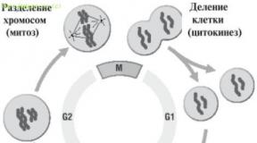
Immediately after conception, the future human is just one cell (zygote) endowed with one original DNA library containing 46 volumes. Among the 46 volumes, always 23 are received from the father, and the other 23 from the mother. The texts of the 23 paternal and 23 maternal volumes, although very similar in general, nevertheless differ in details. For example, in the paternal volume No. 18 on page 253 there is a sentence-prescription (in the form of a gene) that says that the eyes of the child should be brown, and in the same maternal volume on the same page it also says about the color of the eyes, but according to this text color should be blue. The first indication is more strict (dominant) than the second, and as a result, the child's eyes will have brown color. The gene that dictates its rights is called dominant, and the one who cedes his rights - recessive. Blue eyes are only those people who both in the maternal and paternal text contain recessive genes in which there is an indication of blue-eyedness. Then the zygote divides into two cells, each of them divides again, and so on until the appearance of billions of cells. Schematically, the process of cell division is shown in Fig. 8.
At each cell division, the volumes of DNA text contained in the libraries are accurately copied, and practically without errors. An adult human body consists of an average of 1014 cells. For example, there are about 10 billion cells in the brain and liver, and 300 billion cells in the immune system. Throughout a person's life, about 1016 cell divisions occur in his body. The cellular composition of many organs over 70 years of life is updated several times. And each of these cells contains the same 46 volumes of DNA text.
In the late 1960s, an important breakthrough was made in the study of chromosomes. It was due only to the fact that they began to use a special contrast agent - akrikhin-yperite, and then other compounds similar to it, for their coloring. This staining made it possible to reveal a large number of different substructures inside the chromosomes that could not be detected under a microscope without staining. After staining the chromosomes with a specific Giemsa-Romanovsky dye, they look like zebras: transverse light and dark stripes of different intensity are visible along the entire length.

Rice. 8. The main stages of the cell cycle leading to cell division
These bands are called chromosomal G-segments or bands (Fig. 9). The pattern of segmentation is very different for different chromosomes, but the location of chromosome segments is constant for each of the chromosomes in all types of human cells.
The nature of the bands revealed during coloring is not yet completely clear. Now it has only been established that the regions of the chromosomes corresponding to the dark bands (called R-bands) replicate earlier than the light regions (called G-bands). Thus, the banding of chromosomes most likely still has some not yet fully understood meaning.
Staining of chromosomes greatly facilitated their identification, and further contributed to determining the location of genes on them (gene mapping).

Rice. nine. Specific chromosomal G-segments detected by staining of human chromosomes, and the system for their designation according to the decision of the international conference in Paris in 1971. The numbers below the chromosomes indicate their numbers. X and Y - sex chromosomes, p - short arm, q - long arm of chromosomes
Although the detailed processes that occur during staining are not yet completely clear, it is obvious that the staining pattern depends on such a parameter as an increased or decreased content of AT chromosomes or GC pairs in individual bands. And this is another general information about the genome - it is not homogeneous, it has regions enriched with certain pairs of nucleotides.
This, in particular, may be due to the repeatability of certain types of DNA nucleotide sequences in certain regions.
Differential staining of chromosomes has been widely used to detect and identify small individual changes in the genome of a particular person ( polymorphism), in particular, leading to various pathologies. An example of this is the discovery of the so-called Philadelphia chromosome, found in patients with chronic myeloid leukemia. Using chromosome staining, it was found that in patients with this disease, a certain fragment disappears on chromosome 21 and appears at the end of the long arm of chromosome 9 (fragment transfer or translocation, abbreviated as t). Geneticists designate such an event as t (9; 21). Thus, chromosomal analysis indicates that different DNA molecules can exchange individual sections with each other, as a result of which “hybrids” are formed in the genome, consisting of DNA molecules from different chromosomes. An analysis of the already studied properties of chromosomes made it possible to form an idea of the polymorphism of the human genome.
To determine the localization of individual genes on chromosomes (i.e., gene mapping), a whole arsenal of special methods, often very complex in design and execution, is used. One of the main ones is molecular hybridization (formation of a hybrid) of a gene or its fragment with chromosome preparations fixed on a solid substrate, isolated from cells in pure form (this is called hybridization). in situ). The essence of the hybridization method in situ consists in the interaction (hybridization) between denatured (untwisted) DNA strands in chromosomes and complementary nucleotide sequences of chromosomes added to the preparation, individual single-stranded DNA or RNA (they are called probes). In the presence of complementarity between one of the strands of chromosomal DNA and the probe, rather stable molecular hybrids are formed between them. Probes are pre-labeled with different labels (radioactive, fluorescent, etc.). The sites of hybrid formation on chromosomes are identified by the position of these marks on chromosome preparations. So, even before the advent of genetic engineering and DNA sequencing, for example, the location in the human genome of genes encoding large and small ribosomal RNA (rRNA) was found out. The genes of the former turned out to be localized in five different human chromosomes (13, 14, 15, 21, and 22), while the bulk of the small rRNA genes ( 5S RNA) is concentrated in one place on the long arm of chromosome 1.
An example of a pattern obtained by hybridization of probe genes labeled with a fluorescent dye is shown in Fig. 10 on color sticker.

Rice. 10. Hybridization of human chromosomes with probe genes labeled with red and green fluorescent dyes. The arrows indicate the location of the corresponding genes at the ends of two different chromosomes (the picture of hybridizing chromosomes is enlarged at the top right).
Genes located on the same chromosome are defined as linked (linked) genes. If genes are located on different chromosomes, they are inherited independently (independent segregation). When the genes are on the same chromosome (i.e., linked), they are incapable of independent segregation. Occasionally, various changes in chromosomes can occur in germ cells as a result of recombination processes between homologous chromosomes. One of these processes is called crossing over. Due to crossing over, the linkage between genes of the same group is never complete. The closer the linked genes are to each other, the less likely it is that the location of such genes will change in children compared to their parents. The measurement of recombination frequency (crossing over) is used to establish the linear order of genes on a chromosome within a linkage group. Thus, when mapping chromosomes, it is initially established whether these genes are located on the same chromosome, without specifying which one. After at least one of the genes of a given linkage group is localized on a specific chromosome (for example, using hybridization in situ), it becomes clear that all other genes of this linkage group are on the same chromosome.
The first example of the connection of genes with certain chromosomes is the discovery of the linkage of certain inherited traits with the sex chromosomes. To prove the localization of the gene in the male sex Y-chromosome, it is enough to show that this trait is always found only in men and is never found in women. The linkage group of the female X-chromosome is uniquely characterized by the absence of inherited traits transmitted from father to son, and the inheritance of traits from the mother.
Particularly important for the study of the human genome in the early stages of its study was the method called hybridization of somatic cells. When somatic (non-sex) human cells are mixed with cells of other animal species (most often mice or Chinese hamster cells were used for this purpose), in the presence of certain agents, their nuclei can merge (hybridization). During the reproduction of such hybrid cells, some chromosomes are lost. Luckily for experimenters, human-mouse hybrid cells lose most of the human chromosomes. Next, hybrids are selected in which only one human chromosome remains. Studies of such hybrids made it possible to link some of the biochemical characteristics inherent in human cells with certain human chromosomes. Gradually, through the use of selective media, they learned to achieve the preservation or loss of individual human chromosomes that carry certain genes. The selection scheme, although not very simple at first glance, performed quite well in the experiment. So, they came up with a special selective medium on which only those cells in which the thymidine kinase enzyme is synthesized can survive. If mutant mouse cells that do not synthesize thymidine kinase are taken as a partner for hybridization with human cells, then only those hybrids that contain human chromosomes with the thymidine kinase gene will survive. In this way, for the first time, it was possible to establish the localization of the thymidine kinase gene on human chromosome 17.
Read also...
- How to drink Flemoxin before meals or after
- Why dream of levitation To soar in a dream in the air without wings: what does it mean
- Interesting facts about insurance (10 photos)
- "Yarina" or "Janine": which is better, composition of drugs, effectiveness, tips for choosing Change in the date of the onset of menstruation