How much does a child's head MRI cost? MRI for children: how and for what purpose it is performed. How harmful is tomography for a child’s health?
In general, MRI can provide clearer images of brain structures than ultrasound or CT, especially for detecting pathologies of the pituitary gland and brainstem.
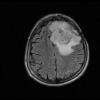
MRI of the brain allows you to detect various brain pathologies: congenital malformations, cystic formations, tumors, bleeding, edema, demyelinating diseases of the nervous system, inflammatory conditions, infections, vascular disorders of brain injury
MRI of the brain is prescribed for children to diagnose the cause of symptoms such as:
- Constant headaches
- Dizziness
- Muscle weakness
- Visual impairment
- Convulsions
- Movement disorders
- Loss of consciousness
Unlike adults, MRI examinations in children have their own characteristics. Small children cannot lie motionless inside the scanner for a long time, especially since during the procedure the scanner makes sounds that can frighten the child.
Therefore, young children are sedated before undergoing MRI to avoid image distortion associated with movement. It is recommended not to feed the child several hours before the procedure to avoid unwanted reactions to drug sedation or contrast injection. After sedation, the child will be connected to equipment to monitor their heart rate, breathing and oxygen levels. The child's parents may be with the child during the study. The effect of sedatives usually wears off within 1-2 hours.
Risks
MRI is absolutely harmless and can be performed more than once. The presence of metal in the body or electronic devices, impaired renal function, and anemia are contraindications for scanning. In addition, given that children often require sedation, consultation with an anesthesiologist is required before the scan.
Examples of MRI examinations

Children are also predisposed to the occurrence of various diseases associated with impaired brain activity. In order to detect a pathological condition, one of the most popular and highly informative techniques is used - magnetic resonance imaging. This method is highly accurate and allows you to detect even the smallest formations that are at the initial stage of their development.
Thanks to the use of contrast, the specialist determines not only the pathology itself, but also its location and the area covered. What are the benefits of MRI for children? Let's look at it in more detail in this article.
This procedure has a wide range of indications, so almost any brain pathology can be diagnosed using this method. MRI of the brain for children can be performed for preventive purposes and for the following pathologies:
- Systematic dizziness and headaches
- For fainting conditions
- Deterioration of visual functionality, decreased visual acuity and auditory dysfunction
- Seizures
- Inhibition of mental development
- Emotional instability (aggression, tearfulness)
- Head injuries, including minor concussions
This type of symptomatology should immediately predispose to carrying out this diagnostic measure. MRI of the brain for a child is most often used to confirm the diagnosis, namely diseases such as:
- Cyst and tumor formations
- Epilepsy
- Hemorrhage into the brain cavity
- Oxygen starvation
- Pituitary gland disease
- Multiple sclerosis

Preparation for the event
Magnetic resonance imaging is a universal and safe method of examining the entire body. Diagnostics are carried out to clarify the diagnosis, identify pathology and for preventive purposes. How is an MRI done on a child at 4 years old? Is it dangerous to do an MRI on a child’s brain? How are newborns and children under 7 years old examined? Let's look at it in the article.
Examination of children
At what age can a child have resonance tomography? This question worries many mothers whose babies suffered from birth injuries. There is no need to worry, since magnetic resonance radiation is not harmful to health. Babies can be examined both in the first year of life and at 5 years.
Children under five years of age are examined in their sleep. First, the baby is euthanized using a mask, then an anesthetic is injected into the body. This necessary maneuver is not caused by the patient’s age, but by his psychological state - a three-year-old child can be very frightened by the capsule of the device and the sounds it makes during operation.
For children aged 6 years, the procedure is performed without anesthesia, so parents should psychologically prepare the child for the peculiarities of tomography. The exception is children with a fear of closed spaces: in this case, the doctor decides what is best to do.
Modern tomographs are adapted for comfortable diagnostics; for example, there are open-type devices. They differ from capsule tomographs in their design - the patient lies on a couch, and a scanning box is located above him.
Designers are improving tomographs every year, and recently devices with feedback have appeared. The child lies with headphones on and can listen to a fairy tale or communicate with his parents, as well as watch a cartoon. In this case, the small patient experiences minimal discomfort during the procedure - he should lie still.
If the baby has passed the examination without anesthesia, the parents can immediately take him home. If anesthesia is given, the little patient is monitored until he fully awakens. Then the doctor monitors his condition, and only after that the parents take the baby home.
MRI of the brain
How are MRIs done for children? Is it dangerous to do an MRI of the head if the child is 2 years old? The impact of the magnetic field on the body is so small that it is not recorded as dangerous. After the procedure there are no complications on the nervous system or decreased immunity.
The tomograph scans the brain in longitudinal and transverse sections every 5 mm, then a computer program creates a three-dimensional image on the display. The doctor can monitor the condition of the organ and identify pathology even in its infancy.
In what cases do pediatricians prescribe brain MRI for children? The doctor will refer you for examination if the baby has disorders of the central nervous system, which is responsible for correct reflexes. This can be determined by the following condition:
- developmental delay;
- decreased vision/hearing;
- tendency to seizures;
- dizziness, fainting;
- other deviations in behavior.
The work of the brain coordinates all physiological processes in the body. If a pathology is detected, the child is urgently examined. It is necessary to examine the baby even after the first seizure appears - they can occur with an increase in body temperature. This condition may not be dangerous, or it may indicate a serious illness.
Causes of pathological abnormalities:
- birth injuries;
- intrauterine pathologies;
- penetration of infection;
- toxic effects;
- head injuries.
Children can be the victim of an accident - a fall or a strong blow. Parents are required to show their child to a doctor if this happens. Fainting and headaches can clearly indicate a concussion, especially if they are accompanied by vomiting. Children under 3 years old cannot tell anything, so mothers should be vigilant and immediately take their child to an appointment with a pediatrician.

How to prepare a baby from 3 years old for the examination procedure? If the child will undergo a tomography without anesthesia, preparation consists of removing metal objects from clothing. For children, crosses and rubber bands with metal connections must be removed; clothing should be loose and comfortable.
Note! Parents have the right to refuse resonance imaging in writing. However, a computer diagnostic (CT) examination is much more dangerous to health than an MRI.
If the child is prescribed anesthesia, you will have to refuse dinner the night before the procedure - anesthesia is administered on an empty stomach.
How can an MRI help? The examination determines:
- disorders of brain structures;
- presence/absence of tumors;
- anomalies in the structure of the skull;
- circulatory disorders;
- other pathologies.
These conditions are very dangerous to health, and it is better to identify them early. The earlier pathological changes are detected in children, the easier they are to correct.
Bottom line
How is an MRI performed on a 4 and 1 year old child? The baby is examined under anesthesia. It is completely safe for children at any age, even for newborns. Using computer processing of the resulting image, the doctor will be able to determine the pathology, if present, and prescribe an adequate course of therapy. Without preliminary diagnosis, it is impossible to make an accurate diagnosis.
Magnetic resonance imaging is a non-invasive medical diagnostic method that visualizes brain diseases. It is based on the effect of a magnetic field on hydrogen protons, which change their position. Magnetic resonance imaging produces a series of layered three-dimensional images of the brain in light and dark shades.
When magnetic tomographs first appeared, the procedure was allowed for children 7 years old and older. Later, when MRI began to be studied in more detail, scanning was allowed for all children, regardless of age.
MRI shows different brain pathologies in children:
- inflammation of infectious and non-infectious nature;
- hemorrhage;
- increased intracranial pressure;
- tumors and cysts;
- injuries and swelling.
Parents do not have to worry about harm from the procedure: the magnetic field does not cause damage to the baby’s body, unlike and.
MRI of the child’s brain is the most suitable option for accurately diagnosing diseases of the central nervous system for several reasons:
- Magnetic resonance imaging is a harmless and painless method. It does not require injections or incisions, which means the child will not worry or worry.
- The method is highly informative and sensitive to the least noticeable pathologies. This means that the method allows children to avoid serious illnesses by identifying pathology at an early stage of development.
- Unlike (ultrasound of the brain for a child 1 year or younger), where the doctor plays a decisive role in deciphering the results (human factor), the electronic computer of a magnetic tomograph deciphers most of the results independently, which means that the influence of the human factor is minimal. Pediatric MRI has a high diagnostic value compared to other neuroimaging methods.

Features of the event
- The child has a cochlear apparatus that has metal parts.
- There are severe heart pathologies in the decompensation stage.
- The body contains prosthetics or electronic devices.
Contrast agents are not administered to the child in the following cases:
- Hemolytic anemia.
- Intolerance to contrast components.
- Acute and chronic kidney and liver failure.
There is no danger to the patient's body from magnetic resonance imaging. You can perform diagnostics an unlimited number of times.
Is MRI with contrast harmful?
It is assumed that frequently performed MR examinations have a negative effect on the human body. In case of malignant neoplasms of the head, spinal column, other organs, parts, or joint lesions, scanning can be repeated (the frequency of procedures is determined by the attending physician). Duplicate sessions help the doctor monitor the restoration of a healthy state and study the activity of the disease.
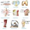
- Control of joint restoration after implantation of prosthetic elements;
- Post-traumatic consequences (fracture, formation of a false joint);
- Limitation of joint mobility;
- Articulation injury, an MRI of the joints is required;
- Joint defects identified by x-ray require an accurate examination to determine the damage;
- Constant pain, treatment does not help;
- The presence of inflammatory processes requiring MRI of the joints;
- Monitoring the status after the operation;
- Age-related disorders.
They resort to the use of contrast when undergoing MRI of the knee joint. Often, to detect spinal defects (displaced vertebral discs, tumors of various types), doctors advise doing a contrast-enhanced examination.
The substance enters the person through a vein. The drug is dispersed among tissues and vascular channels. To examine the brain with contrast tomography, agents consisting of gadolinium are usually used. The drug is not dangerous, improves the ability to analyze the state of the structural elements of the brain, the vascular system, and helps to clarify the structure of formations.
Contraindications to magnetic resonance imaging
Magnetic resonance imaging has limitations:
- The presence in the patient’s body of implanted metal objects, bodies of unknown origin, stimulating devices, devices that improve the perception of sound. Magnetic waves have the property of attraction, so objects will begin to move chaotically inside the body, which causes negative consequences and distorts the resulting images. There are metal structures that are not subject to the influence of the force field, consisting of ferromagnets, magnetic examination becomes possible (MRI with braces);
- Manifestation of hypersensitivity to the contrast agent. Before the session, patients are usually directed to do an allergy test;
- The presence of diseases that prevent MR diagnostics: fear of confined spaces, pathology of the cardiovascular system;
- A person who is intoxicated is not allowed to perform the scan;
- Representatives of the fairer sex do not undergo tomography during the initial stages of fruiting.
You need to be sure that MRI scanning is an absolutely safe research method in order to exclude unfounded mental disorders. Carrying out diagnostics without taking into account contraindications is a risky step.
How harmful is tomography for a child’s health?
Sending a child for magnetic resonance imaging raises concerns among parents due to the possibility of causing damage to the baby’s health.
Is MRI harmful? If necessary, an MRI examination is sent to study the state of the brain, internal parts, and musculoskeletal system. The procedure is absolutely safe. An MRI of the knee joint is often prescribed, since children often suffer injuries to the joints of the lower extremities.
During the scanning process, the presence of closed areas of the tomograph and the noise effect of the operating device can frighten a child. It is possible to remove negative emotions that bother the patient. Sedative drugs are used, through oral administration of the drug or through the substance through a vein. 
The diagnostic session lasts about half an hour. The introduction of a sedative will help the baby not to be nervous during the examination. The attending physician will receive high-quality images. The next day the patient feels absolutely normal.
Reviews from many parents who have had their children scanned disprove all possible concerns regarding the harmfulness of undergoing a nuclear magnetic examination. Emotional breakdowns are allowed to occur. Serious consequences in the form of the development of acute pathologies are absent in practice. It is unlikely to confirm that MRI is very dangerous.
Is MRI harmful for pregnant women?
Is MRI harmful to health during pregnancy? Magnetic tomography is not usually ordered until 12 weeks after conception. During the initial trimester, the embryo develops vital organs. The fetus is in the most vulnerable state to the negative influence of external factors. The absence of clearly defined indications requires that the procedure be performed later (after the first three months of pregnancy).
Magnetic resonance imaging is an ideal and completely non-harmful option for diagnostics. The obvious advantages of the method have been recognized, and the safety of MR imaging for the human body has been approved. The high price limits its applicability, so other types of diagnostic examinations operating on the principle of X-ray radiation continue to be in great demand.
Why is MRI of the spine harmful?
The high price and complexity of the equipment forces the procedure to be performed strictly according to indications: 
- Intervertebral hernia, pathological processes that contribute to disc bulging;
- Oncological processes.
During the procedure, a special couch with the patient constantly moves, and the scanning device reads the signals. Examination of a separate segment of the spinal column lasts about 30 minutes, the full procedure lasts about an hour. The treating doctor will receive an image showing the anomaly in various projections, and it will be possible to make a fairly accurate diagnosis and prescribe the correct treatment.
The method is based on the principle of nuclear magnetic resonance; the patient receives exposure to a powerful force field during MRI of the joints. The possible harmful effect of MR imaging on the human body has not been proven, therefore MR scanning is not dangerous and is considered a highly informative method of examination.
Health risks MRI of the brain
The reasons for prescribing the study can be: pain, loss of orientation in space, and other similar symptoms.
During scanning, a person is placed in a special tomograph capsule that creates powerful magnetic waves. Images of the inside of the head are generated through electromagnetic processes that do not have a harmful effect on the patient.
Side effects may occur due to the use of a contrast agent administered before the session. Therefore, before the procedure, specialists determine the possible presence of an allergic reaction to the components of the contrast amplifier.
On the site you can read reviews from patients who underwent brain research.